
Journal of Research in Dental
and Maxillofacial Sciences

Volume 11, Issue 3 (9-2026)
J Res Dent Maxillofac Sci 2026, 11(3): 213-219 |
Back to browse issues page

Ethics code: IR.IAU.DENTAL.REC.1403.089


Download citation:
BibTeX | RIS | EndNote | Medlars | ProCite | Reference Manager | RefWorks
Send citation to:


BibTeX | RIS | EndNote | Medlars | ProCite | Reference Manager | RefWorks
Send citation to:
Tehrani Z, Alamdari Mahd M, Nemati Anaraki S, Sanei E, Zahedirad S. Comparison of Discoloration of Primary Anterior Teeth Caused by Sideral Gocce versus Lipifer Liposomal Iron Drops: An In Vitro Study. J Res Dent Maxillofac Sci 2026; 11 (3) :213-219
URL: http://jrdms.dentaliau.ac.ir/article-1-1534-en.html
URL: http://jrdms.dentaliau.ac.ir/article-1-1534-en.html


1- Postgraduate Student, Department of Pediatric Dentistry, TeMS.C., Islamic Azad University, Tehran, Iran
2- Department of Pediatric Dentistry, TeMS.C., Islamic Azad University, Tehran, Iran
3- Dentist, Department of Restorative Dentistry, TeMS.c., Islamic Azad University, Tehran, Iran & Restorative Dentistry
4- Dentist, Department of Pediatric Dentistry, TeMS.c., Islamic Azad University, Tehran, Iran
5- Department of Pediatric Dentistry, TeMS.C., Islamic Azad University, Tehran, Iran ,zahedi@iautmu.ac.ir
2- Department of Pediatric Dentistry, TeMS.C., Islamic Azad University, Tehran, Iran
3- Dentist, Department of Restorative Dentistry, TeMS.c., Islamic Azad University, Tehran, Iran & Restorative Dentistry
4- Dentist, Department of Pediatric Dentistry, TeMS.c., Islamic Azad University, Tehran, Iran
5- Department of Pediatric Dentistry, TeMS.C., Islamic Azad University, Tehran, Iran ,
Full-Text [PDF 392 kb]
(3 Downloads)
| Abstract (HTML) (6 Views)
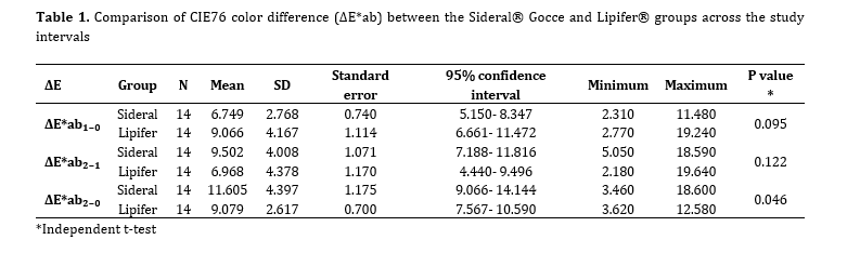
Table 1. Comparison of CIE76 color difference (ΔE*ab) between the Sideral® Gocce and Lipifer® groups across the study intervals
From the first week to the second week (ΔE*ab₂₋₁), color change remained comparable between Sideral® Gocce and Lipifer®. This stability is consistent with the findings of Tayebi et al, [18] which suggested that following initial exposure to liposomal iron supplements, subsequent discoloration might be observed. In contrast, Nazemisalman et al. [6] showed that various iron salts cause greater discoloration in later stages. This discrepancy may be attributed to the use of liposomal formulations in the present study, which exhibit lower destructive and staining effects compared with traditional iron salts.
Based on the reported perceptibility thresholds of approximately 1–2 ΔE*ab units and acceptability thresholds of approximately 3–4 ΔE*ab units for CIE L*a*b* color difference, the mean cumulative ΔE*ab₂₋₀ values in both groups were clearly perceptible and exceeded the acceptability threshold. Nevertheless, the Lipifer® group exhibited significantly less cumulative discoloration than the Sideral® Gocce group after 2 weeks relative to baseline (ΔE*ab₂₋₀). In line with this finding, Babaei et al. [11] demonstrated that liposomal formulations reduce staining via a physical barrier that limits direct iron-enamel contact. The superior performance of Lipifer® may be attributable to its nanoliposomal structure, which likely restricts rapid iron release and minimizes reactions with salivary polyphenols.
Inter-group differences demonstrated statistical significance only in the ΔE*ab₂₋₀ interval, with no significant differences in ΔE*ab₁₋₀ or ΔE*ab₂₋₁. This pattern reflects the cumulative mechanism of iron-induced staining, whereby repeated exposure facilitates progressive ion penetration and pigment formation within enamel [6, 11]. The absence of significant difference in the ΔE*ab₂₋₁ interval suggests uniform staining progression during this phase; however, cumulative assessment from baseline unmasked formulation-specific differences. Overall, these temporal dynamics align with established literature, confirming that iron-induced discoloration is time- and exposure-dependent.
The greater cumulative discoloration observed with Sideral® Gocce may reflect formulation-specific differences between its sucrosomial matrix and the nanoliposomal carrier of Lipifer®. Sucrosomial iron contains ferric pyrophosphate within a phospholipid-sucrose ester matrix, while the effectiveness of lipid-based carriers in retaining iron depends on factors such as particle size, phospholipid-to-iron ratio, encapsulation efficiency, and matrix stability [12, 19]. Repeated exposure may have increased matrix permeability or iron leakage from the sucrosomial formulation, allowing greater iron deposition on enamel, whereas more stable nanoencapsulation may have limited direct iron-enamel contact and discoloration [20].
The lower standard deviation in the Lipifer® group denotes greater uniformity in color outcome, indicating enhanced formulation stability. This contrasts with the findings of Tayebi et al. [18] which reported greater variability with conventional iron drops like Feroglobin. This disparity is likely attributable to the liposomal encapsulation common to both products in the current study.
In line with our findings, Vahedi et al. [2] compared discoloration from four iron supplements, including Sideral®, and reported that the liposomal formulation resulted in significantly less discoloration than ferrous sulfate. Additionally, a recent in vitro study by Nasha et al. [21] demonstrated that a protective enamel surface coating could significantly reduce staining caused by a ferrous sulfate supplement. This aligns with the protective principle of encapsulation technologies used in liposomal supplements, which also aim to shield the enamel from direct contact with free iron ions. While surface coatings represent a promising clinical intervention, their efficacy is dependent on proper application and durability, whereas the protective effect of an encapsulated formulation is intrinsic to the supplement itself [12, 13]. The significant reduction in staining observed with both strategies underscores the importance of preventing direct iron-enamel interaction, whether through advanced delivery systems or topical dental applications.
The present study holds remarkable implications for pediatric dentistry and iron supplementation by suggesting that liposomal iron formulations can mitigate discoloration in primary teeth. However, this study is subject to some limitations. The neutral-pH artificial saliva in the current study likely attenuated staining intensity, as acidic environments accelerate discoloration [7]. Moreover, the daily 3-hour exposure over 14 days may have been insufficient to reveal inter-group differences in all intervals, given that Tayebi et al. [18] observed greater staining with cumulative exposure exceeding 96 hours. Furthermore, the absence of protective enamel coatings may have amplified discoloration in the Sideral® Gocce group, potentially due to less effective iron encapsulation by its sucrosomial matrix compared with Lipifer®’s nanoliposomal system [22]. Another limitation of the present study was the absence of batch-specific measurements of the pH, viscosity, and baseline color of the two iron drops. Future research should employ in vivo designs, extended timelines, variable pH, and larger cohorts to refine these observations and optimize liposomal iron delivery.
Conclusion
The findings indicated that the nanoliposomal iron formulation (Lipifer®) induced less discoloration in primary anterior teeth than the sucrosomial model (Sideral® Gocce), particularly in the cumulative 2-week interval relative to baseline. This disparity may be owing to variations in liposomal design, such as lipid particle size or coating stability. No significant inter-group differences were observed in shorter intervals ΔE*ab₁₋₀ or ΔE*ab₂₋₁. Clinically, liposomal iron drops like Lipifer® and Sideral® Gocce can alleviate esthetic concerns associated with primary tooth staining in children.
Full-Text: (1 Views)
Abstract
Background and Aim: Iron deficiency anemia (IDA) is a prevalent nutritional disorder among children. Oral iron supplements are essential for prevention and treatment of IDA but often cause tooth discoloration. Therefore, this study aimed to compare the color change (ΔE*) induced by Sideral® Gocce and Lipifer® iron drops in extracted primary anterior teeth over a 2-week exposure period.
Materials and Methods: In this in vitro study, 28 sound extracted human primary anterior teeth were randomly divided into two groups (n=14) of Sideral® Gocce (sucrosomial iron) and Lipifer® (nanoliposomal iron). Baseline color was measured with a VITA Easyshade® Compact spectrophotometer using the CIE L*a*b* system. Teeth underwent daily immersion in the respective iron drop, followed by rinsing and storage in artificial saliva. Color assessments were repeated after the first and second weeks. ΔE was calculated using the CIE76 formula for the following intervals: baseline to week 1 (ΔE*ab₁₋₀), week 1 to week 2 (ΔE*ab₂₋₁), and baseline to week 2 (ΔE*ab₂₋₀). Data were analyzed with an independent t-test (alpha=0.05).
Results: No significant differences were found between the groups for ΔE*ab₁₋₀ (P=0.095) or ΔE*ab₂₋₁ (P=0.122). However, Lipifer® caused significantly less cumulative discoloration of primary teeth than Sideral® Gocce over the 2-week exposure period (P=0.046).
Conclusion: The results showed that under the present in vitro conditions, Lipifer® caused significantly less cumulative discoloration of primary anterior teeth than Sideral® Gocce over the 2-week exposure period. No significant differences were observed between the groups during the shorter assessment intervals.
Keywords: Iron compounds; Iron deficiencies; Liposomes; Tooth, Deciduous; Tooth discoloration
Introduction
Dark discoloration of primary anterior teeth due to oral iron supplementation is a frequent esthetic complaint among parents and pediatric dentists. Iron deficiency anemia (IDA) remains the leading nutritional disorder in early childhood [1, 2]. The prevalence of IDA in children under 5 years of age ranges from 18% to 38% in Iran [3]. Untreated IDA impairs cognitive, motor, and behavioral development, with affected children having lower IQ and motor performance than healthy ones [4, 5].
Oral iron supplementation has been widely used in healthcare programs as a key strategy for the prevention and treatment of IDA. Common formulations include ferrous sulphate, fumarate, gluconate, and ferric pyrophosphate [6]. One of the adverse effects of oral iron supplements is extrinsic tooth discoloration, which is primarily caused by the formation of colored iron-polyphenol complexes in saliva and subsequent deposition on the enamel surface. Furthermore, oxidation-reduction reactions between iron ions and these compounds are involved in the formation of dark stains [7]. Staining provokes parental anxiety and reduces treatment adherence [8]. Conversely, the neurodevelopmental sequelae of persistent IDA including impaired learning, growth retardation, and immune compromise, necessitate reliable supplementation [9]. Preventive strategies include diluting drops in vitamin C-rich juices, immediate rinsing, post-dose brushing, and adoption of advanced delivery systems [8].
The physicochemical properties of iron drops, such as pH, viscosity, and color, can influence the absorption of iron ions and their deposition on tooth surfaces. Highly soluble iron forms such as ferrous sulfate are more prone to causing dark stains. In contrast, compounds with lower solubility and a gradual release mechanism, such as ferric pyrophosphate, have a lower potential for discoloration [10, 11]. Liposomal technologies such as Sideral® Gocce (Junia Pharma, Italy) and Lipifer® (Kimia Razi, Iran) are claimed to minimize the potential for tooth discoloration due to their unique formulations [12, 13].
Lipifer® employs nanoliposomal iron encapsulation, whereas Sideral® Gocce utilizes sucrosomial technology which is a liposome-based matrix. Both formulations shield iron from direct enamel contact, thereby preventing discoloration. Furthermore, these coatings create an effective barrier against the reaction of iron ions with salivary compounds, while enhancing iron stability and systemic bioavailability [12, 13]. Despite their theoretical advantage, comparative data on liposomal iron drops and primary tooth discoloration remain limited [11]. Given the importance of high-quality domestic products as well as the esthetic outcomes, the present in vitro study compared CIE76 color change (ΔE*ab) induced by Sideral® Gocce and Lipifer® iron drops on extracted primary anterior teeth over a 2-week exposure protocol.
Materials and Methods
This in vitro study was conducted in 2024 at the Pediatric Dentistry Department of Islamic Azad University, Tehran, Iran (ethics code: IR.IAU.DENTAL.REC.1403.089). The sample size was determined using PASS 11 (two-sample t-test, α=0.05, β=0.2, expected ΔE*ab difference=3, standard deviation=2.7), yielding a minimum of 14 teeth per group [11]. A purposive sampling technique was used, with simple random assignment used to allocate teeth to the study groups.
Sample preparation:
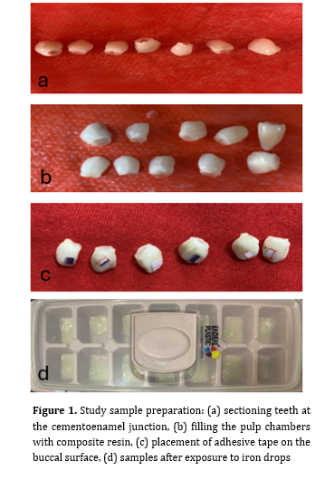
Twenty-eight sound human primary anterior teeth, extracted for orthodontic reasons, were collected. The inclusion criteria were intact buccal enamel without caries, restorations, intrinsic/extrinsic stains, or developmental defects. Teeth were examined under a stereomicroscope (×10 magnification) and stored in 10% formalin for 24 hours for disinfection [14]. Roots were sectioned at the cementoenamel junction, pulp tissue was removed, and pulp chambers were filled with composite resin (Filtek™ Z250; 3M ESPE, USA). A 5 × 5 mm window was created on the buccal surface using adhesive tape; surrounding enamel was coated with nail varnish, and the tape was removed (Figure 1) [11].
Figure 1. Study sample preparation: (a) sectioning teeth at the cementoenamel junction, (b) filling the pulp chambers with composite resin, (c) placement of adhesive tape on the buccal surface, (d) samples after exposure to iron drops
Baseline color measurement:
Teeth were randomly allocated to two groups, as follows:
Group 1 (n=14): Sideral® Gocce sucrosomial iron (Junia Pharma, Italy)
Group 2 (n=14): Lipifer® nanoliposomal iron (Kimia Razi, Iran)
Baseline color was measured using a VITA Easyshade® Compact spectrophotometer (VITA Zahnfabrik, Germany) calibrated per the manufacturer’s protocol (White LED D65, resolution: 25 nm, spectral range: 400-700 nm, ceramic standard calibration). The probe (VITA Zahnfabrik, Bad Säckingen, Germany) was positioned perpendicularly at the center of the buccal window. The spectrophotometer measured the L*, a*, and b* color coordinates of each specimen in the CIE L*a*b* color space at three measurement points. In this system, L* represents perceptual lightness, a* denotes the red-green chromatic spectrum, and b* indicates the yellow-blue chromatic spectrum [11].
During spectrophotometric assessment, each specimen was positioned against a standardized neutral gray background with 18% reflectance. The same background, specimen orientation, and probe position were maintained for all baseline, week-1, and week-2 measurements. The spectrophotometer was calibrated before each measurement session according to the manufacturer’s instructions.
Exposure protocol:
The samples were immersed daily in 10 mL of iron drop for 3 hours at 37°C, then rinsed with distilled water and stored in artificial saliva (1.5 mmol/L Ca, 0.9 mmol/L P, 150 mmol/L KCl, 0.05 mg F/mL, pH 7.0) until the next cycle [11,15]. The two-week regimen simulated chronic exposure.
Secondary color measurement:
After completion of the immersion period, the samples were rinsed with distilled water and air-dried. Color was reassessed using the VITA Easyshade® Compact spectrophotometer at weeks 1 and 2 in a blinded manner. For each tooth, ΔE was calculated using the CIE76 equation:
ΔE*ab = √[(ΔL*)² + (Δa*)² + (Δb*)²]
where ΔL*, Δa*, and Δb* represent the differences in the respective CIE L*a*b* coordinates between the two measurement time points. ΔE*ab was calculated for three intervals: baseline to week 1 (ΔE*ab₁₋₀), week 1 to week 2 (ΔE*ab₂₋₁), and baseline to week 2 (ΔE*ab₂₋₀).
Statistical analysis:
Data normality was assessed using the Shapiro-Wilk test. The ΔE*ab values calculated using the CIE76 equation were compared between the two groups using an independent-samples t-test. A significance level of α = 0.05 was used to determine statistical significance. Analyses were conducted using SPSS version 27.0 (IBM Corp., Armonk, NY, USA).
Results
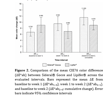
Table 1 presents descriptive statistics for ΔE across all intervals. No significant difference was observed between groups at week 1 (ΔE*ab₁₋₀; P= 0.095) or in the incremental change from week 1 to week 2 (ΔE*ab ₂₋₁; P= 0.122). However, cumulative discoloration over 2 weeks was significantly lower in the Lipifer® group than in the Sideral® Gocce group (ΔE*ab ₂₋₀; P=0.046). The lower standard deviation in the Lipifer® group at ΔE*ab₂₋₀ indicated greater color stability (Figure 2).
Background and Aim: Iron deficiency anemia (IDA) is a prevalent nutritional disorder among children. Oral iron supplements are essential for prevention and treatment of IDA but often cause tooth discoloration. Therefore, this study aimed to compare the color change (ΔE*) induced by Sideral® Gocce and Lipifer® iron drops in extracted primary anterior teeth over a 2-week exposure period.
Materials and Methods: In this in vitro study, 28 sound extracted human primary anterior teeth were randomly divided into two groups (n=14) of Sideral® Gocce (sucrosomial iron) and Lipifer® (nanoliposomal iron). Baseline color was measured with a VITA Easyshade® Compact spectrophotometer using the CIE L*a*b* system. Teeth underwent daily immersion in the respective iron drop, followed by rinsing and storage in artificial saliva. Color assessments were repeated after the first and second weeks. ΔE was calculated using the CIE76 formula for the following intervals: baseline to week 1 (ΔE*ab₁₋₀), week 1 to week 2 (ΔE*ab₂₋₁), and baseline to week 2 (ΔE*ab₂₋₀). Data were analyzed with an independent t-test (alpha=0.05).
Results: No significant differences were found between the groups for ΔE*ab₁₋₀ (P=0.095) or ΔE*ab₂₋₁ (P=0.122). However, Lipifer® caused significantly less cumulative discoloration of primary teeth than Sideral® Gocce over the 2-week exposure period (P=0.046).
Conclusion: The results showed that under the present in vitro conditions, Lipifer® caused significantly less cumulative discoloration of primary anterior teeth than Sideral® Gocce over the 2-week exposure period. No significant differences were observed between the groups during the shorter assessment intervals.
Keywords: Iron compounds; Iron deficiencies; Liposomes; Tooth, Deciduous; Tooth discoloration
Introduction
Dark discoloration of primary anterior teeth due to oral iron supplementation is a frequent esthetic complaint among parents and pediatric dentists. Iron deficiency anemia (IDA) remains the leading nutritional disorder in early childhood [1, 2]. The prevalence of IDA in children under 5 years of age ranges from 18% to 38% in Iran [3]. Untreated IDA impairs cognitive, motor, and behavioral development, with affected children having lower IQ and motor performance than healthy ones [4, 5].
Oral iron supplementation has been widely used in healthcare programs as a key strategy for the prevention and treatment of IDA. Common formulations include ferrous sulphate, fumarate, gluconate, and ferric pyrophosphate [6]. One of the adverse effects of oral iron supplements is extrinsic tooth discoloration, which is primarily caused by the formation of colored iron-polyphenol complexes in saliva and subsequent deposition on the enamel surface. Furthermore, oxidation-reduction reactions between iron ions and these compounds are involved in the formation of dark stains [7]. Staining provokes parental anxiety and reduces treatment adherence [8]. Conversely, the neurodevelopmental sequelae of persistent IDA including impaired learning, growth retardation, and immune compromise, necessitate reliable supplementation [9]. Preventive strategies include diluting drops in vitamin C-rich juices, immediate rinsing, post-dose brushing, and adoption of advanced delivery systems [8].
The physicochemical properties of iron drops, such as pH, viscosity, and color, can influence the absorption of iron ions and their deposition on tooth surfaces. Highly soluble iron forms such as ferrous sulfate are more prone to causing dark stains. In contrast, compounds with lower solubility and a gradual release mechanism, such as ferric pyrophosphate, have a lower potential for discoloration [10, 11]. Liposomal technologies such as Sideral® Gocce (Junia Pharma, Italy) and Lipifer® (Kimia Razi, Iran) are claimed to minimize the potential for tooth discoloration due to their unique formulations [12, 13].
Lipifer® employs nanoliposomal iron encapsulation, whereas Sideral® Gocce utilizes sucrosomial technology which is a liposome-based matrix. Both formulations shield iron from direct enamel contact, thereby preventing discoloration. Furthermore, these coatings create an effective barrier against the reaction of iron ions with salivary compounds, while enhancing iron stability and systemic bioavailability [12, 13]. Despite their theoretical advantage, comparative data on liposomal iron drops and primary tooth discoloration remain limited [11]. Given the importance of high-quality domestic products as well as the esthetic outcomes, the present in vitro study compared CIE76 color change (ΔE*ab) induced by Sideral® Gocce and Lipifer® iron drops on extracted primary anterior teeth over a 2-week exposure protocol.
Materials and Methods
This in vitro study was conducted in 2024 at the Pediatric Dentistry Department of Islamic Azad University, Tehran, Iran (ethics code: IR.IAU.DENTAL.REC.1403.089). The sample size was determined using PASS 11 (two-sample t-test, α=0.05, β=0.2, expected ΔE*ab difference=3, standard deviation=2.7), yielding a minimum of 14 teeth per group [11]. A purposive sampling technique was used, with simple random assignment used to allocate teeth to the study groups.
Sample preparation:
Twenty-eight sound human primary anterior teeth, extracted for orthodontic reasons, were collected. The inclusion criteria were intact buccal enamel without caries, restorations, intrinsic/extrinsic stains, or developmental defects. Teeth were examined under a stereomicroscope (×10 magnification) and stored in 10% formalin for 24 hours for disinfection [14]. Roots were sectioned at the cementoenamel junction, pulp tissue was removed, and pulp chambers were filled with composite resin (Filtek™ Z250; 3M ESPE, USA). A 5 × 5 mm window was created on the buccal surface using adhesive tape; surrounding enamel was coated with nail varnish, and the tape was removed (Figure 1) [11].
Figure 1. Study sample preparation: (a) sectioning teeth at the cementoenamel junction, (b) filling the pulp chambers with composite resin, (c) placement of adhesive tape on the buccal surface, (d) samples after exposure to iron drops
Baseline color measurement:
Teeth were randomly allocated to two groups, as follows:
Group 1 (n=14): Sideral® Gocce sucrosomial iron (Junia Pharma, Italy)
Group 2 (n=14): Lipifer® nanoliposomal iron (Kimia Razi, Iran)
Baseline color was measured using a VITA Easyshade® Compact spectrophotometer (VITA Zahnfabrik, Germany) calibrated per the manufacturer’s protocol (White LED D65, resolution: 25 nm, spectral range: 400-700 nm, ceramic standard calibration). The probe (VITA Zahnfabrik, Bad Säckingen, Germany) was positioned perpendicularly at the center of the buccal window. The spectrophotometer measured the L*, a*, and b* color coordinates of each specimen in the CIE L*a*b* color space at three measurement points. In this system, L* represents perceptual lightness, a* denotes the red-green chromatic spectrum, and b* indicates the yellow-blue chromatic spectrum [11].
During spectrophotometric assessment, each specimen was positioned against a standardized neutral gray background with 18% reflectance. The same background, specimen orientation, and probe position were maintained for all baseline, week-1, and week-2 measurements. The spectrophotometer was calibrated before each measurement session according to the manufacturer’s instructions.
Exposure protocol:
The samples were immersed daily in 10 mL of iron drop for 3 hours at 37°C, then rinsed with distilled water and stored in artificial saliva (1.5 mmol/L Ca, 0.9 mmol/L P, 150 mmol/L KCl, 0.05 mg F/mL, pH 7.0) until the next cycle [11,15]. The two-week regimen simulated chronic exposure.
Secondary color measurement:
After completion of the immersion period, the samples were rinsed with distilled water and air-dried. Color was reassessed using the VITA Easyshade® Compact spectrophotometer at weeks 1 and 2 in a blinded manner. For each tooth, ΔE was calculated using the CIE76 equation:
ΔE*ab = √[(ΔL*)² + (Δa*)² + (Δb*)²]
where ΔL*, Δa*, and Δb* represent the differences in the respective CIE L*a*b* coordinates between the two measurement time points. ΔE*ab was calculated for three intervals: baseline to week 1 (ΔE*ab₁₋₀), week 1 to week 2 (ΔE*ab₂₋₁), and baseline to week 2 (ΔE*ab₂₋₀).
Statistical analysis:
Data normality was assessed using the Shapiro-Wilk test. The ΔE*ab values calculated using the CIE76 equation were compared between the two groups using an independent-samples t-test. A significance level of α = 0.05 was used to determine statistical significance. Analyses were conducted using SPSS version 27.0 (IBM Corp., Armonk, NY, USA).
Results
Table 1 presents descriptive statistics for ΔE across all intervals. No significant difference was observed between groups at week 1 (ΔE*ab₁₋₀; P= 0.095) or in the incremental change from week 1 to week 2 (ΔE*ab ₂₋₁; P= 0.122). However, cumulative discoloration over 2 weeks was significantly lower in the Lipifer® group than in the Sideral® Gocce group (ΔE*ab ₂₋₀; P=0.046). The lower standard deviation in the Lipifer® group at ΔE*ab₂₋₀ indicated greater color stability (Figure 2).
Figure 2. Comparison of the mean CIE76 color difference (ΔE*ab) between Sideral® Gocce and Lipifer® across the evaluated intervals. Bars represent the mean ΔE from baseline to week 1 (ΔE*ab₁₋₀), week 1 to week 2 (ΔE*ab₂₋₁), and baseline to week 2 (ΔE*ab₂₋₀; cumulative change). Error bars indicate 95% confidence intervals
Discussion
The present study compared discoloration caused by Sideral® Gocce and Lipifer® iron drops in primary anterior teeth under in vitro conditions. Tooth discoloration due to iron supplementation is a clinically relevant side effect in pediatric patients, often raising parental concern and compromising treatment adherence. The growing adoption of advanced liposomal delivery systems aims to mitigate such adverse effects.
Pani et al. [16] reported enamel discoloration induced by ferric salts, which is in line with the present findings. Furthermore, Yilmaz et al. [17] also suggested that exposure to ferrous iron and liposomal iron supplements resulted in progressive tooth discoloration. Despite differences in the specific formulations tested, these studies confirm a fundamental tendency for iron-based compounds to cause staining.
The current findings suggested that, at the first week compared to baseline (ΔE*ab₁₋₀), the mean change in color was lower in the Sideral® Gocce group than in the Lipifer® group, but this difference was not statistically significant. This finding aligns with the results of Babaei et al. [11] who reported that initial exposure to liposomal iron drops may yield variable discoloration depending on pH and viscosity, without statistical significance. The neutral pH (6.75) of the artificial saliva used in this study has likely reduced early chemical interactions between iron and enamel, potentially explaining the lack of significance at this stage. Moreover, Yılmaz et al. [7] observed that pH values below 5.5 significantly exacerbate staining, whereas neutral conditions may suppress such reactions.
Discussion
The present study compared discoloration caused by Sideral® Gocce and Lipifer® iron drops in primary anterior teeth under in vitro conditions. Tooth discoloration due to iron supplementation is a clinically relevant side effect in pediatric patients, often raising parental concern and compromising treatment adherence. The growing adoption of advanced liposomal delivery systems aims to mitigate such adverse effects.
Pani et al. [16] reported enamel discoloration induced by ferric salts, which is in line with the present findings. Furthermore, Yilmaz et al. [17] also suggested that exposure to ferrous iron and liposomal iron supplements resulted in progressive tooth discoloration. Despite differences in the specific formulations tested, these studies confirm a fundamental tendency for iron-based compounds to cause staining.
The current findings suggested that, at the first week compared to baseline (ΔE*ab₁₋₀), the mean change in color was lower in the Sideral® Gocce group than in the Lipifer® group, but this difference was not statistically significant. This finding aligns with the results of Babaei et al. [11] who reported that initial exposure to liposomal iron drops may yield variable discoloration depending on pH and viscosity, without statistical significance. The neutral pH (6.75) of the artificial saliva used in this study has likely reduced early chemical interactions between iron and enamel, potentially explaining the lack of significance at this stage. Moreover, Yılmaz et al. [7] observed that pH values below 5.5 significantly exacerbate staining, whereas neutral conditions may suppress such reactions.
Table 1. Comparison of CIE76 color difference (ΔE*ab) between the Sideral® Gocce and Lipifer® groups across the study intervals
From the first week to the second week (ΔE*ab₂₋₁), color change remained comparable between Sideral® Gocce and Lipifer®. This stability is consistent with the findings of Tayebi et al, [18] which suggested that following initial exposure to liposomal iron supplements, subsequent discoloration might be observed. In contrast, Nazemisalman et al. [6] showed that various iron salts cause greater discoloration in later stages. This discrepancy may be attributed to the use of liposomal formulations in the present study, which exhibit lower destructive and staining effects compared with traditional iron salts.
Based on the reported perceptibility thresholds of approximately 1–2 ΔE*ab units and acceptability thresholds of approximately 3–4 ΔE*ab units for CIE L*a*b* color difference, the mean cumulative ΔE*ab₂₋₀ values in both groups were clearly perceptible and exceeded the acceptability threshold. Nevertheless, the Lipifer® group exhibited significantly less cumulative discoloration than the Sideral® Gocce group after 2 weeks relative to baseline (ΔE*ab₂₋₀). In line with this finding, Babaei et al. [11] demonstrated that liposomal formulations reduce staining via a physical barrier that limits direct iron-enamel contact. The superior performance of Lipifer® may be attributable to its nanoliposomal structure, which likely restricts rapid iron release and minimizes reactions with salivary polyphenols.
Inter-group differences demonstrated statistical significance only in the ΔE*ab₂₋₀ interval, with no significant differences in ΔE*ab₁₋₀ or ΔE*ab₂₋₁. This pattern reflects the cumulative mechanism of iron-induced staining, whereby repeated exposure facilitates progressive ion penetration and pigment formation within enamel [6, 11]. The absence of significant difference in the ΔE*ab₂₋₁ interval suggests uniform staining progression during this phase; however, cumulative assessment from baseline unmasked formulation-specific differences. Overall, these temporal dynamics align with established literature, confirming that iron-induced discoloration is time- and exposure-dependent.
The greater cumulative discoloration observed with Sideral® Gocce may reflect formulation-specific differences between its sucrosomial matrix and the nanoliposomal carrier of Lipifer®. Sucrosomial iron contains ferric pyrophosphate within a phospholipid-sucrose ester matrix, while the effectiveness of lipid-based carriers in retaining iron depends on factors such as particle size, phospholipid-to-iron ratio, encapsulation efficiency, and matrix stability [12, 19]. Repeated exposure may have increased matrix permeability or iron leakage from the sucrosomial formulation, allowing greater iron deposition on enamel, whereas more stable nanoencapsulation may have limited direct iron-enamel contact and discoloration [20].
The lower standard deviation in the Lipifer® group denotes greater uniformity in color outcome, indicating enhanced formulation stability. This contrasts with the findings of Tayebi et al. [18] which reported greater variability with conventional iron drops like Feroglobin. This disparity is likely attributable to the liposomal encapsulation common to both products in the current study.
In line with our findings, Vahedi et al. [2] compared discoloration from four iron supplements, including Sideral®, and reported that the liposomal formulation resulted in significantly less discoloration than ferrous sulfate. Additionally, a recent in vitro study by Nasha et al. [21] demonstrated that a protective enamel surface coating could significantly reduce staining caused by a ferrous sulfate supplement. This aligns with the protective principle of encapsulation technologies used in liposomal supplements, which also aim to shield the enamel from direct contact with free iron ions. While surface coatings represent a promising clinical intervention, their efficacy is dependent on proper application and durability, whereas the protective effect of an encapsulated formulation is intrinsic to the supplement itself [12, 13]. The significant reduction in staining observed with both strategies underscores the importance of preventing direct iron-enamel interaction, whether through advanced delivery systems or topical dental applications.
The present study holds remarkable implications for pediatric dentistry and iron supplementation by suggesting that liposomal iron formulations can mitigate discoloration in primary teeth. However, this study is subject to some limitations. The neutral-pH artificial saliva in the current study likely attenuated staining intensity, as acidic environments accelerate discoloration [7]. Moreover, the daily 3-hour exposure over 14 days may have been insufficient to reveal inter-group differences in all intervals, given that Tayebi et al. [18] observed greater staining with cumulative exposure exceeding 96 hours. Furthermore, the absence of protective enamel coatings may have amplified discoloration in the Sideral® Gocce group, potentially due to less effective iron encapsulation by its sucrosomial matrix compared with Lipifer®’s nanoliposomal system [22]. Another limitation of the present study was the absence of batch-specific measurements of the pH, viscosity, and baseline color of the two iron drops. Future research should employ in vivo designs, extended timelines, variable pH, and larger cohorts to refine these observations and optimize liposomal iron delivery.
Conclusion
The findings indicated that the nanoliposomal iron formulation (Lipifer®) induced less discoloration in primary anterior teeth than the sucrosomial model (Sideral® Gocce), particularly in the cumulative 2-week interval relative to baseline. This disparity may be owing to variations in liposomal design, such as lipid particle size or coating stability. No significant inter-group differences were observed in shorter intervals ΔE*ab₁₋₀ or ΔE*ab₂₋₁. Clinically, liposomal iron drops like Lipifer® and Sideral® Gocce can alleviate esthetic concerns associated with primary tooth staining in children.
Type of Study: Original article |
Subject:
pediatric
Send email to the article author
| Rights and permissions | |
 |
This work is licensed under a Creative Commons Attribution-NonCommercial 4.0 International License. |