Journal of Research in Dental
and Maxillofacial Sciences
Volume 10, Issue 4 (12-2025)
J Res Dent Maxillofac Sci 2025, 10(4): 285-291 |
Back to browse issues page

Ethics code: SRMIEC-ST0324-974
Download citation:
BibTeX | RIS | EndNote | Medlars | ProCite | Reference Manager | RefWorks
Send citation to:


BibTeX | RIS | EndNote | Medlars | ProCite | Reference Manager | RefWorks
Send citation to:
Aarasoor Cunniah L R, Nadarajan S, Narayanan V, Narayansamy D, Manjari C V S, Kolanji B. In Vitro Comparison of Morphological and Mechanical Properties, and Antimicrobial Efficacy of Silk Sutures Coated with Curcumin and Honey Extract. J Res Dent Maxillofac Sci 2025; 10 (4) :285-291
URL: http://jrdms.dentaliau.ac.ir/article-1-827-en.html
URL: http://jrdms.dentaliau.ac.ir/article-1-827-en.html
Lakshmi Rathan Aarasoor Cunniah *1 
 , Suryakala Nadarajan2 , Vivek Narayanan2 , Damodharan Narayansamy3 , C V Swapna Manjari4 , Bakyalakshmi Kolanji5
, Suryakala Nadarajan2 , Vivek Narayanan2 , Damodharan Narayansamy3 , C V Swapna Manjari4 , Bakyalakshmi Kolanji5
1- Department of Oral and Maxillofacial Surgery, SRM Kattankulathur Dental College & Hospital, SRM Institute of Science and Technology, SRM Nagar, Potheri, Chengalpattu district, Tamil Nadu State, India , lakshmir5@srmist.edu.in
2- Department of Oral and Maxillofacial Surgery, SRM Kattankulathur Dental College & Hospital, SRM Institute of Science and Technology, SRM Nagar, Potheri, Chengalpattu district, Tamil Nadu State, India
3- Department of Pharmaceutics, SRM College of Pharmacy, SRM Institute of Science and Technology, SRM Nagar, Potheri, Chengalpattu district, Tamil Nadu State, India
4- Department of Microbiology, SRM Medical College & Hospital, SRM Institute of Science and Technology, SRM Nagar, Potheri, Chengalpattu district, Tamil Nadu State, India
5- Department of Public Health Dentistry, Government Dental College & Hospital, Pudukottai
Keywords: Anti-Infective Agents, Plant Extracts, Infection Control, Microscopy, Electron, Scanning, Wound Healing
Full-Text [PDF 502 kb]
(17 Downloads)
| Abstract (HTML) (27 Views)
Full-Text: (7 Views)
Abstract
Background and Aim: Surgical sutures are necessary to achieve hemostasis at the surgical site and promote healing. Currently, antibiotic-coated sutures are used to inhibit bacterial adhesion and proliferation. However, they can lead to bacterial resistance. To overcome this issue, herbal-coated suture materials are employed. This study aimed to assess the morphological and mechanical properties and antimicrobial efficacy of plain silk sutures with suture material coated with curcumin and honey.
Materials and Methods: This in vitro study included three groups (n=3). Group A consisted of plain silk sutures, group B consisted of silk sutures coated with curcumin, and group C consisted of silk sutures coated with honey extract. Morphological assessment was performed using scanning electron microscopy (SEM) and energy dispersive spectroscopy (EDS). Tensile strength was measured using straight-pull and knot-pull tests on a tensile tester, and antimicrobial efficacy was evaluated using antimicrobial culture tests for the three groups. Statistical analysis was carried out using SPSS version 22.0.
Results: The morphology of coated sutures was validated by SEM images and EDS analysis. The tensile strength was not significantly different among the three groups, as assessed by the straight pull test (P=0.494) and knot pull test (P=0.707). Group A demonstrated no antibacterial activity. Group B demonstrated increased resistance to Pseudomonas aeruginosa (P. aeruginosa) (P=0.034), while Group C showed resistance to Escherichia coli (E. coli) (P=0.037).
Conclusion: The results showed that honey-coated sutures were highly resilient and had excellent antimicrobial properties. Curcumin-coated sutures expressed less resilience with better antimicrobial properties.
Keywords: Anti-Infective Agents; Plant Extracts; Infection Control; Microscopy, Electron, Scanning; Wound Healing
Introduction
Surgical sutures are essential for wound healing because they help in minimizing bleeding and reapproximating the tissues in minor oral surgical procedures [1]. Ideal suture materials should provide strong traction resistance, maintain dimensional stability, ensure secure knotting, and offer flexibility to protect the oral mucosa. Currently, antibiotic-coated sutures are widely used in minor oral surgical procedures with the goal of reducing bacterial adhesion and proliferation and preventing infection. However, a major limitation of antibiotic use is the development of bacterial resistance and increased pathogenic virulence [2,3]. As a result, researchers are now exploring alternative bioactive substances with antimicrobial properties, such as triclosan, chlorhexidine, silver nanoparticles, and, recently, aloe vera for suture coating [3,4]. Curcumin has proven to be a potent antimicrobial agent. As the main active compound in turmeric, curcumin is part of a group of substances called curcuminoids, which also include demethoxycurcumin and bisdemethoxycurcumin [5,6]. Curcumin is a multifaceted molecule known for its antibacterial, anti-inflammatory, antioxidant, antimicrobial, and wound-healing properties [7]. Similarly, honey, known for its bioactive compounds, has been thoroughly investigated for its medical applications [8]. This carbohydrate-rich syrup, made by honeybees from floral nectar, is highly effective as a dressing for both infected and non-infected wounds [9]. It aids in granulation, epithelialization, and necrotic tissue removal, accelerates healing, and reduces scarring [10]. Honey and curcumin are used in various minor oral procedures and also as an intra-socket dressing [8]. But there is no evidence of using honey and curcumin as a coating. Thus, this study was conducted aiming to evaluate the morphological and mechanical properties, and antimicrobial activity of plain silk sutures coated with curcumin and honey.
Materials and Methods
Materials and Methods
This study was initiated following institutional ethical committee approval (SRMIEC-ST0324-974). The study was carried out for a period of 6 months from July 2024 to December 2024 in SRM Kattankulathur Dental College & Hospital in Chengalpattu district, Tamil Nadu, India. The sample size was not calculated since it was a pilot study [11]. Hence, three sutures were evaluated in each group. The groups were as follows:
Group A: Plain silk sutures
Group B: Silk sutures coated with curcumin
Group C: Silk sutures coated with honey
Coating of plain silk sutures with curcumin extract and honey extract was carried out under aseptic conditions at SRM College of Pharmacy in 2024.
Suture material:
Non-absorbable black braided silk surgical sutures were used (Ethicon, 2 metrics 3-0; Johnson and Johnson Pvt. Ltd., India). These sutures were supplied in sterile packages and kept in room-temperature storage.
preparation of sutures coated with curcumin and honey:
Commercially available medical-grade curcumin was used in this study. To prepare the solution, 0.1 g of curcumin powder (Hyma Synthesis, Avra Group, India) was dissolved in 10 mL of ethanol (M/S Aasthaa Enterprises, India) and mixed thoroughly until completely dissolved. The coating was applied using the dip-coating technique. After dipping silk sutures in the curcumin solution for 2 minutes, they were dried in a hot air oven set at 50°C for 24 hours [11]. Similarly, medical-grade commercially available honey was used in this study; 10 mL of honey (Nature Nate's Honey & Co., Sweet Harvest Foods, USA) was warmed for 2 minutes before application. Silk sutures were then immersed in honey for 2 minutes and allowed to dry in sunlight for 24 hours [11].
Morphological assessment:
The surface morphology of the sutures was studied using scanning electron microscopy (SEM) and energy dispersive spectroscopy (EDS) [11].
Mechanical properties:
The tensile strength of sutures in the knot pull and straight pull tests was measured by a tensile tester (TKGEC2500; Balancing Instruments and Equipment Pvt. Ltd, India). A clamping pressure with a maximum load of 2500 N was applied after the suture material was cut to a length that went through both grip faces in order to shut the grips for the straight-pull test. In contrast, the knot pull test involved tying a knot in the middle of the suture, placing it against the grip faces, and applying pressure equal to the maximum load of 2500 N to close the grip [11] (Figure 1).
Figure 1. (a) Knot pull test by tensile tester; (b) Straight pull test by tensile tester
Assessment of antimicrobial activity:
Antibacterial activity was evaluated against Escherichia coli (E. coli) and Pseudomonas aeruginosa (P. aeruginosa) (these two are also commonly associated with hospital-acquired infections) [11]. Following preparation of peptone broth inoculums, each microorganism was inoculated separately and incubated at 37°C for 30 minutes. The cultures were then plated onto Mueller-Hinton agar. Following a 24-hour incubation period at 37°C, samples A, B, and C were examined to measure the diameter of the growth inhibition zones using a scale or a vernier caliper [4].
Statistical analysis:
SPSS version 22.0 was used to analyze the data. The Shapiro-Wilk and Kolmogorov-Smirnov tests were used to analyze the normality of data distribution [11]. For normally distributed data, the mean and standard deviation values were reported to characterize the quantitative data and were analyzed by ANOVA. For non-normally distributed data, the median and interquartile range were reported and analyzed by the non-parametric Kruskal-Wallis test. Statistical significance was defined as a P value of less than 0.05.
Results
Morphological assessment:
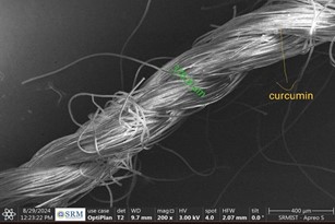
The SEM images of all three groups showed the characteristic multifilament structure of the suture. Group A consisting of plain silk sutures revealed normal multifilament structure of the suture. In group B, sutures had a curcumin deposition with the presence of zinc and sodium atoms as revealed by EDS elemental analysis (Figure 2). Similarly, group C containing honey showed an increase in the thickness of the suture material (Figure 3). Presence of iron and sodium atoms on sutures coated with honey was confirmed by EDS analysis.
Mechanical assessment:
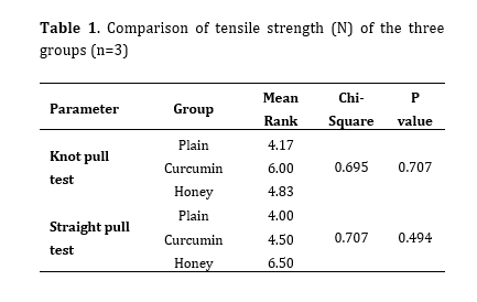
There was no statistically significant difference among the three groups in the knot pull (P=0.707) and straight pull (P=0.494) tests (Table 1).
Antimicrobial assessment:
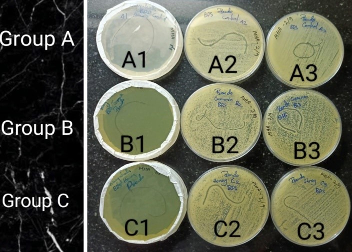
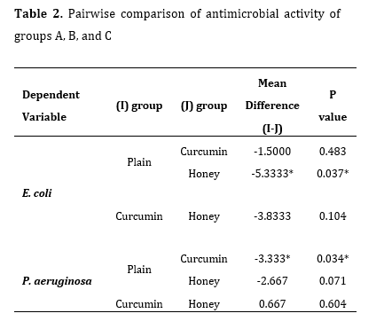
In P. aeruginosa culture, the diameter of the growth inhibition zone was 0 mm in group A, 1 mm, 4 mm, and 5 mm in the three samples of group B, and 1 mm, 3 mm, and 4 mm in the three samples of group C (Figure 4). There was no difference in antibacterial efficacy between the plain and honey groups (P=0.604), but a statistically significant difference was observed between the plain and curcumin groups (P=0.034) in P. aeruginosa culture (Table 2).
Figure 2. SEM image of group B (diameter=376.8 μm)
Figure 3. SEM image of group C (diameter=458.5 μm)
Table 1. Comparison of tensile strength (N) of the three groups (n=3)
Figure 4. Growth inhibition zones for P. aeruginosa
Table 2. Pairwise comparison of antimicrobial activity of groups A, B, and C
Similarly, in E. coli culture, the diameter of the growth inhibition zone was 0 mm in group A, 0.5 mm, 1 mm, and 3 mm in the three samples of group B, and 3 mm, 3 mm, and 10 mm in the three samples of group C. There was a statistically significant difference in antibacterial activity of the plain and honey groups (P=0.037), but not between the plain and curcumin groups (P=0.483) against E. Coli (Table 2).
Discussion
The SEM images of all three groups showed the characteristic multifilament structure of the suture. Group A consisting of plain silk sutures revealed normal multifilament structure of the suture. In group B, sutures had a curcumin deposition with the presence of zinc and sodium atoms as revealed by EDS elemental analysis (Figure 2). Similarly, group C containing honey showed an increase in the thickness of the suture material (Figure 3). Presence of iron and sodium atoms on sutures coated with honey was confirmed by EDS analysis.
Mechanical assessment:
There was no statistically significant difference among the three groups in the knot pull (P=0.707) and straight pull (P=0.494) tests (Table 1).
Antimicrobial assessment:
In P. aeruginosa culture, the diameter of the growth inhibition zone was 0 mm in group A, 1 mm, 4 mm, and 5 mm in the three samples of group B, and 1 mm, 3 mm, and 4 mm in the three samples of group C (Figure 4). There was no difference in antibacterial efficacy between the plain and honey groups (P=0.604), but a statistically significant difference was observed between the plain and curcumin groups (P=0.034) in P. aeruginosa culture (Table 2).
Figure 2. SEM image of group B (diameter=376.8 μm)
Figure 3. SEM image of group C (diameter=458.5 μm)
Table 1. Comparison of tensile strength (N) of the three groups (n=3)
Figure 4. Growth inhibition zones for P. aeruginosa
Table 2. Pairwise comparison of antimicrobial activity of groups A, B, and C
Similarly, in E. coli culture, the diameter of the growth inhibition zone was 0 mm in group A, 0.5 mm, 1 mm, and 3 mm in the three samples of group B, and 3 mm, 3 mm, and 10 mm in the three samples of group C. There was a statistically significant difference in antibacterial activity of the plain and honey groups (P=0.037), but not between the plain and curcumin groups (P=0.483) against E. Coli (Table 2).
Discussion
Most surgical procedures in the oral cavity require primary suturing to achieve hemostasis. Silk sutures are among the most commonly used materials for surgical closure [12]. Silk sutures are natural, multifilament, and non-absorbable, and are available in various sizes. The benefits of using silk sutures include their low cost, ease of use, and strong knot security. However, a major drawback of these materials is accumulation of debris, resulting in subsequent infection [13]. Evidence shows that multifilament sutures exhibit higher levels of microbiological adherence compared to monofilament sutures [1]. Saliva harbors averagely 7.5 x10⁸ CFUs/mL of microorganisms that surround the suture material applied in oral surgical procedures. Since these sutures are partially embedded in oral mucosa [14], extra caution should be used when handling them. As a result, bacteria continuously wick along the suture at the surgical site, prolonging the inflammatory response and requiring further intervention at the sutured site [15].
In order to reduce bacterial adhesion and colonization [16], a number of studies were carried out to produce an antimicrobial suture coated or impregnated with antibacterial agents. In the present study, silk sutures were coated with honey and curcumin extract. The advantages of curcumin include antibacterial, anti-inflammatory, antioxidant, antimicrobial, and wound-healing properties [17,18]. Likewise, honey promotes healing, reduces scarring, and helps with granulation, epithelialization, and elimination of necrotic tissue [19-21]. In this study, SEM analysis was performed to ensure uniform distribution of curcumin and honey extract coatings on suture material. Due to its gel-like nature, honey-coated sutures became thicker. Presence of atomic elements on coated sutures, such as calcium, iron, sodium, zinc, and potassium, was verified by EDS analysis, which confirmed that the suture material was coated with honey [11]. In mechanical assessment using straight pull test and knot pull test, honey-coated silk sutures showed better rigidity and elasticity compared to curcumin-coated and plain silk sutures. However, the difference was not statistically significant. In antimicrobial assessment, evaluation of the diameter of the growth inhibition zones caused by curcumin- and honey-coated suture materials revealed significant growth inhibition of the tested species. According to a study by Kumar [22], curcumin has strong antibacterial activity against Enterococcus faecalis. He demonstrated that 20% curcumin had significant antibacterial activity against Enterococcus faecalis. However, in the present study, group B (curcumin) showed antibacterial activity against P. aeruginosa with the largest size of growth inhibition zone equal to 5 mm. According to Ahmadi-Motamayel et al. [23], Atwa et al. [24], and English et al. [25], honey has antibacterial properties against common pathogenic bacteria such as Lactobacilli, Porphyromonas gingivalis, and Streptococcus mutans, and can even reduce the formation of dental plaque. According to Mandal et al. [26], honey has both bacteriostatic and bactericidal activity.
They reported that the concentration of honey required for growth prevention was 6.5% for E. coli and 7.5% for P. aeruginosa. In the present study, the honey-coated group expressed antibacterial activity against E. coli with a growth inhibition zone of maximally 10 mm. To our knowledge, this is the first study to utilize honey and curcumin extract to coat suture material. However, the main limitation of this study was its in vitro design and small sample size. Further clinical studies are required to assess biocompatibility of these coated suture materials in the oral cavity.
Conclusion
In order to reduce bacterial adhesion and colonization [16], a number of studies were carried out to produce an antimicrobial suture coated or impregnated with antibacterial agents. In the present study, silk sutures were coated with honey and curcumin extract. The advantages of curcumin include antibacterial, anti-inflammatory, antioxidant, antimicrobial, and wound-healing properties [17,18]. Likewise, honey promotes healing, reduces scarring, and helps with granulation, epithelialization, and elimination of necrotic tissue [19-21]. In this study, SEM analysis was performed to ensure uniform distribution of curcumin and honey extract coatings on suture material. Due to its gel-like nature, honey-coated sutures became thicker. Presence of atomic elements on coated sutures, such as calcium, iron, sodium, zinc, and potassium, was verified by EDS analysis, which confirmed that the suture material was coated with honey [11]. In mechanical assessment using straight pull test and knot pull test, honey-coated silk sutures showed better rigidity and elasticity compared to curcumin-coated and plain silk sutures. However, the difference was not statistically significant. In antimicrobial assessment, evaluation of the diameter of the growth inhibition zones caused by curcumin- and honey-coated suture materials revealed significant growth inhibition of the tested species. According to a study by
They reported that the concentration of honey required for growth prevention was 6.5% for E. coli and 7.5% for P. aeruginosa. In the present study, the honey-coated group expressed antibacterial activity against E. coli with a growth inhibition zone of maximally 10 mm. To our knowledge, this is the first study to utilize honey and curcumin extract to coat suture material. However, the main limitation of this study was its in vitro design and small sample size. Further clinical studies are required to assess biocompatibility of these coated suture materials in the oral cavity.
Conclusion
The results showed that honey-coated sutures were highly resilient and had excellent antimicrobial properties. Curcumin-coated sutures expressed less resilience with better antimicrobial properties.
Type of Study: Original article |
Subject:
Oral & maxillofacial surgery
References
1. Li Y, Meng Q, Chen S, Ling P, Kuss MA, Duan B, et al. Advances, challenges, and prospects for surgical suture materials. Acta Biomater. 2023 Sep 15;168:78-112. [DOI:10.1016/j.actbio.2023.07.041] [PMID]
2. Mankodi H. Performance of ecofriendly nano coated suture. Int J Sci Eng Res. 2015 Feb;6(2):268-72.
3. Ravishankar PL, Vijayan V, Rao SK, Vadivelu SA, Narayanaswamy D, Teja S. In Vitro Antibacterial Efficacy of Sutures Coated With Aloe vera and Ciprofloxacin: A Comparative Evaluation. J Pharm Bioallied Sci. 2019 May;11(Suppl 2):S164-8. [DOI:10.4103/JPBS.JPBS_284_18] [PMID] []
4. Kaur B, Sekhar V, Sharma P, Malhotra S, Jain A. A comparative assessment of antibacterial properties of neem oil coated sutures: An in vitro study. J Indian Soc Periodontol. 2023 Sep- Oct;27(5):487-91. [DOI:10.4103/jisp.jisp_475_22] [PMID] []
5. Hidayah RN, Santosaningsih D, Muti'ah R. Bioinformatics study of curcumin, demethoxycurcumin, bisdemethoxycurcumin and cyclocurcumin compounds in Curcuma longa as an antiviral agent via nucleocapsid on SARS- CoV-2 inhibition. InAIP Conference Proceedings 2024 Feb 6 (Vol. 3055, No. 1). AIP Publishing. [DOI:10.1063/5.0197724]
6. Kumar A, Chetia H, Sharma S, Kabiraj D, Talukdar NC, Bora U. Curcumin Resource Database. Database (Oxford). 2015 Jul 27;2015:bav070. [DOI:10.1093/database/bav070] [PMID] []
7. Sha AM, Garib BT. Antibacterial Effect of Curcumin against Clinically Isolated Porphyromonas gingivalis and Connective Tissue Reactions to Curcumin Gel in the Subcutaneous Tissue of Rats. Biomed Res Int. 2019 Sep 30;2019:6810936. [DOI:10.1155/2019/6810936] [PMID] []
8. Israili ZH. Antimicrobial properties of honey. Am J Ther. 2014 Jul-Aug;21(4):304-23. [DOI:10.1097/MJT.0b013e318293b09b] [PMID]
9. Passi D, Singh G, Dutta S, Sharma S, Mishra S, Gupta C. RETRACTED ARTICLE: Honey Extract as Medicament for
10. Treatment of Dry Socket: An Ancient Remedy Rediscovered- Case Series and Literature Review. J Maxillofac Oral Surg. 2016 Sep;15(3):345-8. [DOI:10.1007/s12663-014-0692-y] [PMID] []
11. Pleeging CC, Wagener FA, De Rooster H, Cremers NA. Revolutionizing non-conventional wound healing using honey by simultaneously targeting multiple molecular mechanisms. Drug Resist Updat. 2022 May;62:100834. [DOI:10.1016/j.drup.2022.100834] [PMID]
12. Ramani SA, Senthil SU, Rajaram VI, Kumari BN, Ravi NI, Mahendra JA, Namasivayam AM. Comparison of mechanical, antibacterial and morphological properties of silk sutures coated with silver nanoparticles and aloe Vera herbal extract: An in-vitro study. J Clin Diagn Res. 2023 Oct 1;17(10):ZC18-22. [DOI:10.7860/JCDR/2023/62524.18576]
13. Kandathil AM, Aslam SA, Abidha R, Cherian MP, Sudarsanan
14. M. Evaluation of microbial adherence on antibacterial suture materials during intraoral wound healing: a prospective comparative study. J Contemp Dent Pract. 2023 Sep 1;24(8):515- 20. [DOI:10.5005/jp-journals-10024-3553] [PMID]
15. Chua RA, Lim SK, Chee CF, Chin SP, Kiew LV, Sim KS, et al. Surgical site infection and development of antimicrobial sutures: a review. Eur Rev Med Pharmacol Sci. 2022 Feb;26(3):1010-21.
16. Banche G, Roana J, Mandras N, Amasio M, Gallesio C, Allizond V, et al. Microbial adherence on various intraoral suture materials in patients undergoing dental surgery. J Oral Maxillofac Surg. 2007 Aug;65(8):1503-7. [DOI:10.1016/j.joms.2006.10.066] [PMID]
17. Seydibeyog˘lu EA, Işık A. The effect of natural antimicrobial agents on the characteristics of surgical sutures. Dokuz Eylül Üniversitesi Mühendislik Fakültesi Fen ve Mühendislik Dergisi. 2020;22(64):11-20.
18. Li H, Wang Z, Robledo-Lara JA, He J, Huang Y, Cheng F. Antimicrobial surgical sutures: Fabrication and application of infection prevention and wound healing. Fibers Polym. 2021 Sep;22(9):2355-67. [DOI:10.1007/s12221-021-0026-x]
19. Wu X, Zheng X, Tang H, Zhao L, He C, Zou Y, et al. A network pharmacology approach to identify the mechanisms and molecular targets of curcumin against Alzheimer disease. Medicine. 2022 Aug 26;101(34):e30194. [DOI:10.1097/MD.0000000000030194] [PMID] []
20. Jurenka JS. Anti-inflammatory properties of curcumin, a major constituent of Curcuma longa: a review of preclinical and clinical research. Altern Med Rev. 2009 Jun;14(2):141-53.
21. Faraz A, Fernando WB, Williams M, Jayasena V. Effects of different processing methods on the antioxidant and antimicrobial properties of honey: a review. Int J Food Sci Technol. 2023 Jul;58(7):3489-501. [DOI:10.1111/ijfs.16460]
22. Pleeging CC, De Rooster H, Van Wijk B, Wagener FA, Cremers NA. Intra-socket application of medical-grade honey after tooth extraction attenuates inflammation and promotes healing in cats. J Feline Med Surg.2022 Dec;24(12):e618-27. [DOI:10.1177/1098612X221125772] [PMID] []
23. Abu-Mostafa N, Al-Daghamin S, Al-Anazi A, Al-Jumaah N, Alnesafi A. The influence of intra-alveolar application of honey versus Chlorhexidine rinse on the incidence of Alveolar Osteitis following molar teeth extraction. A randomized clinical parallel trial. J Clin Exp Dent. 2019 Oct 1;11(10):e871-e876. [DOI:10.4317/jced.55743] [PMID] []
24. Kumar H. An in vitro evaluation of the antimicrobial efficacy of Curcuma longa, Tachyspermum ammi, chlorhexidine gluconate, and calcium hydroxide on Enterococcus faecalis. J Conserv Dent. 2013 Mar-Apr;16(2):144-7. [DOI:10.4103/0972-0707.108197] [PMID] []
25. Ahmadi-Motamayel F, Hendi SS, Alikhani MY, Khamverdi Z. Antibacterial activity of honey on cariogenic bacteria J Dent (Tehran). 2013;10:10-5
26. Atwa AD, AbuShahba RY, Mostafa M, Hashem MI. Effect of honey in preventing gingivitis and dental caries in patients undergoing orthodontic treatment Saudi Dent J. 2014;26:108- 14 [DOI:10.1016/j.sdentj.2014.03.001] [PMID] []
27. English HK, Pack AR, Molan PC. The effects of Manuka honey on plaque and gingivitis: A pilot study J Int Acad Periodontol. 2004;6:63-7
28. Mandal S, DebMandal M, Pal NK, Saha K. Antibacterial activity of honey against clinical isolates of Escherichia coli, Pseudomonas aeruginosa and Salmonella enterica serovar Typhi. Asian Pac J Trop Med. 2010 Dec;3(12):961-4 [DOI:10.1016/S1995-7645(11)60009-6]
Send email to the article author
| Rights and permissions | |
 |
This work is licensed under a Creative Commons Attribution-NonCommercial 4.0 International License. |