

BibTeX | RIS | EndNote | Medlars | ProCite | Reference Manager | RefWorks
Send citation to:
URL: http://jrdms.dentaliau.ac.ir/article-1-184-en.html


2- Dentist
3- Assistant professor, Department of Orthodontics, Dental Branch of Tehran, ,
Abstract
Background and Aim: This study aimed to evaluate the effect of repeated bonding by self-etching primers (SEPs) and a conventional phosphoric acid-etchant on shear bond strength (SBS), adhesive remnant index (ARI), and enamel morphology at different debonding time points.
Materials and Methods: In this experimental study, 120 premolars were randomly divided into six groups of 20. In the first three groups, the brackets were bonded by Transbond XT, Transbond Plus, and Beauty Ortho Bond, and were debonded after 30 minutes. Adhesive remnants were removed from the enamel surface by a tungsten carbide bur. Rebonding was done with new brackets as described. The remaining three groups were debonded after aging. The SBS, ARI, and enamel surface morphology were evaluated. The SBS data were analyzed by two-way analysis of variance (ANOVA). The ARI scores were compared by using Mann-U-Whitney and Kruskal-Wallis tests.
Results: The SBS of Transbond XT in the first debonding was significantly higher than that of Transbond Plus. Transbond Plus showed a higher SBS than Beauty Ortho Bond. In the second debonding, the SBS values of Transbond XT and Transbond Plus were not significantly different, but their SBS values were significantly higher than that of Beauty Ortho Bond. SEPs showed a higher bond strength in the second bonding compared to the first bonding. Scanning electron microscopy (SEM) showed more porosity in the enamel surface before the second bonding compared to the first bonding. The SBS of Beauty Ortho Bond significantly decreased after aging, and SEM images showed a gap at the resin-enamel interface.
Conclusion: SEPs are recommended for secondary bonding in the clinical setting due to a decreased chair time, less damage to enamel, and an adequate bond strength.
Keywords: Dental Bonding, Orthodontic Brackets, Self-Etch Adhesive, Aging
Introduction
Bracket debonding from the tooth surface is a problem encountered by clinicians in orthodontic dentistry. This usually occurs as the result of inaccuracy in the process of bonding and due to inadequate moisture control by dentists. However, in some cases, this may be due to excessive occlusal forces. Bracket debonding can prolong the course of treatment, increase the chair time, and damage the dental enamel. (1-4) On the other hand, in many cases, orthodontists have to rebond the bracket in order to correct its position, especially in the preadjusted system where the correct position of brackets is the most important factor for treatment success. Thus, achieving an acceptably strong rebond between the bracket and tooth that has lost part of its fluoride-rich enamel due to primary bonding and removal of resin remnants is an important goal for clinicians. (5,6)
Since 1950, when etching was first introduced by Buonocore, great advancements have been made in the field of bonding. (7) Researchers seek to achieve an effective bond in the shortest time possible and with the least damage to the dental enamel. By the recent introduction of self-etching primers (SEPs) into the market, as an alternative to etch-and-rinse techniques, the post-etch rinsing and drying steps have been eliminated, and the etchant and the primer are used simultaneously in one combined step. This decreases the chair time for bracket bonding and reduces the risk of perioperative errors by clinicians. Studies have shown an adequately high bond strength achieved by the use of the mentioned technique. (8-10) In this method, iatrogenic damages to the enamel and its demineralization are prevented as well; (8-11) this is especially important in rebonding. Thus, self-etching adhesives are recommended for this purpose to save time and to prevent demineralization and loss of enamel. (12-14)
The bond strength and efficacy of SEPs have been the subject of numerous investigations; however, the success rate of rebonding by the use of self-etching adhesives is still questionable. Studies on this subject are scarce, and the available ones by Nicolas et al, (13) Montasser et al, (14) and Bishara et al (15) are controversial, indicating the need for further investigations. Bishara et al reported the rebond strength to be lower than that in primary bonding. (15) Montasser et al reported this rate to be equal to that in primary bonding, (14) while Nicolas et al found no significant difference between the two values. (13) Similar to the primary bond, a rebond has the risk of immediate failure after the bonding and placement of the wire in the bracket or late failure due to occlusal forces. In this study, we tried to simulate the clinical setting and to assess the rebond strength of brackets after 30 minutes and after aging (storage of specimens in artificial saliva at 37°C for 3 months and subsequent thermocycling). (16,17)
This study sought to assess the effect of rebonding with self-etching adhesives on the shear bond strength (SBS) of metal orthodontic brackets in 2016-2017.
Materials and Methods
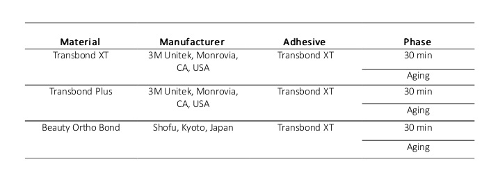
In this experimental study, 132 human premolars were evaluated. After extraction, the teeth were washed, immersed in 0.2% thymol solution for one week, and then, they were stored in normal saline until the experiment. In this study, 252 premolar brackets (standard edgewise, Discovery® Series, Dentaurum, Ispringen, Germany) were used, out of which, 132 were used for primary bonding, and 120 were used for rebonding. In the primary bond groups, two more teeth were included considering the risk of exclusion from the study. For rebonding, new brackets were used. The specimens were divided into three groups in such a way that three teeth extracted from the same patient were allocated to one group. One type of adhesive was used in each group. Table 1 summarizes the characteristics of the adhesives.
Table 1. Characteristics of the studied materials
In group 1, the brackets were bonded to the teeth by using Transbond XT (3M Unitek, Monrovia, CA, USA). The teeth were etched with 35% phosphoric acid (3M Unitek, Monrovia, CA, USA) for 15 seconds, rinsed for 20 seconds, and air-dried with oil- and moisture-free air drier until they gained a dull and frosty appearance. A thin coat of Transbond XT was applied to the enamel, which was gently air-dried from a 30-cm distance and was cured for 10 seconds. Transbond XT adhesive was applied to the bracket base, and the bracket was placed on the buccal surface of the tooth in the middle of the clinical crown, exactly on the height of contour. Care was taken to ensure the placement of the bracket slot perpendicular to the long axis of the tooth. The bracket was pressed on the tooth surface by using a gauge with a 300-g load and was light-cured for 40 seconds (10 seconds from each side) by using OptiLux 501 light-curing unit (Kerr, Orange, CA, USA) with 600 mW/cm2 intensity.
In group 2, Transbond Plus adhesive (3M Unitek, Monrovia, CA, USA) was used. The SEP was applied to the enamel surface and was gently air-dried from a 30-cm distance. The adhesive was applied to the bracket surface, the same as in group 1, and was light-cured.
In group 3, Beauty Ortho Bond adhesive (Shofu, Kyoto, Japan) was used. One drop of primer A was mixed with one drop of primer B and was applied to enamel surface for 3 seconds. The next steps were similar to those described for group 1. For correct mounting of teeth into cylindrical molds, a 0.18×0.25-mm stainless steel wire, parallel to the horizon and fixed at a specific height, was passed through the brackets, and then, the roots of the teeth (with brackets and wires on their surfaces) were mounted and fixed into a self-curing acrylic resin. The bracket slot was parallel to the horizon, and during the SBS testing, the load was applied perpendicular to the bracket base. The specimens in each group were divided into two subgroups of 22 each. The bond strength in one subgroup was measured after 30 minutes and in the other after aging. The specimens in the aging subgroup were stored in artificial saliva at 37°C for 3 months, and then, they were subjected to thermocycling for 6000 cycles between 5-55°C according to the method described by Faltermeier et al. (18) The SBS was measured in a Zwick universal testing machine (Instron M500, Ulm, Germany) with a 1-kN load cell attached to a metal rod at a crosshead speed of 0.5 mm/minute. The teeth were fixed on the base of the device, and the shear load was applied by the 30° beveled tip of the metal rod at the bracket-tooth interface along the incisogingival axis and parallel to the tooth surface until fracture (Figure 1). The amount of the applied load in Newton (N) was divided by the bracket’s cross-sectional area in mm2 (12.33) to calculate the bond strength in Megapascal (MPa).
Figure 1. Specimen under shear bond strength testing
The Adhesive Remnant Index (ARI) was evaluated according to the method described by Artun and Bergland (19) and by using a digital stereomicroscope (Motic DMW-143-Triple Output, Hong Kong) at ×10 magnification and was scored from 0 to 3 based on the amount of adhesive remnant on the enamel surface:
0: No adhesive remnant on the enamel surface.
1: Less than 50% resin on the enamel surface.
2: More than 50% resin on the enamel surface.
3: The entire adhesive left on the enamel surface. (19)
After debonding, adhesive remnants were removed from the enamel surface by using a carbide bur (Komet FGH 22 GK016, Germany) until no adhesive remnant was seen on the surface with the naked eye. Bonding in each group was repeated as before. In the subgroups that had undergone the primary bond strength measurement after 30 minutes, the second bond strength measurement was done after 30 minutes. The same was applied to the subgroups that underwent aging. The ARI was evaluated again as well.
Preparation of the specimens for scanning electron microscopy (SEM):
In order to observe the cross-section of the resin-enamel interface, one specimen from each group was mounted in acrylic resin, sectioned by a slow-speed diamond saw (IsoMet; Buehler, Lake Bluff, IL, USA) into occlusal and cervical halves, and then, it was sliced. During the next phase, the specimens were ground and stored in an incubator at 37°C for 10 days. All the specimens were gold plated and evaluated under an SEM at ×3000 magnification (Figure 2).
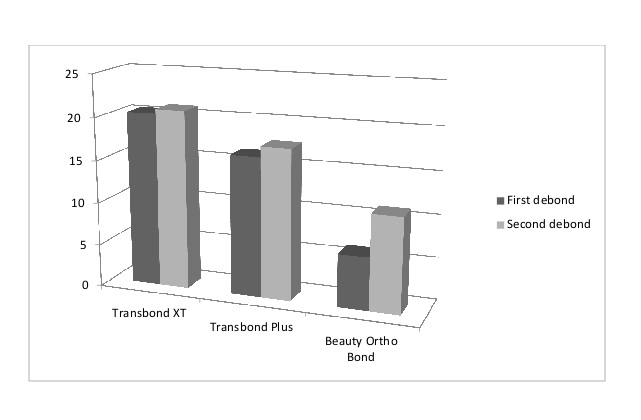
figure 2-Primary and Secondary shear bond strengths in the three group 30 minutes after bonding the orthodontic brackets.
For observation of the enamel surface after preparation, six premolars were used. The crowns were cut from the roots. The enamel surface was prepared with a phosphoric acid-etchant in the first specimen and with the two SEPs in the second and third specimens. Acetone was used for eliminating the primer in the SEPs. After debonding and removal of resin remnants with the carbide bur, the next three specimens were prepared the same as the first three specimens, were stored in an incubator for 10 days, and were observed under the SEM at ×2000 magnification (Figure 3).
Figure 3. Primary and secondary shear bond strengths in the three groups after aging
Statistical analysis:
Two-way analysis of variance (ANOVA) was used to assess the variables related to the adhesive system, the phases of debonding, and the time of debonding, which indicated significant interactions between the variables. Thus, one-way ANOVA was applied whenever required followed by Scheffe post-hoc test for multiple comparisons. Kruskal-Wallis test or Mann-U-Whitney and Dunn's tests, as a post-hoc test, were also applied for the comparison of the ARI and the mode of failure among the three adhesive groups, between the first and second debonding processes, and after 30 minutes and thermocycling.
Results
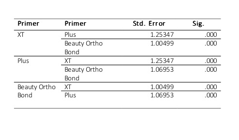
Univariate ANOVA revealed significant differences in the first debonding among the three adhesives and also between different debonding time points (P<0.05). However, the interaction of the adhesives and different time points was not significant (P=0.06), which means that the differences among the adhesives 30 minutes after debonding were similar to those after aging. As observed in Table 2, the post-hoc test demonstrated that in the primary bond strength testing, the SBS of Transbond XT was significantly higher than that of Transbond Plus. The SBS of Transbond Plus was also significantly higher than that of Beauty Ortho Bond.
Table 2. Comparison of the primary shear bond strength of the adhesives
Univariate ANOVA showed significant differences among the adhesives at different time points and in the interaction of the adhesives and the time points in the second debonding (P<0.05). The differences among the adhesives are shown in Table 3 (the post-hoc test). At 30 minutes after debonding, the bond strengths of Transbond XT and Transbond Plus were significantly higher than that of Beauty Ortho Bond, whereas the bond strengths of Transbond XT and Transbond Plus were not significantly different (P=0.208).
The situation was the same after thermocycling. However, it should be noted that the changes in the bond strengths of Transbond XT and Transbond Plus were not significant (P=0.995). However, the obtained values were more similar to one another compared to the values after 30 minutes; this can explain the significant interaction of the two variables.
Moreover, each adhesive was separately evaluated:
Transbond XT:
The secondary bond strength of Transbond XT was not significantly different from its primary bond strength (P=0.43). The interaction of the first and second bondings and the time points was not significant either (P=0.23). In other words, the secondary bond of Transbond XT was not significantly different from its primary bond after 30 minutes or after aging. The interaction with the time points was not significant either (P=0.26). The bond strength of Transbond XT was not significantly different after 30 minutes and after aging.
Transbond Plus:
The secondary bond strength of Transbond Plus was significantly greater than its primary bond strength (P<0.05). The interaction of the bonding phase and the time points was not significant (P=0.25). After 30 minutes and after aging, the secondary bond strength of Transbond Plus was higher than its primary bond strength. The interaction with the time points was not significant (P=0.59). The bond strength of Transbond Plus after aging was not significantly different from the value after 30 minutes. Thus, Transbond XT and Transbond Plus maintain their bond strength after aging.
Beauty Ortho Bond:
The secondary bond strength of Beauty Ortho Bond after 30 minutes was significantly higher than its primary bond strength; however, after aging, no significant difference was found between the primary and secondary bond strengths (both were too low).
The bond strength of Beauty Ortho Bond significantly decreased after thermocycling. Thus, for this adhesive, the effects of time (P<0.05) and debonding phase (P<0.05) and the interaction between them were significant (P<0.05).
Comparison of the variables of time and primer in the first and second bondings is shown in Figures 3 and 4.
ARI:
Dunn’s test demonstrated that in the primary and secondary bondings, the ARI in the Transbond XT group was significantly higher than that in the Beauty Ortho Bond group after 30 minutes and after aging (P<0.017). After aging, Transbond Plus showed a higher ARI than Beauty Ortho Bond (P<0.017). However, these two SEPs showed no significant difference 30 minutes after the first (P=0.06) and second (P=0.77) bondings.
The ARI was not significantly different in the first and second bondings and at different time points in the Transbond Plus and Transbond XT groups (P>0.017). Comparison of the 30-minute and post-aging time points in the three groups of adhesives with Mann-U-Whitney test showed no significant differences in this respect after the first and second bondings (P>0.017), which means that the amounts of adhesive remnants on the enamel were not significantly different after 30 minutes or post-aging.
The Wilcoxon test compared the first and second bonds and showed that the ARI of Transbond XT at two different time points was not significantly different between the first and second bonds (P=0.083). The ARI of Transbond Plus at 30 minutes (P=0.083) or post-aging (P=0.317) was not significantly different between the first and second bonds. However, in the Beauty Ortho Bond group, the ARI at 30 minutes after the second bonding was significantly higher than that at 30 minutes after the first bonding (P=0.008). This finding is in complete agreement with the bond strength test results and SEM images. However, after thermocycling, the bond strength of this adhesive was so weak. There was little adhesive remnant on the enamel surface after the first and second debondings, and the values were not significantly different (P=0.705).
Table 3. Comparison of the secondary shear bond strength of the adhesives at two time points
Discussion
Cleaning and preparation of enamel surface for new bracket bonding not only increase the clinician’s working time but also traumatize the enamel. Cleaning the composite remnants from enamel surface by the use of burs and rotary instruments results in the loss of 11.3-19.2 μm of the enamel surface. Re-etching leads to further loss of 10-50 μm of the enamel and changes the surface structure to the depth of 200 μm. (20,21) The shear rebond strength tests aim to improve the bonding system, decrease the damage to enamel, and reduce the clinical working time. For rebonding, in addition to a conventional adhesive (Transbond XT), two SEPs, namely Transbond Plus and Beauty Ortho Bond were used in this study. The latter has the ability to release and uptake fluoride. (22) For a comprehensive evaluation of these adhesives, aging was done before and after debonding of brackets.
The results showed that in primary bonding, Transbond XT with a separate etchant had a higher bond strength than Transbond Plus which is a SEP. Transbond Plus also showed a higher bond strength than Beauty Ortho Bond. These results are in accord with the findings of Aljubouri et al (23) and Grubisa et al (24) who believed that adhesives with separate etchants have a higher bond strength than SEPs. The authors believe that the higher bond strength of total-etch adhesives is attributed to the greater penetration of resin into the enamel demineralized through separate-etching. SEPs form a hybrid layer and decrease the penetration of resin tags due to the higher pH of the acid and simultaneous etching and priming. (23,24) These results are in contrast to those of Scougall Vilchis et al (25) and Iijima et al (9) who reported the bond strength of SEPs to be equal to that of conventional etching systems. These authors believe that SEPs are as effective as phosphoric acid for dissolving the enamel and that they create a shallower etched pattern. However, due to simultaneous etching and priming, the penetration of the primer is increased, which creates an excellent mechanical retention.
In comparison of the two SEPs, Transbond Plus showed a higher bond strength than Beauty Ortho Bond, which confirms the findings of Endo et al (22) and Iijima et al. (9) Although these two adhesives are both SEPs, as observed in SEM images, Beauty Ortho Bond adhesive, due to a higher pH than Transbond Plus, creates less demineralization and a smoother enamel surface, which justify its weak bond to enamel. However, it should be noted that it has a bond strength of 6 MPa, which is within the clinically acceptable range. (26,27)
Our study showed that the bond strength of Transbond XT adhesive was not significantly different in primary and secondary bondings. This result is in accord with the findings of Endo et al, (22) Nicolas et al, (13) and Grunheid and Larson, (28) and in contrast with the findings of Bishara et al who reported that there was a significant decrease in the SBS of Transbond XT between the debonding sequences 1 and 2. (29)
An interesting point is that Transbond Plus had a higher strength in the secondary bond than in the primary bond, equal to that of Transbond XT and the conventional bonding system. Beauty Ortho Bond had a lower bond strength compared to the mentioned two adhesives. However, its secondary bond strength was greater than its primary bond strength.
The intact enamel surface is rich in minerals and has a greater fluoride content than ground enamel. After tooth eruption, some changes occur in the enamel's outer surface, and the saliva saturated with calcium phosphate hypermineralizes the enamel. Fluoride ions convert hydroxyapatite to fluoroapatite. (30) Thus, the prismless enamel may prevent the penetration of SEPs, which may leave some enamel areas unetched, decreasing the penetration of resin into the microporosities of the intact enamel. (31) Resin tags are short and barely detectable. They are structurally incomplete. Since bond strength is due to micromechanical retention, a decreased resin penetration decreases the bond strength. (32) In a comprehensive study by Kanemura et al in 1999, SEPs showed greater penetration into prepared enamel compared to intact enamel. (33) By removing the superficial fluoride-rich enamel, the penetration of SEPs was enhanced, leading to their greater bond strength to a more porous surface. (33)
In 2010, Karan et al quantitatively evaluated the enamel roughness after debonding by an atomic force microscope (AFM) and showed that the use of a tungsten carbide bur for removal of adhesive remnants caused enamel surface roughness. (34) In our study, a low-speed tungsten carbide bur was used for removal of adhesive remnants. This bur also removes a small amount of fluoride-rich enamel in addition to adhesive remnants, enhancing the penetration of SEPs and increasing the secondary bond strength. This explains the higher secondary bond strength of Beauty Ortho Bond and Transbond Plus, which are both SEPs, compared to their primary bond strengths. This finding is in accord with that of Montasser et al. (14)
However, this finding is in contrast to that of Nicolas et al (13) and Endo et al (22) who reported that the secondary bond strength was not significantly different from the primary bond strength. Such differences in bond strength may be attributed to the technique of the operator and to the methodology of the study. Sample size may also play a role in this regard. Nicolas et al (13) used bovine teeth, which are different from human teeth.
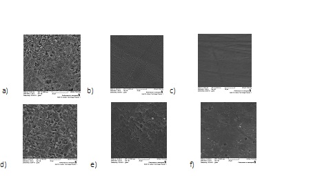
Figure 5 shows the SEM images of the enamel surface; a, b, and c show enamel surfaces before the first bond, while d, e, and f show them before the second bond. As expected and observed, the porosity of enamel was greater before the second bond because the enamel surface was ground by the carbide bur for removal of adhesive remnants. Figures 5a and 5d show the enamel surface after etching with 37% phosphoric acid (conventional technique). The clear etched pattern and honeycombing are evident. Figures 5b and 5e show the enamel surface structure after treatment with Transbond Plus SEP. The etched pattern is irregular and less prominent in the second bond compared to the first bond. Images 3c and 3f show the enamel surface treated with Beauty Ortho Bond. The surface is smooth without adequate retention, explaining a weak enamel bond.
Another factor influencing the bond strength is the adhesive remnants on the enamel surface. Resin remnants decrease the porosity and roughness of enamel surfaces (35,36) and can form a chemical bond with the new resin. (14) In our study, we cleaned the enamel surface from the remnants by the use of a carbide bur in such a way that no resin remnants were visible on the surface with the naked eye. In order to ensure the absence of resin remnants, the enamel surface after debonding was evaluated under the SEM by using silicon (Si) and carbon (C) mapping.
Figure 4. Evaluation of enamel surface with scanning electron microscopy (SEM) at ×2000 magnification. (a) Etched with phosphoric acid, (b) Treated with Transbond Plus, (c) Treated with Beauty Ortho Bond before the first bond. (d) Etched with phosphoric acid, (e) Treated with Transbond Plus, (f) Treated with Beauty Ortho Bond before the second bond.
Figure 5 shows that although resin remnants were removed by the carbide bur, some resin remnants were still present on the enamel surface. In Figure 2a, the etched enamel before bonding was searched for elements. The diagram only shows the presence of calcium (Ca) and phosphate (P). Figures 2b, 2c, and 2d illustrate the enamel surface after debonding of Transbond XT, Transbond Plus, and Beauty Ortho Bond, respectively, and after resin removal by the carbide bur. In all three groups, in addition to Ca and P, C and Si were found in different percentages; this indicates the presence of resin remnants which are invisible to the naked eye.
Figure 5. Searching for elements on the enamel surface by scanning electron microscopy (SEM) at ×2000 magnification. (a) Etched surface. (b) Enamel surface after the removal of Transbond XT resin by a carbide bur. (c) Enamel surface after the removal of Transbond Plus resin by a carbide bur. (d) Enamel surface after the removal of Beauty Ortho Bond resin by a carbide bur. Red points are Carbon and green points are Silicon.
In this study, in order to simulate the clinical setting, the bond strength was measured 30 minutes after bonding (the time of load application to brackets in the clinic) and after aging. Many adhesives undergo structural changes due to thermal alterations after exposure to the oral cavity. A comprehensive study should compare the bond strength while taking these conditions into account. (16,37,38)
The results of our study demonstrated that the bond strengths of Transbond Plus and Transbond XT, whether in the first or second bond, were not significantly different at 30 minutes and after aging. However, the bond strength of Beauty Ortho Bond significantly decreased after aging following both the first and second bonds. These results are in agreement with those of Yuasa et al in 2009 regarding Transbond XT and Transbond Plus. (16) Yuasa et al mentioned leakage at the enamel-adhesive interface following aging in the Beauty Ortho Bond group. (16)
The decreased bond strength in the Beauty Ortho Bond group after aging may be attributed to the hydrolysis and degradation of interface components. (39) Also, water sorption may weaken the polymer matrix. (40)
Figure 6 shows SEM images of the resin-enamel interface. The gap formed at the resin-enamel interface after aging in the Beauty Ortho Bond group can be seen following the first and second bonds, explaining the significant reduction in the bond strength.
Evaluation of the ARI:
In restorative dentistry, the highest bond strength to tooth structure is desirable; however, in orthodontics, the bond strength should be high enough to maintain the bracket on the tooth surface and at the same time has to be low enough to allow for easy cleaning of enamel following removal of brackets. (4,9,10)
The results of our study demonstrated that the adhesive remnants on the enamel after 30 minutes and after aging were significantly greater in the Transbond XT group than in the Beauty Ortho Bond group.
The weak bond of Beauty Ortho Bond adhesive leads to bond failure at the adhesive-enamel interface, whereas Transbond XT with separate etching forms a strong bond to enamel, and bond failure mostly occurs at the bracket-adhesive interface. (9,22) Moreover, based on the results, Transbond Plus and Beauty Ortho Bond were not significantly different in terms of the ARI at 30 minutes after debonding since they are both SEPs. However, after aging, the amount of adhesive remnants in the Beauty Ortho Bond group was insignificantly greater compared to that in the Transbond Plus group, indicating the weak bond and insufficient retention of Beauty Ortho Bond after aging.
Transbond XT and Transbond Plus were not significantly different in terms of adhesive remnants, and this finding is in agreement with the results of previous studies. (9,13,14,22,27) Transbond Plus is a SEP that forms a bond equal in strength to that of Transbond XT.
Transbond XT and Transbond Plus were not significantly different in terms of the ARI in comparing the first and second bonds; however, in the Beauty Ortho Bond group, at 30 minutes after debonding, the ARI following the second bond was greater than that after the first bond. This finding can be attributed to the increased enamel roughness due to resin removal by a carbide bur leading to a significant increase in the second bond strength. However, the Beauty Ortho Bond group showed no significant difference in the bond strength in comparing the two bonds after aging, and practically no adhesive was left on the enamel in the two groups after debonding.
Figure 6. Resin-enamel interface in the studied groups at ×3000 magnification.Note the gap at the resin-enamel interface in the Beauty Ortho Bond group in the
first and second bonds
Conclusion
SEPs are recommended for secondary bonding in the clinical setting due to a decreased chair time, less damage to enamel, and an adequate bond strength.
| Rights and permissions | |
 |
This work is licensed under a Creative Commons Attribution-NonCommercial 4.0 International License. |