
Journal of Research in Dental
and Maxillofacial Sciences

Volume 11, Issue 3 (9-2026)
J Res Dent Maxillofac Sci 2026, 11(3): 220-231 |
Back to browse issues page

Ethics code: IR.TUMS.DENTISTRY.REC.1404.086


Download citation:
BibTeX | RIS | EndNote | Medlars | ProCite | Reference Manager | RefWorks
Send citation to:


BibTeX | RIS | EndNote | Medlars | ProCite | Reference Manager | RefWorks
Send citation to:
Moshkbouy E, Mokhtari S, Baniebrahimi G, Davaie S, Kharazifard M J. Comparative Effects of Silver Diamine Fluoride and Self-Assembling Peptide on Microhardness, Qualitative Surface Morphology, and Remineralization of Incipient Enamel Lesions in Primary Teeth: An In Vitro Study. J Res Dent Maxillofac Sci 2026; 11 (3) :220-231
URL: http://jrdms.dentaliau.ac.ir/article-1-1570-en.html
URL: http://jrdms.dentaliau.ac.ir/article-1-1570-en.html
Elham Moshkbouy1 
 , Saeedeh Mokhtari1 , Ghazaleh Baniebrahimi *2 , Sotoudeh Davaie3 , Mohammad Javad Kharazifard4
, Saeedeh Mokhtari1 , Ghazaleh Baniebrahimi *2 , Sotoudeh Davaie3 , Mohammad Javad Kharazifard4
1- Department of Pediatric Dentistry, School of Dentistry, Tehran University of Medical Sciences, Tehran, Iran.
2- Department of Pediatric Dentistry, School of Dentistry, Tehran University of Medical Sciences, Tehran, Iran. ,ghazalebaniebrahimi@gmail.com
3- Department of Dental Materials, School of Dentistry, Tehran University of Medical Sciences, Tehran, Iran. & Research Center for Science and Technology in Medicine, Advanced Medical Technologies and Equipment Institute, Tehran University of Medical Sciences, Tehran, Iran.
4- Dental Research Center, Dentistry Research Institute, Tehran University of Medical Sciences, Tehran, Iran
2- Department of Pediatric Dentistry, School of Dentistry, Tehran University of Medical Sciences, Tehran, Iran. ,
3- Department of Dental Materials, School of Dentistry, Tehran University of Medical Sciences, Tehran, Iran. & Research Center for Science and Technology in Medicine, Advanced Medical Technologies and Equipment Institute, Tehran University of Medical Sciences, Tehran, Iran.
4- Dental Research Center, Dentistry Research Institute, Tehran University of Medical Sciences, Tehran, Iran
Keywords: Dental enamel, Hardness, Microscopy, Electron, Scanning, Silver Diamine Fluoride, Tooth, Deciduous, Tooth remineralization
Full-Text [PDF 737 kb]
(5 Downloads)
| Abstract (HTML) (15 Views)
Table 1. Mean microhardness values in the three groups at baseline, after demineralization, and after remineralization
Figure 1. ESEM micrographs of enamel surface at x500 magnification: (A) sound enamel; (B) demineralized enamel; (C) control group; (D) SAP group; (E) SDF group
Figure 2. ESEM micrographs of enamel surface at x2000 magnification: (A) sound enamel; (B) demineralized enamel; (C) control group; (D) SAP group; (E) SDF group
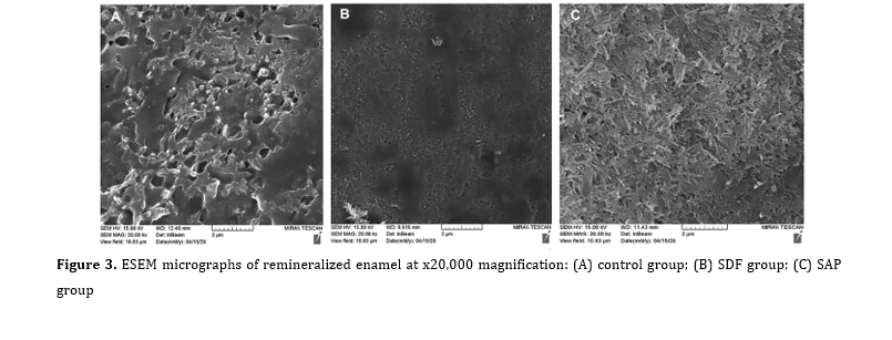
Figure 3. ESEM micrographs of remineralized enamel at x20,000 magnification: (A) control group; (B) SDF group; (C) SAP group
Discussion
Full-Text: (1 Views)
Abstract
Background and Aim: This study compared the effects of silver diamine fluoride (SDF) and self-assembling peptide (SAP) on microhardness, qualitative surface morphology, and remineralization of artificially induced incipient enamel lesions in primary teeth.
Materials and Methods: Sixty extracted primary molars were included in this in vitro study. Thirty-six teeth were selected for microhardness testing and exposed to a demineralizing solution for 36 hours to induce incipient enamel lesions. The samples were randomly assigned to SDF, SAP, and untreated control groups (n=12 each) and were subsequently stored in artificial saliva under pH-cycling conditions for 45 days. Vickers microhardness was recorded at baseline, after lesion formation, and after the remineralization period. Twenty-four other teeth underwent environmental scanning electron microscopy (ESEM) to examine their enamel surface morphology after the demineralization/remineralization cycles. Statistical analysis was performed with one-way ANOVA and a repeated-measures general linear model (alpha=0.05).
Results: Baseline microhardness did not differ significantly among the groups (P=0.564). Demineralization significantly reduced enamel microhardness in all groups, without a significant intergroup difference (P=0.778). Following remineralization, microhardness differed significantly among the groups; both SAP (P<0.001) and SDF (P=0.004) produced higher values than the control. Time (P<0.001) and interaction of time and group (P<0.001) significantly affected the microhardness. ESEM observations showed the most organized mineral/crystal reformation in the SAP-treated samples, followed by those treated with SDF.
Conclusion: In this in vitro study, SDF and SAP comparably improved the microhardness of demineralized primary enamel. ESEM findings suggested a more favorable structural reorganization and remineralization pattern after SAP application.
Keywords: Dental enamel; Hardness; Microscopy, Electron, Scanning; Silver Diamine Fluoride; Tooth, Deciduous; Tooth remineralization
Materials and Methods: Sixty extracted primary molars were included in this in vitro study. Thirty-six teeth were selected for microhardness testing and exposed to a demineralizing solution for 36 hours to induce incipient enamel lesions. The samples were randomly assigned to SDF, SAP, and untreated control groups (n=12 each) and were subsequently stored in artificial saliva under pH-cycling conditions for 45 days. Vickers microhardness was recorded at baseline, after lesion formation, and after the remineralization period. Twenty-four other teeth underwent environmental scanning electron microscopy (ESEM) to examine their enamel surface morphology after the demineralization/remineralization cycles. Statistical analysis was performed with one-way ANOVA and a repeated-measures general linear model (alpha=0.05).
Results: Baseline microhardness did not differ significantly among the groups (P=0.564). Demineralization significantly reduced enamel microhardness in all groups, without a significant intergroup difference (P=0.778). Following remineralization, microhardness differed significantly among the groups; both SAP (P<0.001) and SDF (P=0.004) produced higher values than the control. Time (P<0.001) and interaction of time and group (P<0.001) significantly affected the microhardness. ESEM observations showed the most organized mineral/crystal reformation in the SAP-treated samples, followed by those treated with SDF.
Conclusion: In this in vitro study, SDF and SAP comparably improved the microhardness of demineralized primary enamel. ESEM findings suggested a more favorable structural reorganization and remineralization pattern after SAP application.
Keywords: Dental enamel; Hardness; Microscopy, Electron, Scanning; Silver Diamine Fluoride; Tooth, Deciduous; Tooth remineralization
Introduction
Dental caries remains highly prevalent, particularly among socioeconomically disadvantaged children. If untreated, incipient enamel lesions of primary teeth may progress to dentinal and pulpal involvement, causing pain, infection, and impaired oral function [1]. Since primary teeth are essential for mastication, speech, esthetics, arch-space maintenance, and quality of life, contemporary caries management emphasizes minimally invasive approaches that promote lesion arrest and remineralization rather than restorative treatment alone [2-4].
Silver diamine fluoride (SDF) is a simple, inexpensive, and effective caries-control agent with combined antimicrobial and remineralizing actions. Its silver and fluoride ions inhibit cariogenic microorganisms, promote mineral deposition, and increase resistance to acid dissolution [5-9]. However, SDF may cause black discoloration of treated lesions and generally requires periodic reapplication [6,10].
Self-assembling peptides (SAPs), particularly P11-4, represent a biomimetic alternative for non-cavitated enamel lesions. By penetrating subsurface porosities and forming a three-dimensional scaffold, these peptides facilitate calcium-phosphate deposition and guided crystal nucleation [3,4,11-15]. Although SDF and SAP act through different mechanisms, direct comparative evidence in primary enamel remains limited. Therefore, this study compared their effects on microhardness, qualitative surface morphology, and remineralization of artificially induced incipient enamel lesions in primary teeth. The null hypothesis was that SDF and SAP would not significantly affect these outcomes.
Materials and Methods
Silver diamine fluoride (SDF) is a simple, inexpensive, and effective caries-control agent with combined antimicrobial and remineralizing actions. Its silver and fluoride ions inhibit cariogenic microorganisms, promote mineral deposition, and increase resistance to acid dissolution [5-9]. However, SDF may cause black discoloration of treated lesions and generally requires periodic reapplication [6,10].
Self-assembling peptides (SAPs), particularly P11-4, represent a biomimetic alternative for non-cavitated enamel lesions. By penetrating subsurface porosities and forming a three-dimensional scaffold, these peptides facilitate calcium-phosphate deposition and guided crystal nucleation [3,4,11-15]. Although SDF and SAP act through different mechanisms, direct comparative evidence in primary enamel remains limited. Therefore, this study compared their effects on microhardness, qualitative surface morphology, and remineralization of artificially induced incipient enamel lesions in primary teeth. The null hypothesis was that SDF and SAP would not significantly affect these outcomes.
Materials and Methods
This in vitro study was carried out on 60 extracted primary molars that had at least one intact enamel wall. The teeth were collected from children aged 3-7 years presenting to the Pediatric Dentistry Department, School of Dentistry, Tehran University of Medical Sciences, in 2025. Ethical approval was obtained from the university ethics committee (IR.TUMS. DENTISTRY.REC.1404.086). All included teeth were primary molars extracted for therapeutic indications. Parental consent was obtained for the use of the extracted teeth for research purposes.
Eligibility criteria:
The inclusion criteria were extracted primary molars of children aged 3-7 years with at least one intact, smooth, caries-free enamel surface and parental consent for research use. Teeth were excluded if they lacked sufficient remaining enamel, or exhibited enamel hypoplasia, fluorosis, developmental enamel defects, restorations involving the selected enamel surface, visible cracks or fractures, or other structural abnormalities. The teeth were examined clinically and radiographically before inclusion, and eligibility was confirmed by the responsible postgraduate student and the study supervisors.
Sample size:
For the microhardness outcome, the required sample size was 12 specimens per group. This number was obtained using one-way ANOVA power analysis of PASS 11, with alpha=0.05, beta=0.2, an estimated microhardness standard deviation of 49, and an effect size of 0.55 based on Al-Qahtani et al. [16]. For the qualitative environmental scanning electron microscopy (ESEM) assessment, 8 teeth per group were included according to a study by Magalhães et al. [17].
Specimen preparation:
After collection, the teeth were disinfected in 0.1% chloramine-T trihydrate (Merck KGaA, Darmstadt, Germany) at room temperature for 24 hours [18]. They were then cleaned with fluoride-free prophylaxis paste (Golchai Co., Tehran, Iran). Debris, calculus, and soft tissue remnants were removed, and the teeth were kept in distilled water until use. Existing carious tissue was removed until at least one intact, smooth, caries-free enamel surface remained; this constituted the endpoint for specimen preparation. The selected enamel surface was assessed clinically and radiographically to confirm the absence of residual caries and structural defects. Before specimen selection, a calibration session was conducted between the responsible postgraduate student and the supervising faculty member to standardize the eligibility criteria and specimen assessment. The specimens were assigned to the SDF, SAP, or control group by a random-number table.
Microhardness assessment:
Thirty-six teeth were used for microhardness testing. Each specimen was embedded in self-cure acrylic resin (Acropars; Marlic Medical Industries Co., Tehran, Iran), sequentially polished under gentle water irrigation with silicon carbide waterproof abrasive papers (Matador 991A; Starcke GmbH & Co. KG, Melle, Germany) with P800, P1000, and P1500 grits, and coded before testing. The baseline microhardness was determined using a Vickers hardness tester (160 V-Test II; Baresis, Germany). At every measurement point, three vertical indentations were made on each enamel surface, at least 100 µm apart, using a 300-g load for 15 seconds. These test parameters were selected to reproduce the previously published Vickers microhardness protocol of Azadi et al, [18] for primary enamel and to facilitate methodological comparability. The mean of the three readings was recorded as the microhardness value.
Demineralization process:
A reduction of approximately 30%-40% in hardness is generally considered suitable for producing early enamel lesions in primary teeth [18,19]. Therefore, before the main experiment, two additional primary molars that met the same eligibility criteria as the main study samples were used in a pilot test to determine the exposure time needed to achieve this reduction. These pilot samples were separate from the 60 teeth included in the main experiment and were not included in the final statistical analyses. The demineralizing solution contained 2.2 mM CaCl2, 2.2 mM NaH2PO4, and 0.05 M acetic acid; the pH was adjusted to 5.5 with 1 M KOH. All reagents were of analytical grade and obtained from Merck KGaA (Darmstadt, Germany) [18]. The 36-hour demineralization period was determined empirically in the pilot phase as the time required to achieve the predefined 30%-40% reduction in enamel microhardness. The pilot procedure followed previously described methods for artificial enamel lesion induction and microhardness assessment [18,19]. Accordingly, the 36 microhardness specimens were immersed in this solution for 36 hours and were then re-tested for microhardness. The assigned interventions were subsequently performed as follows:
Control group (n=12): The samples received no active remineralizing treatment.
SDF group (n=12): After air drying, one drop of 38% SDF (Biodinamica, Brazil) was placed on the demineralized enamel and actively rubbed for 10 seconds. The material was left undisturbed for 3 minutes to allow penetration, after which the surface was dried, and the excess material was removed with a cotton swab (Q-Tips, USA) [20,21].
SAP group (n=12): Following drying, the enamel was etched with 35% phosphoric acid gel (Ultra-Etch; Ultradent Products Inc., South Jordan, UT, USA) for 20 seconds in accordance with the manufacturer's instructions. The teeth were rinsed and air-dried, and SAP (Curodont™ Repair; Credentis AG, Switzerland) was applied to the demineralized enamel with a sponge applicator. The agent was allowed to penetrate for 1-5 minutes, with an average application time of approximately 3 minutes [15,22].
To evaluate the response of treated enamel to repeated acidic challenges, all samples were kept in artificial saliva consisting of sodium chloride, potassium phosphate monobasic, potassium chloride, potassium thiocyanate, and urea. The solution was prepared using analytical-grade chemicals (Merck KGaA, Darmstadt, Germany), and its pH was adjusted to 7.0. The samples underwent pH cycling at 37°C for 45 days to simulate alternating acidic and neutral oral conditions. During each 24-hour cycle, teeth were exposed to the demineralizing solution for 3 hours and to the remineralizing solution for 21 hours. The daily 3-hour demineralization and 21-hour remineralization schedule was selected according to a previously described pH-cycling protocol [23]. The 45-day duration was selected with reference to a previous in vitro study that evaluated microhardness changes in primary dentin at 24 hours and 45 days following fluoride-based treatments under pH-cycling conditions [24]. This extended period was intended to assess cumulative post-treatment changes after repeated acidic challenges. At the end of the 45 days, microhardness was measured again. All measurements were performed by the same technician, who was blinded to the participants’ group assignments.
ESEM assessments:
Twenty-four teeth (8 per group) were prepared for ESEM. Each tooth was sectioned to obtain the largest possible sound enamel surface. The samples were mounted using thermoplastic mounting wax (Agar Scientific Ltd., Essex, UK). Artificial caries induction and group-specific treatment were performed as described for the microhardness samples. After completion of the 45-day cycling protocol, the samples were sputter-coated with gold using a gold sputter coater (Q150R S; Quorum Technologies Ltd., Laughton, East Sussex, UK) and examined using ESEM (MIRA3; TESCAN, Czech Republic) at different magnifications to qualitatively evaluate surface morphology, visible porosity, surface irregularity, and morphological features consistent with remineralization. No image-analysis software was used to derive quantitative surface roughness measurements from the ESEM images, and no numerical surface roughness parameters (such as Ra, Rq, or Rz) were obtained. The ESEM images were independently reviewed by two evaluators for the qualitative morphological features described above. When their interpretations differed, the images were jointly reassessed and discussed until consensus was reached. Since no predefined categorical scoring system was used, a kappa coefficient was not calculated.
Four additional samples, not included in the main analysis, were used as microscopic references for sound and demineralized enamel. Two samples with sound enamel were left untreated to show the normal horizontal organization of enamel under the microscope. Two other samples were examined after immersion in the demineralizing solution without any subsequent material application, providing reference images for the artificially induced incipient enamel lesions.
Statistical analysis:
Quantitative data were first checked for normality using the Kolmogorov-Smirnov test, Q-Q plots, and histograms. Since the microhardness data were normally distributed, one-way ANOVA was used for comparisons among the three groups. A repeated-measures general linear model assessed the combined effects of measurement time (baseline, after demineralization, and after remineralization) and group (SAP, SDF, or control) on enamel microhardness. Analyses were conducted in SPSS version 21 (SPSS Inc., IL, USA), and P<0.05 was considered statistically significant.
Results
Eligibility criteria:
The inclusion criteria were extracted primary molars of children aged 3-7 years with at least one intact, smooth, caries-free enamel surface and parental consent for research use. Teeth were excluded if they lacked sufficient remaining enamel, or exhibited enamel hypoplasia, fluorosis, developmental enamel defects, restorations involving the selected enamel surface, visible cracks or fractures, or other structural abnormalities. The teeth were examined clinically and radiographically before inclusion, and eligibility was confirmed by the responsible postgraduate student and the study supervisors.
Sample size:
For the microhardness outcome, the required sample size was 12 specimens per group. This number was obtained using one-way ANOVA power analysis of PASS 11, with alpha=0.05, beta=0.2, an estimated microhardness standard deviation of 49, and an effect size of 0.55 based on Al-Qahtani et al. [16]. For the qualitative environmental scanning electron microscopy (ESEM) assessment, 8 teeth per group were included according to a study by Magalhães et al. [17].
Specimen preparation:
After collection, the teeth were disinfected in 0.1% chloramine-T trihydrate (Merck KGaA, Darmstadt, Germany) at room temperature for 24 hours [18]. They were then cleaned with fluoride-free prophylaxis paste (Golchai Co., Tehran, Iran). Debris, calculus, and soft tissue remnants were removed, and the teeth were kept in distilled water until use. Existing carious tissue was removed until at least one intact, smooth, caries-free enamel surface remained; this constituted the endpoint for specimen preparation. The selected enamel surface was assessed clinically and radiographically to confirm the absence of residual caries and structural defects. Before specimen selection, a calibration session was conducted between the responsible postgraduate student and the supervising faculty member to standardize the eligibility criteria and specimen assessment. The specimens were assigned to the SDF, SAP, or control group by a random-number table.
Microhardness assessment:
Thirty-six teeth were used for microhardness testing. Each specimen was embedded in self-cure acrylic resin (Acropars; Marlic Medical Industries Co., Tehran, Iran), sequentially polished under gentle water irrigation with silicon carbide waterproof abrasive papers (Matador 991A; Starcke GmbH & Co. KG, Melle, Germany) with P800, P1000, and P1500 grits, and coded before testing. The baseline microhardness was determined using a Vickers hardness tester (160 V-Test II; Baresis, Germany). At every measurement point, three vertical indentations were made on each enamel surface, at least 100 µm apart, using a 300-g load for 15 seconds. These test parameters were selected to reproduce the previously published Vickers microhardness protocol of Azadi et al, [18] for primary enamel and to facilitate methodological comparability. The mean of the three readings was recorded as the microhardness value.
Demineralization process:
A reduction of approximately 30%-40% in hardness is generally considered suitable for producing early enamel lesions in primary teeth [18,19]. Therefore, before the main experiment, two additional primary molars that met the same eligibility criteria as the main study samples were used in a pilot test to determine the exposure time needed to achieve this reduction. These pilot samples were separate from the 60 teeth included in the main experiment and were not included in the final statistical analyses. The demineralizing solution contained 2.2 mM CaCl2, 2.2 mM NaH2PO4, and 0.05 M acetic acid; the pH was adjusted to 5.5 with 1 M KOH. All reagents were of analytical grade and obtained from Merck KGaA (Darmstadt, Germany) [18]. The 36-hour demineralization period was determined empirically in the pilot phase as the time required to achieve the predefined 30%-40% reduction in enamel microhardness. The pilot procedure followed previously described methods for artificial enamel lesion induction and microhardness assessment [18,19]. Accordingly, the 36 microhardness specimens were immersed in this solution for 36 hours and were then re-tested for microhardness. The assigned interventions were subsequently performed as follows:
Control group (n=12): The samples received no active remineralizing treatment.
SDF group (n=12): After air drying, one drop of 38% SDF (Biodinamica, Brazil) was placed on the demineralized enamel and actively rubbed for 10 seconds. The material was left undisturbed for 3 minutes to allow penetration, after which the surface was dried, and the excess material was removed with a cotton swab (Q-Tips, USA) [20,21].
SAP group (n=12): Following drying, the enamel was etched with 35% phosphoric acid gel (Ultra-Etch; Ultradent Products Inc., South Jordan, UT, USA) for 20 seconds in accordance with the manufacturer's instructions. The teeth were rinsed and air-dried, and SAP (Curodont™ Repair; Credentis AG, Switzerland) was applied to the demineralized enamel with a sponge applicator. The agent was allowed to penetrate for 1-5 minutes, with an average application time of approximately 3 minutes [15,22].
To evaluate the response of treated enamel to repeated acidic challenges, all samples were kept in artificial saliva consisting of sodium chloride, potassium phosphate monobasic, potassium chloride, potassium thiocyanate, and urea. The solution was prepared using analytical-grade chemicals (Merck KGaA, Darmstadt, Germany), and its pH was adjusted to 7.0. The samples underwent pH cycling at 37°C for 45 days to simulate alternating acidic and neutral oral conditions. During each 24-hour cycle, teeth were exposed to the demineralizing solution for 3 hours and to the remineralizing solution for 21 hours. The daily 3-hour demineralization and 21-hour remineralization schedule was selected according to a previously described pH-cycling protocol [23]. The 45-day duration was selected with reference to a previous in vitro study that evaluated microhardness changes in primary dentin at 24 hours and 45 days following fluoride-based treatments under pH-cycling conditions [24]. This extended period was intended to assess cumulative post-treatment changes after repeated acidic challenges. At the end of the 45 days, microhardness was measured again. All measurements were performed by the same technician, who was blinded to the participants’ group assignments.
ESEM assessments:
Twenty-four teeth (8 per group) were prepared for ESEM. Each tooth was sectioned to obtain the largest possible sound enamel surface. The samples were mounted using thermoplastic mounting wax (Agar Scientific Ltd., Essex, UK). Artificial caries induction and group-specific treatment were performed as described for the microhardness samples. After completion of the 45-day cycling protocol, the samples were sputter-coated with gold using a gold sputter coater (Q150R S; Quorum Technologies Ltd., Laughton, East Sussex, UK) and examined using ESEM (MIRA3; TESCAN, Czech Republic) at different magnifications to qualitatively evaluate surface morphology, visible porosity, surface irregularity, and morphological features consistent with remineralization. No image-analysis software was used to derive quantitative surface roughness measurements from the ESEM images, and no numerical surface roughness parameters (such as Ra, Rq, or Rz) were obtained. The ESEM images were independently reviewed by two evaluators for the qualitative morphological features described above. When their interpretations differed, the images were jointly reassessed and discussed until consensus was reached. Since no predefined categorical scoring system was used, a kappa coefficient was not calculated.
Four additional samples, not included in the main analysis, were used as microscopic references for sound and demineralized enamel. Two samples with sound enamel were left untreated to show the normal horizontal organization of enamel under the microscope. Two other samples were examined after immersion in the demineralizing solution without any subsequent material application, providing reference images for the artificially induced incipient enamel lesions.
Statistical analysis:
Quantitative data were first checked for normality using the Kolmogorov-Smirnov test, Q-Q plots, and histograms. Since the microhardness data were normally distributed, one-way ANOVA was used for comparisons among the three groups. A repeated-measures general linear model assessed the combined effects of measurement time (baseline, after demineralization, and after remineralization) and group (SAP, SDF, or control) on enamel microhardness. Analyses were conducted in SPSS version 21 (SPSS Inc., IL, USA), and P<0.05 was considered statistically significant.
Results
Between-group comparisons:
The mean microhardness values at baseline, after demineralization, and after remineralization are summarized in Table 1. The groups were comparable at baseline (F=0.582, P=0.564) and remained comparable after demineralization (F=0.253, P=0.778). After the remineralization phase, however, the intergroup difference became significant (F=13.115, P<0.001). The greatest recovery in microhardness was observed in the SAP group, followed by the SDF group, whereas the control group showed the least recovery. Pairwise comparisons after remineralization showed significantly higher microhardness in the SAP group than in the control group (P<0.001), and also in the SDF group compared with the control group (P=0.004). The SAP and SDF groups did not differ significantly from each other (P=0.315).
The repeated-measures general linear model demonstrated the significant effect of time (P<0.001) and the significant interaction effect of time and group (P<0.001) on microhardness. The main effect of group was not significant (P=0.337). Since Mauchly's test was significant (P<0.001), the sphericity assumption was not met, and the Greenhouse-Geisser correction was applied. The results remained statistically significant after correction (P<0.001). These findings indicated that the overall hardness levels were not different when group was considered alone, but the trajectory of hardness change across time varied significantly among the groups.
Within-group comparisons:
SAP group: Microhardness changed significantly over time in the SAP group (P<0.001). Pairwise comparisons showed a significant reduction after demineralization compared with baseline (P<0.001). After the remineralization period, microhardness increased significantly compared with the post-demineralization value (P<0.001), although it remained significantly below the original baseline value (P<0.001).
SDF group: A significant time effect was also found in the SDF group (P<0.001). Mauchly's test was significant (P=0.038), indicating violation of the sphericity assumption; therefore, the Greenhouse-Geisser correction was used. The corrected analysis remained significant (P<0.001). Post-hoc comparisons showed that demineralization significantly decreased microhardness relative to baseline (P<0.001), while remineralization significantly increased the value compared with the demineralized state (P<0.001). Nevertheless, the final value was still significantly lower than baseline (P<0.001).
Control group: In the control group, the time effect was significant (P<0.001). Mauchly's test was also significant (P=0.006); thus, the Greenhouse-Geisser correction was applied, and the corrected results remained significant (P<0.001). Microhardness decreased significantly after demineralization compared with baseline (P<0.001). The value after the remineralization phase was still significantly lower than baseline (P<0.001), and the difference between the post-demineralization and post-remineralization values was not significant (P=0.235).
ESEM findings:
ESEM examination demonstrated surface changes across the different experimental stages and treatment groups. All observations were qualitative and were not interpreted as quantitative measurements of surface roughness.
Sound enamel at baseline showed a dense, orderly surface with well-organized hydroxyapatite crystals. The intact, smooth, and homogeneous appearance indicated a continuous mineralized surface with minimal visible surface irregularity.
After demineralization, this organized crystalline pattern was markedly disrupted. The enamel surface became irregular and porous, with wider spaces between crystals and loss of the regular mineralized arrangement. These findings reflected mineral loss and structural breakdown. An irregular, porous surface with a fish-scale or honeycomb-like appearance was also evident, consistent with dissolution of the prismatic core.
During the post-remineralization stage, group-dependent differences were apparent. In the SDF group, the enamel surface showed partial structural recovery. Areas of mineral deposition and newly formed crystal-like structures were visible near the porous regions; however, their arrangement was not as uniform as sound enamel, and residual irregularities were still present. The less visible porosity gave the surface a more uniform appearance than the demineralized condition, although complete restoration was not observed.
The SAP-treated samples showed more pronounced morphological improvement. Their enamel crystals appeared denser and more regularly arranged, and the surface pattern was closer to the baseline morphology. This suggested the stronger capacity of SAP to promote remineralization and microscopic structural repair. Qualitatively, the reduction in visible porosity and surface irregularity made the demineralized surface appear more similar to intact enamel.
In the control specimens, the irregular and porous pattern produced by demineralization largely persisted because no remineralizing agent had been applied. Crystal reconstruction was not evident, and the ESEM images showed no clear qualitative improvement in visible surface morphology or irregularity.
Overall, the qualitative ESEM findings indicated less visible porosity and surface irregularity after both active treatments than in the control group. The SAP group showed the most favorable apparent restoration of structural order and enamel crystal arrangement, followed by the SDF group (Figures 1-3).
The mean microhardness values at baseline, after demineralization, and after remineralization are summarized in Table 1. The groups were comparable at baseline (F=0.582, P=0.564) and remained comparable after demineralization (F=0.253, P=0.778). After the remineralization phase, however, the intergroup difference became significant (F=13.115, P<0.001). The greatest recovery in microhardness was observed in the SAP group, followed by the SDF group, whereas the control group showed the least recovery. Pairwise comparisons after remineralization showed significantly higher microhardness in the SAP group than in the control group (P<0.001), and also in the SDF group compared with the control group (P=0.004). The SAP and SDF groups did not differ significantly from each other (P=0.315).
The repeated-measures general linear model demonstrated the significant effect of time (P<0.001) and the significant interaction effect of time and group (P<0.001) on microhardness. The main effect of group was not significant (P=0.337). Since Mauchly's test was significant (P<0.001), the sphericity assumption was not met, and the Greenhouse-Geisser correction was applied. The results remained statistically significant after correction (P<0.001). These findings indicated that the overall hardness levels were not different when group was considered alone, but the trajectory of hardness change across time varied significantly among the groups.
Within-group comparisons:
SAP group: Microhardness changed significantly over time in the SAP group (P<0.001). Pairwise comparisons showed a significant reduction after demineralization compared with baseline (P<0.001). After the remineralization period, microhardness increased significantly compared with the post-demineralization value (P<0.001), although it remained significantly below the original baseline value (P<0.001).
SDF group: A significant time effect was also found in the SDF group (P<0.001). Mauchly's test was significant (P=0.038), indicating violation of the sphericity assumption; therefore, the Greenhouse-Geisser correction was used. The corrected analysis remained significant (P<0.001). Post-hoc comparisons showed that demineralization significantly decreased microhardness relative to baseline (P<0.001), while remineralization significantly increased the value compared with the demineralized state (P<0.001). Nevertheless, the final value was still significantly lower than baseline (P<0.001).
Control group: In the control group, the time effect was significant (P<0.001). Mauchly's test was also significant (P=0.006); thus, the Greenhouse-Geisser correction was applied, and the corrected results remained significant (P<0.001). Microhardness decreased significantly after demineralization compared with baseline (P<0.001). The value after the remineralization phase was still significantly lower than baseline (P<0.001), and the difference between the post-demineralization and post-remineralization values was not significant (P=0.235).
ESEM findings:
ESEM examination demonstrated surface changes across the different experimental stages and treatment groups. All observations were qualitative and were not interpreted as quantitative measurements of surface roughness.
Sound enamel at baseline showed a dense, orderly surface with well-organized hydroxyapatite crystals. The intact, smooth, and homogeneous appearance indicated a continuous mineralized surface with minimal visible surface irregularity.
After demineralization, this organized crystalline pattern was markedly disrupted. The enamel surface became irregular and porous, with wider spaces between crystals and loss of the regular mineralized arrangement. These findings reflected mineral loss and structural breakdown. An irregular, porous surface with a fish-scale or honeycomb-like appearance was also evident, consistent with dissolution of the prismatic core.
During the post-remineralization stage, group-dependent differences were apparent. In the SDF group, the enamel surface showed partial structural recovery. Areas of mineral deposition and newly formed crystal-like structures were visible near the porous regions; however, their arrangement was not as uniform as sound enamel, and residual irregularities were still present. The less visible porosity gave the surface a more uniform appearance than the demineralized condition, although complete restoration was not observed.
The SAP-treated samples showed more pronounced morphological improvement. Their enamel crystals appeared denser and more regularly arranged, and the surface pattern was closer to the baseline morphology. This suggested the stronger capacity of SAP to promote remineralization and microscopic structural repair. Qualitatively, the reduction in visible porosity and surface irregularity made the demineralized surface appear more similar to intact enamel.
In the control specimens, the irregular and porous pattern produced by demineralization largely persisted because no remineralizing agent had been applied. Crystal reconstruction was not evident, and the ESEM images showed no clear qualitative improvement in visible surface morphology or irregularity.
Overall, the qualitative ESEM findings indicated less visible porosity and surface irregularity after both active treatments than in the control group. The SAP group showed the most favorable apparent restoration of structural order and enamel crystal arrangement, followed by the SDF group (Figures 1-3).
Table 1. Mean microhardness values in the three groups at baseline, after demineralization, and after remineralization
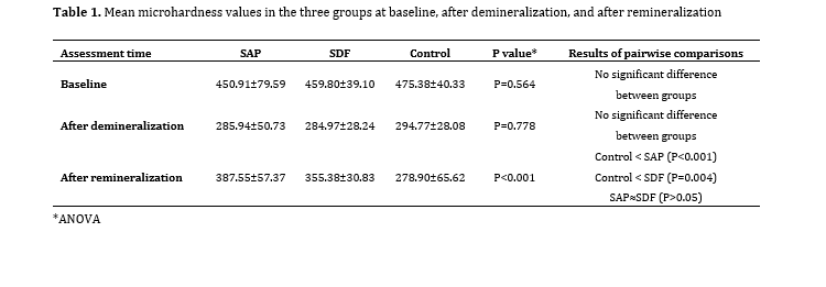
Figure 1. ESEM micrographs of enamel surface at x500 magnification: (A) sound enamel; (B) demineralized enamel; (C) control group; (D) SAP group; (E) SDF group
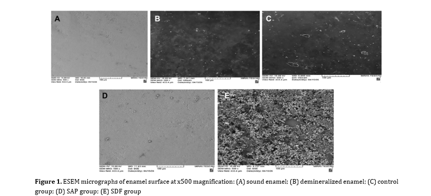
Figure 2. ESEM micrographs of enamel surface at x2000 magnification: (A) sound enamel; (B) demineralized enamel; (C) control group; (D) SAP group; (E) SDF group
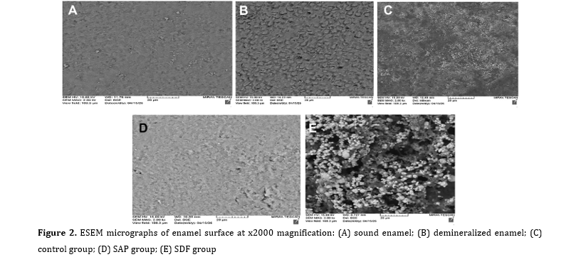
Figure 3. ESEM micrographs of remineralized enamel at x20,000 magnification: (A) control group; (B) SDF group; (C) SAP group
Discussion
This study evaluated how SDF and SAP affected microhardness, qualitative surface morphology, and remineralization of artificially induced incipient enamel lesions in primary teeth. Baseline and post-demineralization microhardness values were comparable among the three groups, whereas a significant difference was detected after the remineralization phase. The significant effects of time and time-by-group interaction, together with the non-significant main effect of group, suggest that the treatments differed mainly in how microhardness changed across the experimental stages rather than in initial microhardness levels. In the final comparison, both SAP and SDF were superior to the control; therefore, the null hypothesis was rejected. Although SAP produced numerically higher values than SDF, this difference was not statistically significant. Microhardness recovery was 61.6% in the SAP group and 40.3% in the SDF group, whereas the control group showed no real recovery and had a further numerical loss of -8.8%. At the remineralization stage, SAP and SDF presented approximately 39% and 27% higher microhardness, respectively, than the control, in agreement with Kumar et al. [25] and Shetty et al. [26].
SDF has stronger clinical establishment in pediatric dentistry than SAP. Its caries-arresting and mineralizing effects are attributed to the combined action of silver, fluoride-mediated mineralization, and physicochemical changes within the lesion. SAP, whose closest research counterpart is mainly P11-4, acts through a biomimetic scaffold and is particularly relevant for early non-cavitated enamel lesions. Thus, under the conditions of this in vitro study, both agents showed significant remineralizing potential compared with no treatment. However, despite the greater numerical improvement seen with SAP in microhardness and surface morphology, the two interventions did not differ significantly from each other.
The proposed action of SDF is multi-factorial [16,20,26,27]. Reviews and physicochemical investigations suggest that SDF should not be viewed simply as a topical fluoride agent; instead, its therapeutic effect involves both silver and fluoride. SDF interacts with hydroxyapatite and organic components of dental tissues, resulting in denser areas and mineral/metallic deposits and modifying the lesion response to later acid exposure. These mechanisms may explain the significant increase in microhardness observed for SDF compared with the control in this study [16,26]. With SDF application, both fluoride and silver ions are released and may deposit within porous regions, partially blocking intercrystalline spaces. This explains the microscopic reduction in pores and surface irregularities observed in the SDF group.
For incipient enamel lesions, however, the effect of SDF may be more closely related to lesion arrest and protection than to full recovery of the original mechanical properties. This pattern is consistent with the present finding that SDF improved microhardness but remained numerically lower than SAP [20]. Because of their low-viscosity monomeric structure, SAPs can diffuse into the lesion body, and in a calcium- and phosphate-rich environment such as artificial saliva, SAP molecules can form a scaffold within enamel porosities and attract mineral ions. As mineral accumulation continues, new hydroxyapatite crystals may grow and fill empty spaces, thereby reducing porosity, increasing structural density, and improving microhardness.
Most available evidence on SAPs relates to P11-4. Mechanistic and biomimetic studies indicate that P11-4 can diffuse into the lesion body, assemble within the demineralized zone, and create a three-dimensional framework that supports new hydroxyapatite nucleation and growth. Accordingly, SAPs are expected to contribute to subsurface remodeling rather than only surface hardening. This biological rationale supports the higher numerical performance of SAP in the present study, even though statistical superiority was not demonstrated in this sample [22]. However, the SAP protocol included phosphoric-acid etching, whereas the SDF protocol did not. Since phosphoric-acid conditioning can itself modify enamel surface morphology and facilitate material penetration, the greater numerical improvement observed with SAP cannot be attributed solely to the peptide.
The substrate used here was primary enamel rather than permanent enamel, which is also important for interpretation. Micro-computed tomography and SEM studies have shown that primary enamel tends to have lower mineral density and different structural characteristics than permanent enamel and may also display greater microporosity. These features may influence both lesion formation and treatment response, and could explain why neither SDF nor SAP restored hardness to baseline despite producing a positive treatment effect [28].
Compared with SDF, clinical evidence for SAPs in primary enamel is still relatively limited and heterogeneous. Much of the published SAP literature concerns P11-4, Curodont/analogues, permanent teeth, orthodontic white spot lesions, or combined treatment protocols. The present results are consistent with studies reporting remineralization after SAP and fluoride-based treatments. Kaur et al. [23] found improved remineralization with SAP and fluoride compounds compared with untreated controls, while Sezici et al. [29] reported numerically higher values for SAP without a significant difference between interventions. Similarly, Alcorn et al. [20] and Krishnamoorthi et al. [30] reported remineralizing effects for SDF and SAP, respectively.
The microhardness findings can be interpreted in relation to enamel crystal density and structural integrity. Although Vickers testing is applied to the surface, the indenter penetrates several micrometers into the tissue. Therefore, greater subsurface integrity and crystal density are expected to increase the hardness value. SAP may enhance this effect by supporting mineral deposition within the lesion body, whereas SDF appears to act more strongly at the outer portion of the lesion. ESEM observations supported this interpretation: specimens treated with SAP and SDF showed less porosity and surface irregularity than controls, and SAP-treated specimens displayed more marked mineral coverage and a more homogeneous surface. The fish-scale/honeycomb pattern seen after demineralization was largely reversed in the SAP group, resulting in a flatter surface compared with the other groups.
The control group had the least apparent remineralization and the greatest visible surface irregularity, indicating that artificial saliva alone was not sufficient to seal the pores or reverse the demineralized surface pattern. As expected, mineral loss during demineralization involved dissolution of hydroxyapatite and removal of calcium and phosphate ions from subsurface enamel, producing microscopic voids. These qualitative observations agree with previous reports [30,31].
A 38% SDF product containing approximately 44800 ppm fluoride was selected in this study to provide a high fluoride concentration for primary enamel lesions. The specimens in the SDF group developed dark discoloration over time, whereas the SAP-treated specimens maintained their original light appearance.
The principal limitation of this study was the relatively small sample size for directly comparing SAP with SDF. Although the data were adequate to show that both active treatments performed better than the control, the study may have lacked power to detect smaller differences between the two materials. A second limitation was the in vitro design, which allowed experimental control but could not reproduce biofilm activity, salivary clearance, natural pH changes, patient cooperation, or behavioral influences. Third, microhardness and qualitative ESEM imaging were used to infer remineralization without direct compositional analysis. Consequently, the observed microhardness recovery and morphological changes should be interpreted as findings consistent with remineralization rather than direct chemical confirmation. The Ca/P ratio, fluoride uptake, and silver deposition were not measured. Future studies should therefore incorporate energy-dispersive X-ray spectroscopy and complementary methods, such as micro-computed tomography and Fourier-transform infrared or Raman spectroscopy, to characterize the composition, depth, and distribution of mineral deposition. Fourth, direct evidence regarding SAP in primary teeth remains limited, because many P11-4 studies have been conducted on permanent teeth, orthodontic white spot lesions, or combined protocols. Finally, peptide-based materials are more expensive than fluoride-containing alternatives. Although two evaluators independently reviewed the ESEM images and resolved differences by consensus, we did not calculate a formal inter-rater reliability coefficient because the assessment was descriptive and did not use a predefined categorical scoring system. In addition, the SAP group underwent phosphoric-acid etching as part of the manufacturer's application protocol, whereas the SDF group did not. Because no acid-etched control or acid-etched SDF group was included, we could not separate the independent contribution of etching from the effect of SAP; therefore, interpret the between-treatment morphological differences cautiously.
Future research should proceed in three main directions. First, in vivo investigations followed by large randomized clinical trials are needed for non-cavitated lesions in primary teeth, with follow-up periods of 6, 12, and 18 months. Second, protocol-response studies should examine concentration, application time, frequency of use, and combined approaches such as SAP plus fluoride or SDF plus potassium iodide. Third, multimodal mechanochemical studies are needed to determine whether the microhardness gain in primary enamel reflects deep remineralization or mainly superficial rehardening.
Conclusion
SDF has stronger clinical establishment in pediatric dentistry than SAP. Its caries-arresting and mineralizing effects are attributed to the combined action of silver, fluoride-mediated mineralization, and physicochemical changes within the lesion. SAP, whose closest research counterpart is mainly P11-4, acts through a biomimetic scaffold and is particularly relevant for early non-cavitated enamel lesions. Thus, under the conditions of this in vitro study, both agents showed significant remineralizing potential compared with no treatment. However, despite the greater numerical improvement seen with SAP in microhardness and surface morphology, the two interventions did not differ significantly from each other.
The proposed action of SDF is multi-factorial [16,20,26,27]. Reviews and physicochemical investigations suggest that SDF should not be viewed simply as a topical fluoride agent; instead, its therapeutic effect involves both silver and fluoride. SDF interacts with hydroxyapatite and organic components of dental tissues, resulting in denser areas and mineral/metallic deposits and modifying the lesion response to later acid exposure. These mechanisms may explain the significant increase in microhardness observed for SDF compared with the control in this study [16,26]. With SDF application, both fluoride and silver ions are released and may deposit within porous regions, partially blocking intercrystalline spaces. This explains the microscopic reduction in pores and surface irregularities observed in the SDF group.
For incipient enamel lesions, however, the effect of SDF may be more closely related to lesion arrest and protection than to full recovery of the original mechanical properties. This pattern is consistent with the present finding that SDF improved microhardness but remained numerically lower than SAP [20]. Because of their low-viscosity monomeric structure, SAPs can diffuse into the lesion body, and in a calcium- and phosphate-rich environment such as artificial saliva, SAP molecules can form a scaffold within enamel porosities and attract mineral ions. As mineral accumulation continues, new hydroxyapatite crystals may grow and fill empty spaces, thereby reducing porosity, increasing structural density, and improving microhardness.
Most available evidence on SAPs relates to P11-4. Mechanistic and biomimetic studies indicate that P11-4 can diffuse into the lesion body, assemble within the demineralized zone, and create a three-dimensional framework that supports new hydroxyapatite nucleation and growth. Accordingly, SAPs are expected to contribute to subsurface remodeling rather than only surface hardening. This biological rationale supports the higher numerical performance of SAP in the present study, even though statistical superiority was not demonstrated in this sample [22]. However, the SAP protocol included phosphoric-acid etching, whereas the SDF protocol did not. Since phosphoric-acid conditioning can itself modify enamel surface morphology and facilitate material penetration, the greater numerical improvement observed with SAP cannot be attributed solely to the peptide.
The substrate used here was primary enamel rather than permanent enamel, which is also important for interpretation. Micro-computed tomography and SEM studies have shown that primary enamel tends to have lower mineral density and different structural characteristics than permanent enamel and may also display greater microporosity. These features may influence both lesion formation and treatment response, and could explain why neither SDF nor SAP restored hardness to baseline despite producing a positive treatment effect [28].
Compared with SDF, clinical evidence for SAPs in primary enamel is still relatively limited and heterogeneous. Much of the published SAP literature concerns P11-4, Curodont/analogues, permanent teeth, orthodontic white spot lesions, or combined treatment protocols. The present results are consistent with studies reporting remineralization after SAP and fluoride-based treatments. Kaur et al. [23] found improved remineralization with SAP and fluoride compounds compared with untreated controls, while Sezici et al. [29] reported numerically higher values for SAP without a significant difference between interventions. Similarly, Alcorn et al. [20] and Krishnamoorthi et al. [30] reported remineralizing effects for SDF and SAP, respectively.
The microhardness findings can be interpreted in relation to enamel crystal density and structural integrity. Although Vickers testing is applied to the surface, the indenter penetrates several micrometers into the tissue. Therefore, greater subsurface integrity and crystal density are expected to increase the hardness value. SAP may enhance this effect by supporting mineral deposition within the lesion body, whereas SDF appears to act more strongly at the outer portion of the lesion. ESEM observations supported this interpretation: specimens treated with SAP and SDF showed less porosity and surface irregularity than controls, and SAP-treated specimens displayed more marked mineral coverage and a more homogeneous surface. The fish-scale/honeycomb pattern seen after demineralization was largely reversed in the SAP group, resulting in a flatter surface compared with the other groups.
The control group had the least apparent remineralization and the greatest visible surface irregularity, indicating that artificial saliva alone was not sufficient to seal the pores or reverse the demineralized surface pattern. As expected, mineral loss during demineralization involved dissolution of hydroxyapatite and removal of calcium and phosphate ions from subsurface enamel, producing microscopic voids. These qualitative observations agree with previous reports [30,31].
A 38% SDF product containing approximately 44800 ppm fluoride was selected in this study to provide a high fluoride concentration for primary enamel lesions. The specimens in the SDF group developed dark discoloration over time, whereas the SAP-treated specimens maintained their original light appearance.
The principal limitation of this study was the relatively small sample size for directly comparing SAP with SDF. Although the data were adequate to show that both active treatments performed better than the control, the study may have lacked power to detect smaller differences between the two materials. A second limitation was the in vitro design, which allowed experimental control but could not reproduce biofilm activity, salivary clearance, natural pH changes, patient cooperation, or behavioral influences. Third, microhardness and qualitative ESEM imaging were used to infer remineralization without direct compositional analysis. Consequently, the observed microhardness recovery and morphological changes should be interpreted as findings consistent with remineralization rather than direct chemical confirmation. The Ca/P ratio, fluoride uptake, and silver deposition were not measured. Future studies should therefore incorporate energy-dispersive X-ray spectroscopy and complementary methods, such as micro-computed tomography and Fourier-transform infrared or Raman spectroscopy, to characterize the composition, depth, and distribution of mineral deposition. Fourth, direct evidence regarding SAP in primary teeth remains limited, because many P11-4 studies have been conducted on permanent teeth, orthodontic white spot lesions, or combined protocols. Finally, peptide-based materials are more expensive than fluoride-containing alternatives. Although two evaluators independently reviewed the ESEM images and resolved differences by consensus, we did not calculate a formal inter-rater reliability coefficient because the assessment was descriptive and did not use a predefined categorical scoring system. In addition, the SAP group underwent phosphoric-acid etching as part of the manufacturer's application protocol, whereas the SDF group did not. Because no acid-etched control or acid-etched SDF group was included, we could not separate the independent contribution of etching from the effect of SAP; therefore, interpret the between-treatment morphological differences cautiously.
Future research should proceed in three main directions. First, in vivo investigations followed by large randomized clinical trials are needed for non-cavitated lesions in primary teeth, with follow-up periods of 6, 12, and 18 months. Second, protocol-response studies should examine concentration, application time, frequency of use, and combined approaches such as SAP plus fluoride or SDF plus potassium iodide. Third, multimodal mechanochemical studies are needed to determine whether the microhardness gain in primary enamel reflects deep remineralization or mainly superficial rehardening.
Conclusion
Within the limitations of this in vitro study, both SAP and SDF significantly improved the microhardness of demineralized primary enamel compared with the control. SAP showed greater numerical recovery and more favorable qualitative ESEM morphology, but it was not statistically superior to SDF. Further clinical and compositional studies are needed to confirm these findings in primary teeth.
Type of Study: Original article |
Subject:
pediatric
References
1. Maklennan A, Borg-Bartolo R, Wierichs RJ, Esteves-Oliveira M, Campus G. A systematic review and meta-analysis on early-childhood-caries global data. BMC Oral Health. 2024 Jul;24(1):835. [DOI:10.1186/s12903-024-04605-y] [PMID] [PMCID]
2. Colak H, Dülgergil CT, Dalli M, Hamidi MM. Early childhood caries update: a review of causes, diagnoses, and treatments. J Nat Sci Biol Med. 2013 Jan;4(1):29-38. [DOI:10.4103/0976-9668.107257] [PMID] [PMCID]
3. Gurunathan D, Somasundaram S, Kumar S. Casein phosphopeptide-amorphous calcium phosphate: a remineralizing agent of enamel. Aust Dent J. 2012 Dec;57(4):404-8. [DOI:10.1111/adj.12006] [PMID]
4. Zhang S. Discovery and design of self-assembling peptides. Interface Focus. 2017 Dec;7(6):20170028. [DOI:10.1098/rsfs.2017.0028] [PMID] [PMCID]
5. Bagis EE, Derelioglu SS, Sengül F, Yılmaz S. The effect of the treatment of severe early childhood caries on growth-development and quality of life. Children (Basel). 2023 Feb;10(2):411. [DOI:10.3390/children10020411] [PMID] [PMCID]
6. Lam PP, Chua H, Ekambaram M, Lo EC, Yiu CK. Risk predictors of early childhood caries increment: a systematic review and meta-analysis. J Evid Based Dent Pract. 2022 Sep;22(3):101732. [DOI:10.1016/j.jebdp.2022.101732] [PMID]
7. Zheng FM, Yan IG, Duangthip D, Gao SS, Lo ECM, Chu CH. Silver diamine fluoride therapy for dental care. Jpn Dent Sci Rev. 2022 Nov;58:249-57. [DOI:10.1016/j.jdsr.2022.08.001] [PMID] [PMCID]
8. Surendranath P, Krishnappa S, Srinath S. Silver diamine fluoride in preventing caries: a review of current trends. Int J Clin Pediatr Dent. 2022;15(Suppl 2):S247-51. [DOI:10.5005/jp-journals-10005-2167] [PMID] [PMCID]
9. Yan IG, Zheng FM, Yin IX, Sun IG, Lo ECM, Chu CH. Stability of silver and fluoride contents in silver diamine fluoride solutions. Int Dent J. 2023 Dec;73(6):840-6. [DOI:10.1016/j.identj.2023.05.001] [PMID] [PMCID]
10. Jiang CM, Duangthip D, Chan AKY, Tamrakar M, Lo ECM, Chu CH. Global research interest regarding silver diamine fluoride in dentistry: a bibliometric analysis. J Dent. 2021 Oct;113:103778. [DOI:10.1016/j.jdent.2021.103778] [PMID]
11. Shi P, He X, Cong H, Yu B, Shen Y. Preparation and properties of self-assembling polypeptide hydrogels and their application in biomedicine. ACS Mater Lett. 2024 Mar;6(5):1649-77. [DOI:10.1021/acsmaterialslett.3c01546]
12. La Manna S, Di Natale C, Onesto V, Marasco D. Self-assembling peptides: from design to biomedical applications. Int J Mol Sci. 2021 Nov;22(23):12662. [DOI:10.3390/ijms222312662] [PMID] [PMCID]
13. de Souza Araújo IJ, Guimarães GN, Machado RA, Bertassoni LE, Davies RPW, Puppin-Rontani RM. Self-assembly peptide P11-4 induces mineralization and cell-migration of odontoblast-like cells. J Dent. 2022 Jun;121:104111. [DOI:10.1016/j.jdent.2022.104111] [PMID] [PMCID]
14. De Sousa J, Carvalho R, Barbosa-Martins L, Torquato R, Mugnol K, Nascimento F, et al. The self-assembling peptide P11-4 prevents collagen proteolysis in dentin. J Dent Res. 2019 Mar;98(3):347-54. [DOI:10.1177/0022034518817351] [PMID]
15. Atteya SM, Amer HA, Saleh SM, Safwat Y. Self-assembling peptide and nano-silver fluoride in remineralizing early enamel carious lesions: randomized controlled clinical trial. BMC Oral Health. 2023 Aug;23(1):577. [DOI:10.1186/s12903-023-03269-4] [PMID] [PMCID]
16. Al-Qahtani AS, Almutairi B, Al-Hamdan RS, Alzahrani KM. Remineralizing pretreatment using casein phosphopeptide-amorphous calcium phosphate fluoride, self-assembling peptide, and nanohydroxyapatite gel activation via invisible infrared light on dentin microhardness and micro-shear bond strength to composite restoration. Photodiagnosis Photodyn Ther. 2024 Jun;47:104210. [DOI:10.1016/j.pdpdt.2024.104210] [PMID]
17. Magalhães GAP, Fraga MAA, de Souza Araújo IJ, Pacheco RR, Correr AB, Puppin-Rontani RM. Effect of a self-assembly peptide on surface roughness and hardness of bleached enamel. J Funct Biomater. 2022 Jun;13(2):79. [DOI:10.3390/jfb13020079] [PMID] [PMCID]
18. Azadi P, Sarlak H, Nourmohammadi S. Comparing enamel microhardness in deciduous teeth with primary carious lesions after applying three fluoride-containing materials in vitro. J Arak Univ Med Sci. 2021 May;24(2):256-67. [DOI:10.32598/jams.24.2.6175.1]
19. Moecke SE, Silva AGCS, Andrade ACM, Borges AB, Torres CRG. Efficacy of S-PRG filler varnishes on enamel caries remineralization. J Dent. 2022 Apr;119:104074. [DOI:10.1016/j.jdent.2022.104074] [PMID]
20. Alcorn A, Al Dehailan L, Cook NB, Tang Q, Lippert F. Longitudinal in vitro effects of silver diamine fluoride on early enamel caries lesions. Oper Dent. 2022 May;47(3):309-19. [DOI:10.2341/20-237-L] [PMID]
21. Valizade K, Seraj B, Ghadimi S, Kharazifard MJ, Chiniforush N. Effect of silver diamine fluoride combined with photodynamic therapy on primary enamel microhardness. Photodiagnosis Photodyn Ther. 2024 Oct;49:104274. [DOI:10.1016/j.pdpdt.2024.104274] [PMID]
22. Alkilzy M, Qadri G, Splieth CH, Santamaría RM. Biomimetic enamel regeneration using self-assembling peptide P11-4. Biomimetics (Basel). 2023 Jul;8(3):290. [DOI:10.3390/biomimetics8030290] [PMID] [PMCID]
23. Kaur S, Bhola M, Bajaj N, Brar GS. Comparative evaluation of the remineralizing potential of silver diamine fluoride, casein phosphopeptide-amorphous calcium phosphate, and fluoride varnish on the enamel surface of primary and permanent teeth: an in vitro study. Int J Clin Pediatr Dent. 2023 Aug;16(Suppl 1):S91-6. [DOI:10.5005/jp-journals-10005-2622] [PMID] [PMCID]
24. Rafiei S, Jafari Ghavam Abad A, Paryab M, Tayebi G, Younespour S. Comparison of the microhardness of primary dentin with artificially induced caries following the application of sodium fluoride varnish with the intensive technique versus fluoride therapy with silver diamine fluoride. Int J Dent. 2026;2026(1):7229454. [DOI:10.1155/ijod/7229454] [PMID] [PMCID]
25. Kumar VLN, Itthagarun A, King NM. The effect of casein phosphopeptide-amorphous calcium phosphate on remineralization of artificial caries-like lesions: an in vitro study. Aust Dent J. 2008 Mar;53(1):34-40. [DOI:10.1111/j.1834-7819.2007.00006.x] [PMID]
26. Shetty S, Hegde MN, Bopanna TP. Enamel remineralization assessment after treatment with three different remineralizing agents using surface microhardness: an in vitro study. J Conserv Dent. 2014 Jan;17(1):49-52. [DOI:10.4103/0972-0707.124136] [PMID] [PMCID]
27. Zhao IS, Gao SS, Hiraishi N, Burrow MF, Duangthip D, Mei ML, et al. Mechanisms of silver diamine fluoride on arresting caries: a literature review. Int Dent J. 2018 Apr;68(2):67-76. [DOI:10.1111/idj.12320] [PMID]
28. Hayashi-Sakai S, Sakamoto M, Hayashi T, Kondo T, Sugita K, Sakai J, et al. Evaluation of permanent and primary enamel and dentin mineral density using micro-computed tomography. Oral Radiol. 2019 Jan;35(1):29-34. [DOI:10.1007/s11282-018-0315-2] [PMID]
29. Sezici YL, Yetkiner E, Yetkiner AA, Eden E, Attin R. Comparative evaluation of fluoride varnishes, self-assembling peptide-based remineralization agent, and enamel matrix protein derivative on artificial enamel remineralization in vitro. Prog Orthod. 2021 Jan;22(1):4. [DOI:10.1186/s40510-020-00345-1] [PMID] [PMCID]
30. Krishnamoorthi A, Shanbhog RS, Godhi BS, Sundaravadivelu M. Efficacy of self-assembling peptide P11-4 in remineralizing in vitro caries-like lesions in primary enamel samples in combination with calcium phosphate-based remineralization agents. Int J Clin Pediatr Dent. 2024 May;17(5):552-7. [DOI:10.5005/jp-journals-10005-2845] [PMID] [PMCID]
31. Zhang Q, Zou J, Yang R, Zhou X. Remineralization effects of casein phosphopeptide-amorphous calcium phosphate creme on artificial early enamel lesions of primary teeth. Int J Paediatr Dent. 2011 Sep;21(5):374-81. [DOI:10.1111/j.1365-263X.2011.01135.x] [PMID]
Send email to the article author
| Rights and permissions | |
 |
This work is licensed under a Creative Commons Attribution-NonCommercial 4.0 International License. |