
Journal of Research in Dental
and Maxillofacial Sciences

Volume 11, Issue 3 (9-2026)
J Res Dent Maxillofac Sci 2026, 11(3): 259-267 |
Back to browse issues page

Ethics code: IR.SBMU.DRC.REC.1402.087


Download citation:
BibTeX | RIS | EndNote | Medlars | ProCite | Reference Manager | RefWorks
Send citation to:


BibTeX | RIS | EndNote | Medlars | ProCite | Reference Manager | RefWorks
Send citation to:
Mojtahedzadeh K, Panahandeh N, Nikaein M. Comparative Effects of Two Fluoride Varnishes on Microhardness of Demineralized Enamel: An In Vitro Study. J Res Dent Maxillofac Sci 2026; 11 (3) :259-267
URL: http://jrdms.dentaliau.ac.ir/article-1-1438-en.html
URL: http://jrdms.dentaliau.ac.ir/article-1-1438-en.html


1- Dental Research Center, Research Institute of Dental Sciences, School of Dentistry, Shahid Beheshti University of Medical Sciences, Tehran, Iran.
2- Department of Restorative Dentistry, School of Dentistry, Shahid Beheshti University of Medical Sciences, Tehran, Iran. ,Mahsanikaeen1994@gmail.com
2- Department of Restorative Dentistry, School of Dentistry, Shahid Beheshti University of Medical Sciences, Tehran, Iran. ,
Keywords: Dental Enamel, Fluorides, Topical, Hardness, Tooth Demineralization, Tooth Remineralization
| Abstract (HTML) (16 Views)
Full-Text: (3 Views)
Abstract
Background and Aim: Different fluoride varnish formulations may exhibit varying effects on the recovery of demineralized enamel. This study aimed to compare the effects of two fluoride varnishes on the microhardness of demineralized enamel.
Materials and Methods: In this in vitro experimental study, 40 sound enamel specimens were prepared and assigned to four groups (n=10): sound enamel, demineralized enamel, remineralized enamel using AV varnish (Avijeh Teb Parsian), and remineralized enamel using Enamelast (Ultradent). In each group, the baseline enamel microhardness was first measured at the surface and then from the most superficial point towards the dentinoenamel junction at 10 µm intervals. To measure the depth of demineralization and remineralization, the specimens were mesiodistally sectioned by a thin sectioner. A standard acid with a pH of 4.4 was used for enamel demineralization. Microhardness was measured by the Vickers microhardness tester. Data were analyzed by repeated measures ANOVA and Tukey’s HSD test (alpha=0.05).
Results: Application of fluoride varnish effectively enhanced enamel remineralization, and the efficacy of the two varnishes was significantly different for enamel remineralization both at the enamel surface and also at different depths (P<0.05).
Conclusion: The results showed that both Enamelast and AV fluoride varnishes increased demineralized enamel microhardness. Enamelast was more effective than AV for enhancement of superficial enamel microhardness while AV was more effective in enhancement of deep enamel microhardness. None of the varnishes could increase demineralized enamel microhardness to the level of sound enamel.
Keywords: Dental Enamel; Fluorides, Topical; Hardness; Tooth Demineralization; Tooth Remineralization
Introduction
Materials and Methods: In this in vitro experimental study, 40 sound enamel specimens were prepared and assigned to four groups (n=10): sound enamel, demineralized enamel, remineralized enamel using AV varnish (Avijeh Teb Parsian), and remineralized enamel using Enamelast (Ultradent). In each group, the baseline enamel microhardness was first measured at the surface and then from the most superficial point towards the dentinoenamel junction at 10 µm intervals. To measure the depth of demineralization and remineralization, the specimens were mesiodistally sectioned by a thin sectioner. A standard acid with a pH of 4.4 was used for enamel demineralization. Microhardness was measured by the Vickers microhardness tester. Data were analyzed by repeated measures ANOVA and Tukey’s HSD test (alpha=0.05).
Results: Application of fluoride varnish effectively enhanced enamel remineralization, and the efficacy of the two varnishes was significantly different for enamel remineralization both at the enamel surface and also at different depths (P<0.05).
Conclusion: The results showed that both Enamelast and AV fluoride varnishes increased demineralized enamel microhardness. Enamelast was more effective than AV for enhancement of superficial enamel microhardness while AV was more effective in enhancement of deep enamel microhardness. None of the varnishes could increase demineralized enamel microhardness to the level of sound enamel.
Keywords: Dental Enamel; Fluorides, Topical; Hardness; Tooth Demineralization; Tooth Remineralization
Introduction
Dental caries is the most common oral disease worldwide [1]. In today’s world, more attention has been directed to preventive measures and conservative approaches in healthcare provision [2]. Dental caries is a dynamic process composed of repeated cycles of tooth demineralization and remineralization [3]. Food fermentation by the activity of cariogenic bacteria, subsequent acid production, and dissolution of tooth minerals is the main cause of the development of carious lesions [4]. Enamel is composed of 96wt% minerals and 4wt% organic materials and water [3]. Dental caries initiates in the enamel and has a slow progression rate at first, because enamel is more resistant to demineralization compared with deeper tooth structures [5]. Organic acids, produced as a byproduct by the activity of bacteria, decrease the biofilm pH. Long periods of low pH (the critical threshold for enamel demineralization is 5.5) can convert the microbiome to acidogenic and acidophilic bacteria (aciduric mutans and non-mutans streptococci) and increase the acidity of the environment. To reach an equilibrium in a low pH, calcium and phosphate ions are released from the tooth surface into the biofilm, causing tooth demineralization. Calcium and phosphate ions are the structural blocks required for the remineralization process, which are found in the saliva, and have different saturation rates in different individuals [6].
At present, several materials are available in the market for enhancement of remineralization and prevention of demineralization; however, their comparative efficacy is in need of further investigations. The effects of fluoride therapy on enamel microhardness have been the topic of many previous investigations [7-10]. It has been well confirmed that fluoride therapy enhances enamel remineralization and increases enamel resistance to demineralization [3].
Several products are available for the application of fluoride such as fluoride toothpastes, mouthwashes, gels, and varnishes. Immediately after the application of fluoride, a relatively high concentration of fluoride can be detected in oral fluids and dental plaque. Nonetheless, the fluoride concentration significantly decreases with time. Thus, repeated applications of relatively low concentrations of fluoride are recommended to prevent demineralization and enhance remineralization [11]. Considering the need for an effective low-cost, domestically manufactured fluoride varnish, and the confirmed efficacy of Enamelast for enamel remineralization [12,13], this study aimed to compare the effects of AV fluoride varnish and Enamelast fluoride varnish on enamel microhardness and enhancement of remineralization. The effects of varnishes on remineralization at different depths of enamel were also evaluated and compared. The null hypothesis of the study was that no significant difference would be found between the abovementioned two fluoride varnishes regarding their effect on enamel microhardness.
Materials and Methods
At present, several materials are available in the market for enhancement of remineralization and prevention of demineralization; however, their comparative efficacy is in need of further investigations. The effects of fluoride therapy on enamel microhardness have been the topic of many previous investigations [7-10]. It has been well confirmed that fluoride therapy enhances enamel remineralization and increases enamel resistance to demineralization [3].
Several products are available for the application of fluoride such as fluoride toothpastes, mouthwashes, gels, and varnishes. Immediately after the application of fluoride, a relatively high concentration of fluoride can be detected in oral fluids and dental plaque. Nonetheless, the fluoride concentration significantly decreases with time. Thus, repeated applications of relatively low concentrations of fluoride are recommended to prevent demineralization and enhance remineralization [11]. Considering the need for an effective low-cost, domestically manufactured fluoride varnish, and the confirmed efficacy of Enamelast for enamel remineralization [12,13], this study aimed to compare the effects of AV fluoride varnish and Enamelast fluoride varnish on enamel microhardness and enhancement of remineralization. The effects of varnishes on remineralization at different depths of enamel were also evaluated and compared. The null hypothesis of the study was that no significant difference would be found between the abovementioned two fluoride varnishes regarding their effect on enamel microhardness.
Materials and Methods
This in vitro experimental study was conducted on sound human premolar teeth (with no cracks, caries, or hypocalcification) extracted due to periodontal problems or as part of orthodontic treatment. The study protocol was approved by the ethics committee of the university (IR.SBMU.DRC.REC.1402.087).
Sample size:
The sample size was calculated to be 9 in each group using PASS 21, assuming α=0.05, β=0.1 (study power of 90%), mean values of µ1=350, µ2=387, µ3=447, and µ4=510, and a common standard deviation value of 90 for all, extracted from a pilot study. To increase accuracy, 10 specimens were considered in each group.
Pilot study:
First, a pilot study with a small sample size was conducted to find the best duration of application of varnishes and immersion in acid, best mounting position, and best method of measurement of microhardness. Initial assessments revealed that immersion of specimens in acid for 96 hours did not provide sufficient depth of demineralization for evaluation of changes. Thus, the demineralization time was increased to 14 days.
Specimen preparation:
Forty sound premolars with no caries or enamel defects were used in this study. The teeth were cleaned of debris and periodontal tissue residues and were disinfected in 0.5% chloramine T solution (Halamid®, Axcentive Co., France) for 24 hours. The buccal surface of the teeth was polished with silicon carbide abrasive papers (220-, 400-, 800-, and 1200-grit), and the teeth were randomly assigned to 4 groups (n=10):
Group 1 (control): The teeth were decoronated, and their cross-section was sealed with nail varnish. Each tooth was sectioned into a buccal and a lingual half through a mesiodistal section, and the sectioned surface was polished with abrasive paper. The enamel microhardness of the buccal surface was measured by a Vickers hardness tester (Zwick Roell, Ulm, Germany) by application of 50 g load for 10 seconds [14]. Also, after mounting the specimens in acrylic resin such that their buccal surface remained exposed, the internal specimen surface was used to measure the microhardness from the superficial to deep areas at 10-µm intervals.
Group 2 (demineralized): The teeth were decoronated, and their cross-section was sealed with nail varnish. They were then immersed in a demineralizing solution used in a study by Ozgul et al. [15] at 37°C for 14 days. The demineralizing solution was composed of 2.2 mM CaCl2, 2.2 mM NaH2PO4, 0.05 M acetic acid, and 1 M KOH to adjust the pH to 4.4 at 37°C [15]. Next, the teeth were rinsed under running water and were immersed in artificial saliva composed of 1.5 mM CaCl2, 0.9 mM NaH2PO4, and 0.15 M KCl with a pH of 7 for 24 hours [15]. The next procedures were the same as those explained for group 1.
Group 3: Enamelast varnish (Ultradent, USA): The teeth were decoronated, and their cross-section was sealed with nail varnish. They were then immersed in the aforementioned demineralizing solution at 37°C for 14 days. The teeth were rinsed under running water and Enamelast varnish was applied on the enamel surface of the teeth according to the manufacturer’s instructions. The teeth were then immersed in artificial saliva for 24 hours. Excess varnish was removed from the surface using acetone. Sectioning and microhardness assessment were performed as explained for group 1.
Group 4: AV varnish (Avijeh Teb Parsian, Iran): The teeth were decoronated, and their cross-section was sealed with nail varnish. They were then immersed in the aforementioned demineralizing solution at 37°C for 14 days. The teeth were then rinsed under running water, and AV varnish was applied to the enamel surface of the teeth according to the manufacturer’s instructions. The teeth were then immersed in artificial saliva for 24 hours. Excess varnish was removed from the surface by using acetone. Sectioning and microhardness assessment were performed as explained for group 1.
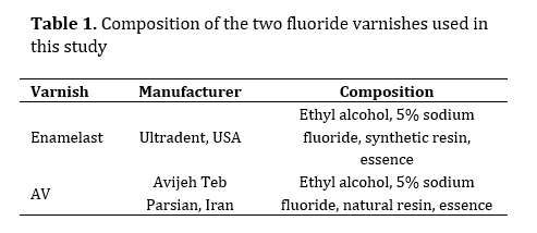
Table 1 presents the composition of the two fluoride varnishes used in this study.
Table 1. Composition of the two fluoride varnishes used in this study
Statistical analysis:
Normal distribution of data was confirmed by the Shapiro-Wilk test (P>0.05). Accordingly, two-way repeated measures ANOVA was applied to analyze the main and interaction effects of depth and group on microhardness, with depth as the within-subject factor and group as the between-subject factor. Since their interaction effect was found to be significant, subgroup analysis was performed. For this purpose, the mean microhardness at different depths was compared within each group with one-way repeated measures ANOVA followed by pairwise comparisons with the Bonferroni test. Moreover, the mean microhardness of the groups was compared at each depth by one-way ANOVA, followed by pairwise comparisons with Tukey’s HSD test. All statistical analyses were performed using SPSS version 26 (SPSS Inc., IL, USA) at 0.05 level of significance.
Results
Sample size:
The sample size was calculated to be 9 in each group using PASS 21, assuming α=0.05, β=0.1 (study power of 90%), mean values of µ1=350, µ2=387, µ3=447, and µ4=510, and a common standard deviation value of 90 for all, extracted from a pilot study. To increase accuracy, 10 specimens were considered in each group.
Pilot study:
First, a pilot study with a small sample size was conducted to find the best duration of application of varnishes and immersion in acid, best mounting position, and best method of measurement of microhardness. Initial assessments revealed that immersion of specimens in acid for 96 hours did not provide sufficient depth of demineralization for evaluation of changes. Thus, the demineralization time was increased to 14 days.
Specimen preparation:
Forty sound premolars with no caries or enamel defects were used in this study. The teeth were cleaned of debris and periodontal tissue residues and were disinfected in 0.5% chloramine T solution (Halamid®, Axcentive Co., France) for 24 hours. The buccal surface of the teeth was polished with silicon carbide abrasive papers (220-, 400-, 800-, and 1200-grit), and the teeth were randomly assigned to 4 groups (n=10):
Group 1 (control): The teeth were decoronated, and their cross-section was sealed with nail varnish. Each tooth was sectioned into a buccal and a lingual half through a mesiodistal section, and the sectioned surface was polished with abrasive paper. The enamel microhardness of the buccal surface was measured by a Vickers hardness tester (Zwick Roell, Ulm, Germany) by application of 50 g load for 10 seconds [14]. Also, after mounting the specimens in acrylic resin such that their buccal surface remained exposed, the internal specimen surface was used to measure the microhardness from the superficial to deep areas at 10-µm intervals.
Group 2 (demineralized): The teeth were decoronated, and their cross-section was sealed with nail varnish. They were then immersed in a demineralizing solution used in a study by Ozgul et al. [15] at 37°C for 14 days. The demineralizing solution was composed of 2.2 mM CaCl2, 2.2 mM NaH2PO4, 0.05 M acetic acid, and 1 M KOH to adjust the pH to 4.4 at 37°C [15]. Next, the teeth were rinsed under running water and were immersed in artificial saliva composed of 1.5 mM CaCl2, 0.9 mM NaH2PO4, and 0.15 M KCl with a pH of 7 for 24 hours [15]. The next procedures were the same as those explained for group 1.
Group 3: Enamelast varnish (Ultradent, USA): The teeth were decoronated, and their cross-section was sealed with nail varnish. They were then immersed in the aforementioned demineralizing solution at 37°C for 14 days. The teeth were rinsed under running water and Enamelast varnish was applied on the enamel surface of the teeth according to the manufacturer’s instructions. The teeth were then immersed in artificial saliva for 24 hours. Excess varnish was removed from the surface using acetone. Sectioning and microhardness assessment were performed as explained for group 1.
Group 4: AV varnish (Avijeh Teb Parsian, Iran): The teeth were decoronated, and their cross-section was sealed with nail varnish. They were then immersed in the aforementioned demineralizing solution at 37°C for 14 days. The teeth were then rinsed under running water, and AV varnish was applied to the enamel surface of the teeth according to the manufacturer’s instructions. The teeth were then immersed in artificial saliva for 24 hours. Excess varnish was removed from the surface by using acetone. Sectioning and microhardness assessment were performed as explained for group 1.
Table 1 presents the composition of the two fluoride varnishes used in this study.
Table 1. Composition of the two fluoride varnishes used in this study
Statistical analysis:
Normal distribution of data was confirmed by the Shapiro-Wilk test (P>0.05). Accordingly, two-way repeated measures ANOVA was applied to analyze the main and interaction effects of depth and group on microhardness, with depth as the within-subject factor and group as the between-subject factor. Since their interaction effect was found to be significant, subgroup analysis was performed. For this purpose, the mean microhardness at different depths was compared within each group with one-way repeated measures ANOVA followed by pairwise comparisons with the Bonferroni test. Moreover, the mean microhardness of the groups was compared at each depth by one-way ANOVA, followed by pairwise comparisons with Tukey’s HSD test. All statistical analyses were performed using SPSS version 26 (SPSS Inc., IL, USA) at 0.05 level of significance.
Results
Table 2 shows the mean microhardness at different depths from the enamel surface in the four groups. Two-way repeated measures ANOVA showed a significant difference in microhardness at different depths (P<0.001). Also, the mean microhardness was significantly different among the four groups (P<0.001). As shown in Figure 1, the interaction effect of group and depth on microhardness was significant (P<0.001); in other words, in the demineralized group, microhardness increased with greater depths. However, it decreased with increasing depth in the remaining three groups. In total, the microhardness in Enamelast and AV groups was greater than that in the demineralized group at 60 and 70 µm depths. Considering the significant interaction effect of group and depth on microhardness, subgroup analysis was performed, such that the effect of depth on microhardness was separately assessed within each group. Also, the microhardness of the four groups was compared separately at each depth. In all four groups, the association of microhardness change by an increase in depth was significant (P<0.001 for all four). Also, as shown in Figure 1, the change in microhardness was variable in different groups by an increase in depth. Table 3 shows pairwise comparisons of microhardness at different depths within each group. Also, the microhardness of the four groups was compared at each depth using one-way ANOVA, which revealed significant differences among the four groups at all depths (P<0.001 for all 11 comparisons). Thus, pairwise comparison of the microhardness of the groups was performed separately at each depth (Table 4). The microhardness of superficial enamel (H0) in the Enamelast group was significantly higher than that in the AV group (P<0.05).
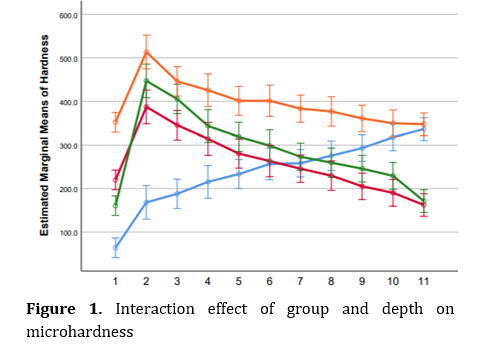
Table 2. Mean microhardness (VHN) at different depths from the enamel surface in the four groups
Table 3. Pairwise comparison of microhardness at different depths within each group
Table 4. Pairwise comparison of the microhardness of the groups at each depth
Discussion
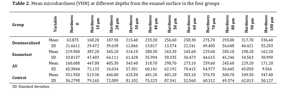
Table 3. Pairwise comparison of microhardness at different depths within each group
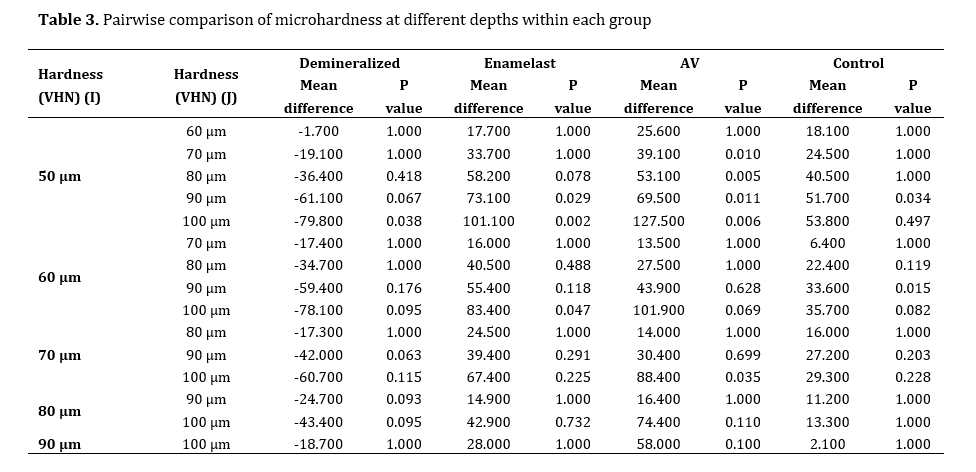
Table 4. Pairwise comparison of the microhardness of the groups at each depth
Discussion
This study compared the effects of AV fluoride varnish and Enamelast fluoride varnish on enamel microhardness and enhancement of remineralization. The effects of varnishes on remineralization at different depths of enamel were also evaluated and compared. The null hypothesis of the study was that no significant difference would be found between the abovementioned two fluoride varnishes regarding their effect on enamel microhardness. The results showed that application of fluoride varnish effectively enhanced enamel remineralization, and the efficacy of the two varnishes was significantly different for enamel remineralization both at the enamel surface and at different depths. Thus, the null hypothesis of the study was rejected.
The microhardness of sound enamel was 330 to 390 VHN in the present study, which was expected and close to the values reported in similar studies [1,15]. Also, as expected, the microhardness of sound enamel decreased from the surface towards deeper areas as approximating the dentinoenamel junction, which was also in agreement with previous findings [6,16].
In line with previous findings [14,17], the current results showed a reduction in enamel surface microhardness due to the effect of demineralization. The acidic compounds used for enamel demineralization are variable in the literature. The pH of the acidic solution used for this purpose in the present study was 4.4. In a study by Ozgul et al. [15], the specimens were immersed for 4 days in an acidic solution with a pH of 4.4, and they reported the resultant demineralization depth to be 60 µm. Considering their results and the findings of the pilot study, the specimens were immersed in the acidic solution for 14 days (to be able to assess the changes at deeper areas), yielding a 100-110 µm demineralization depth in the present study. The enamel surface microhardness after demineralization in the current study was lower than that in some other studies, which can be due to differences in demineralizing solutions or duration of immersion of the specimens.
Enamel surface microhardness is a suitable measure for evaluation of the efficacy of fluoride varnishes and other remineralizing agents [14]. In the present study, both varnishes increased enamel microhardness. The greatest microhardness for both varnishes was measured at 10 µm depth; the mean VHN at this point was 390 VHN for Enamelast and 440 VHN for AV, which were still lower than the baseline (sound) enamel microhardness at this point. At neutral pH, formation of fluorohydroxyapatite occurs too slow to compensate for the usual wear of the tooth surface. Therefore, despite daily use of fluoridated toothpaste, toothbrushing decreases the fluoride content of the buccal surface enamel over the years [18]. Since the specimens were obtained from the buccal surface enamel in the present study and were polished with abrasive paper prior to microhardness testing, the superficial enamel microhardness in all groups was lower than that of subsurface enamel. Also, considering this limitation, for measurement of enamel microhardness at different depths, unpolished proximal surfaces were used.
Godoi et al. [12] reported that the mean enamel surface microhardness following the application of Enamelast varnish was significantly higher than that in the Duraphat and Clinpro varnish groups. Also, Enamelast provided a higher concentration of soluble and insoluble fluoride compared with the other two varnishes. They added that none of the tested varnishes could effectively enhance subsurface enamel remineralization. In the present study, both Enamelast and AV varnishes effectively increased enamel surface microhardness; while, Enamelast was effective to a depth of 60 µm, and AV varnish was effective to 70 µm depth for microhardness enhancement. This increase in the subsurface enamel microhardness was different from the results of Godoi et al. [12]. Since the method of demineralization was different in the two studies, and they did not measure the depth of demineralization, difference in the results can be due to different demineralization patterns.
In the current study, the microhardness values in subsurface areas were greater in the AV varnish group than the Enamelast group, and it appears that in subsurface enamel and deeper areas, the AV varnish had a superior performance compared to Enamelast. AV varnish has natural resin containing calcium in its composition, which appears to be effective for remineralization. The presence of modified tricalcium phosphate in a varnish can enhance enamel remineralization potential, especially at greater depths [12].
Resin, sodium fluoride, and solvent are the main constituents of varnishes. No consensus exists regarding the precise durability of varnishes on the tooth surface; however, this time varies from 4 hours to a couple of days [19]. The main advantage of varnish compared with other remineralizing agents is the increased contact time of fluoride with the tooth surface. Thus, manufacturers are searching for strategies to prolong the durability of varnish on the tooth surface. They particularly worked on the resin components of varnishes for their further stabilization on the tooth surface. In the current study, removal of AV resin was challenging. Removal of fluoride varnish resin for microhardness assessment in vitro requires frequent acetone irrigation. Nonetheless, despite frequent rinsing with acetone, removal of the varnish might have not been performed completely, which can explain the observed differences in microhardness at the enamel surface [12]. Enamelast contains synthetic resin as thickener in its composition, which is probably removed more easily with acetone solvents, explaining lower enamel microhardness in the AV varnish group. Both varnishes had 5% NaF, and the solvent of both is ethyl alcohol.
In general, it is believed that fluoride release from varnishes is weak compared with other methods of fluoride therapy due to the presence of hydrophobic resin in the composition of varnishes [20]. Another study also reported higher efficacy of other remineralizing agents such as MI Paste and ICON compared with fluoride varnish, which may be due to the absence of resin in their composition. In other words, although resin increases the contact time of fluoride with the tooth surface, resin-free compounds and even natural saliva have greater potential for deposition of minerals compared with resin compounds [21]. In the current study, the microhardness at the deepest point evaluated did not reach that of sound enamel in any of the varnish groups; while at the same depth, the microhardness of demineralized enamel was almost comparable to that of sound enamel. This finding can be attributed to the presence of resin in the composition of varnishes, which serves as a barrier against the penetration of remineralizing molecules into the enamel structure. In the clinical setting, optimal oral conditions for the formation of fluorapatite crystals includes super-saturation of saliva, oral fluids, and dental plaque with fluoride, which usually occurs at a pH > 4.5 [18]. On the other hand, availability of phosphate, calcium, and even fluoride ions in the saliva and oral environment is necessary to enhance remineralization. Thus, it may be concluded that when demineralization depth exceeds 60 µm, application of fluoride varnish prevents the penetration of saliva calcium and phosphate into the enamel and impedes natural remineralization, and is therefore, not suitable for enhancement of remineralization. Remineralization of superficial enamel also decreases the porosities and subsequent remineralization at deeper areas.
This study had several limitations such as an in vitro design, which limits the generalizability of the findings to the clinical setting, and lack of microscopic observation of the enamel surface. Future studies are recommended to assess the changes in enamel structure following application of different varnishes using a scanning electron microscope. Also, the efficacy of AV varnish should be compared with other remineralizing agents.
Conclusion
The microhardness of sound enamel was 330 to 390 VHN in the present study, which was expected and close to the values reported in similar studies [1,15]. Also, as expected, the microhardness of sound enamel decreased from the surface towards deeper areas as approximating the dentinoenamel junction, which was also in agreement with previous findings [6,16].
In line with previous findings [14,17], the current results showed a reduction in enamel surface microhardness due to the effect of demineralization. The acidic compounds used for enamel demineralization are variable in the literature. The pH of the acidic solution used for this purpose in the present study was 4.4. In a study by Ozgul et al. [15], the specimens were immersed for 4 days in an acidic solution with a pH of 4.4, and they reported the resultant demineralization depth to be 60 µm. Considering their results and the findings of the pilot study, the specimens were immersed in the acidic solution for 14 days (to be able to assess the changes at deeper areas), yielding a 100-110 µm demineralization depth in the present study. The enamel surface microhardness after demineralization in the current study was lower than that in some other studies, which can be due to differences in demineralizing solutions or duration of immersion of the specimens.
Enamel surface microhardness is a suitable measure for evaluation of the efficacy of fluoride varnishes and other remineralizing agents [14]. In the present study, both varnishes increased enamel microhardness. The greatest microhardness for both varnishes was measured at 10 µm depth; the mean VHN at this point was 390 VHN for Enamelast and 440 VHN for AV, which were still lower than the baseline (sound) enamel microhardness at this point. At neutral pH, formation of fluorohydroxyapatite occurs too slow to compensate for the usual wear of the tooth surface. Therefore, despite daily use of fluoridated toothpaste, toothbrushing decreases the fluoride content of the buccal surface enamel over the years [18]. Since the specimens were obtained from the buccal surface enamel in the present study and were polished with abrasive paper prior to microhardness testing, the superficial enamel microhardness in all groups was lower than that of subsurface enamel. Also, considering this limitation, for measurement of enamel microhardness at different depths, unpolished proximal surfaces were used.
Godoi et al. [12] reported that the mean enamel surface microhardness following the application of Enamelast varnish was significantly higher than that in the Duraphat and Clinpro varnish groups. Also, Enamelast provided a higher concentration of soluble and insoluble fluoride compared with the other two varnishes. They added that none of the tested varnishes could effectively enhance subsurface enamel remineralization. In the present study, both Enamelast and AV varnishes effectively increased enamel surface microhardness; while, Enamelast was effective to a depth of 60 µm, and AV varnish was effective to 70 µm depth for microhardness enhancement. This increase in the subsurface enamel microhardness was different from the results of Godoi et al. [12]. Since the method of demineralization was different in the two studies, and they did not measure the depth of demineralization, difference in the results can be due to different demineralization patterns.
In the current study, the microhardness values in subsurface areas were greater in the AV varnish group than the Enamelast group, and it appears that in subsurface enamel and deeper areas, the AV varnish had a superior performance compared to Enamelast. AV varnish has natural resin containing calcium in its composition, which appears to be effective for remineralization. The presence of modified tricalcium phosphate in a varnish can enhance enamel remineralization potential, especially at greater depths [12].
Resin, sodium fluoride, and solvent are the main constituents of varnishes. No consensus exists regarding the precise durability of varnishes on the tooth surface; however, this time varies from 4 hours to a couple of days [19]. The main advantage of varnish compared with other remineralizing agents is the increased contact time of fluoride with the tooth surface. Thus, manufacturers are searching for strategies to prolong the durability of varnish on the tooth surface. They particularly worked on the resin components of varnishes for their further stabilization on the tooth surface. In the current study, removal of AV resin was challenging. Removal of fluoride varnish resin for microhardness assessment in vitro requires frequent acetone irrigation. Nonetheless, despite frequent rinsing with acetone, removal of the varnish might have not been performed completely, which can explain the observed differences in microhardness at the enamel surface [12]. Enamelast contains synthetic resin as thickener in its composition, which is probably removed more easily with acetone solvents, explaining lower enamel microhardness in the AV varnish group. Both varnishes had 5% NaF, and the solvent of both is ethyl alcohol.
In general, it is believed that fluoride release from varnishes is weak compared with other methods of fluoride therapy due to the presence of hydrophobic resin in the composition of varnishes [20]. Another study also reported higher efficacy of other remineralizing agents such as MI Paste and ICON compared with fluoride varnish, which may be due to the absence of resin in their composition. In other words, although resin increases the contact time of fluoride with the tooth surface, resin-free compounds and even natural saliva have greater potential for deposition of minerals compared with resin compounds [21]. In the current study, the microhardness at the deepest point evaluated did not reach that of sound enamel in any of the varnish groups; while at the same depth, the microhardness of demineralized enamel was almost comparable to that of sound enamel. This finding can be attributed to the presence of resin in the composition of varnishes, which serves as a barrier against the penetration of remineralizing molecules into the enamel structure. In the clinical setting, optimal oral conditions for the formation of fluorapatite crystals includes super-saturation of saliva, oral fluids, and dental plaque with fluoride, which usually occurs at a pH > 4.5 [18]. On the other hand, availability of phosphate, calcium, and even fluoride ions in the saliva and oral environment is necessary to enhance remineralization. Thus, it may be concluded that when demineralization depth exceeds 60 µm, application of fluoride varnish prevents the penetration of saliva calcium and phosphate into the enamel and impedes natural remineralization, and is therefore, not suitable for enhancement of remineralization. Remineralization of superficial enamel also decreases the porosities and subsequent remineralization at deeper areas.
This study had several limitations such as an in vitro design, which limits the generalizability of the findings to the clinical setting, and lack of microscopic observation of the enamel surface. Future studies are recommended to assess the changes in enamel structure following application of different varnishes using a scanning electron microscope. Also, the efficacy of AV varnish should be compared with other remineralizing agents.
Conclusion
The results showed that both Enamelast and AV fluoride varnishes increased demineralized enamel microhardness. Enamelast was more effective than AV for enhancement of superficial enamel microhardness while AV was more effective in enhancement of deep enamel microhardness. None of the varnishes could increase demineralized enamel microhardness to the level of sound enamel.
Type of Study: Original article |
Subject:
Restorative Dentistry
References
1. Dhillon SN, Deshpande AN, Macwan C, Patel KS, Shah YS, Jain AA. Comparative evaluation of microhardness and enamel solubility of treated surface enamel with resin infiltrant, fluoride varnish, and casein phosphopeptide-amorphous calcium phosphate: An in vitro study. Int J Clin Pediatr Dent. 2020;13(Suppl 1):S14-25. [DOI:10.5005/jp-journals-10005-1833] [PMID] [PMCID]
2. Maden EA, Altun C, Polat GG, Basak F. The in vitro evaluation of the effect of xyliwhite, probiotic, and the conventional toothpastes on the enamel roughness and microhardness. Niger J Clin Pract. 2018 Mar;21(3):306-11. [DOI:10.4103/njcp.njcp_431_16] [PMID]
3. Alkattan R, Lippert F, Tang Q, Eckert GJ, Ando M. The influence of hardness and chemical composition on enamel demineralization and subsequent remineralization. J Dent. 2018 Aug;75:34-40. [DOI:10.1016/j.jdent.2018.05.002] [PMID]
4. Featherstone JD. Dental caries: a dynamic disease process. Aust Dent J. 2008 Sep;53(3):286-91. [DOI:10.1111/j.1834-7819.2008.00064.x] [PMID]
5. Basir L, Meshki R, Aghababa H, Rakhshan V. Effects of three commercial toothpastes incorporating" chitosan, casein phosphopeptide-amorphous calcium phosphate, sodium monofluorophosphate, and sodium fluoride" on remineralization of incipient enamel caries in the primary dentition: A preliminary in vitro study. J Dent Res. 2020 Dec;17(6):433-8. [DOI:10.4103/1735-3327.302895] [PMID] [PMCID]
6. Zandoná AG, Ritter AV, Eidson RS. Dental caries: etiology, clinical characteristics, risk assessment, and management. InSturdevant's Art and Science of Operative Dentistry 2018 Jan. Elsevier. pp. 40-94. [DOI:10.1016/B978-0-323-47833-5.00002-2]
7. Rashid S, ElSalhy M. Efficacy of MI Paste® on Bleaching-Related Sensitivity: Randomized Clinical Trial. Int J Dent. 2021 Jun;2021:9963823. [DOI:10.1155/2021/9963823] [PMID] [PMCID]
8. Hashemikamangar SS, Merati H, Valizadeh S, Saberi S. Effects of lasers and fluoride varnish on microhardness and calcium and phosphorus content of demineralized enamel. Front Dent. 2024 Jul;21:27. [DOI:10.18502/fid.v21i27.16142] [PMID] [PMCID]
9. Kooshki F, Fatemi SM, Darvishghaderi S, Vahedi P. Comparison of the effects of fluoride varnish containing silver nanoparticles and conventional fluoride varnish on the surface microhardness of tooth enamel. Dent Med Probl. 2024 Mar-Apr;61(2):241-7. [DOI:10.17219/dmp/151702] [PMID]
10. Abd El Halem S. Effect of diode laser and fluoride application on micro-hardness of demineralized enamel (an in vitro study). Ahram Can Dent J. 2024 Oct;3(4):89-97. [DOI:10.21608/acdj.2024.396560]
11. Dionysopoulos D, Koliniotou-Koumpia E, Tolidis K, Gerasimou P. Effect of fluoride treatments on bleached enamel microhardness and surface morphology. Oral Health Prev Dent. 2017;15(2):169-75.
12. Godoi FA, Carlos NR, Bridi EC, Amaral FL, França FM, Turssi CP, et al. Remineralizing effect of commercial fluoride varnishes on artificial enamel lesions. Braz Oral Res 2019 May;33:e044. [DOI:10.1590/1807-3107bor-2019.vol33.0044] [PMID]
13. Tulumbaci F, Gungormus M. In vitro remineralization of primary teeth with a mineralization-promoting peptide containing dental varnish. J Appl Oral Sci. 2020 Sep;28:e20200259. [DOI:10.1590/1678-7757-2020-0259] [PMID] [PMCID]
14. Kooshki F, Fatemi SM, Darvishghaderi S, Vahedi P. Comparison of the effects of fluoride varnish containing silver nanoparticles and conventional fluoride varnish on the surface microhardness of tooth enamel. Dent Med Probl. 2024 Mar-Apr;61(2):241-7. [DOI:10.17219/dmp/151702] [PMID]
15. Ozgul BM, Orhan K, Oz FT. Micro-computed tomographic analysis of progression of artificial enamel lesions in primary and permanent teeth after resin infiltration. Journal of Oral Science. 2015;57(3):177-83.. [DOI:10.2334/josnusd.57.177] [PMID]
16. Punhagui MF, Jussiani EI, Andrello AC, Favaro JC, Guiraldo RD, Lopes MB, et al. Effect of application time and concentration of silver diamine fluoride on the enamel remineralization. J Clin Exp Dent. 2021 Jul;13(7):e653-8. [DOI:10.4317/jced.58318] [PMID] [PMCID]
17. Kooshki F, Pajoohan S, Kamareh S. Effects of treatment with three types of varnish remineralizing agents on the microhardness of demineralized enamel surface. J Clin Exp Dent. 2019 Jul;11(7):e630-5. [DOI:10.4317/jced.55611] [PMID] [PMCID]
18. Fejerskov O, Nyvad B, Kidd E, editors. Dental caries: the disease and its clinical management. John Wiley & Sons; 2015 May 26. Chapter 14, p.283.
19. Giacaman R, Fernández C, Muñoz-Sandoval C, Fuentes N. Longer retention time of fluoridated varnishes enhances enamel remineralisation in vitro. Oral Health Prev Dent. 2017;15(6):569-73.
20. Fernández CE, Tenuta LM, Zárate P, Cury JA. Insoluble NaF in Duraphat® may prolong fluoride reactivity of varnish retained on dental surfaces. Braz Dent J. 2014;25(2):160-4. [DOI:10.1590/0103-6440201302405] [PMID]
21. Behrouzi P, Heshmat H, Ganjkar MH, Tabatabaei SF, Kharazifard MJ. Effect of two methods of remineralization and resin infiltration on surface hardness of artificially induced enamel lesions. J Dent. 2020 Mar;21(1):12-7.
Send email to the article author
| Rights and permissions | |
 |
This work is licensed under a Creative Commons Attribution-NonCommercial 4.0 International License. |