Journal of Research in Dental
and Maxillofacial Sciences
Volume 11, Issue 2 (6-2026)
J Res Dent Maxillofac Sci 2026, 11(2): 107-113 |
Back to browse issues page

Download citation:
BibTeX | RIS | EndNote | Medlars | ProCite | Reference Manager | RefWorks
Send citation to:


BibTeX | RIS | EndNote | Medlars | ProCite | Reference Manager | RefWorks
Send citation to:
Abbasnejad S, Abbasi F, Araghi S, Tour Savadkouhi S. Marginal Adaptation of GuttaFlow 2, GuttaFlow Bioseal, and AH Plus in the Apical Third of Human Teeth Obturated by the Single-Cone Technique. J Res Dent Maxillofac Sci 2026; 11 (2) :107-113
URL: http://jrdms.dentaliau.ac.ir/article-1-1348-en.html
URL: http://jrdms.dentaliau.ac.ir/article-1-1348-en.html


1- Department of Endodontics, Dental School, Islamic Azad University of Medical Sciences, Tehran, Iran , Sayeh.abbasnejad@gmail.com
2- Department of Endodontics, Dental School, Islamic Azad University of Medical Sciences, Tehran, Iran
2- Department of Endodontics, Dental School, Islamic Azad University of Medical Sciences, Tehran, Iran
Keywords: Dental Marginal Adaptation, Epoxy Resin-Based Root Canal Sealer, GuttaFlow, Root Canal Therapy
Full-Text [PDF 670 kb]
(4 Downloads)
| Abstract (HTML) (7 Views)
Full-Text: (3 Views)
Abstract
Background and Aim: This study compared the marginal adaptation of GuttaFlow 2, GuttaFlow Bioseal, and AH Plus in the apical third of human single-canal teeth obturated by the single-cone technique.
Materials and Methods: This in vitro study evaluated 51 eligible extracted single-canal maxillary incisors. The teeth underwent biomechanical preparation with BioRaCe rotary system to #40/4%, and were then randomly divided into 3 experimental groups (n=17) of GuttaFlow 2, GuttaFlow Bioseal, and AH Plus. The canals were rinsed with 17% EDTA for 1 minute, 5.25% NaOCl for 30 seconds, and 2 mL of distilled water for smear layer removal, and were obturated with the single-cone obturation technique. The roots were then transversely sectioned at 3 and 6 mm from the apex, and the gap at the root filling material-canal wall interface was measured under a scanning electron microscope (SEM). Data were analyzed with the Kruskal-Wallis and Mann-Whitney U tests (alpha=0.05).
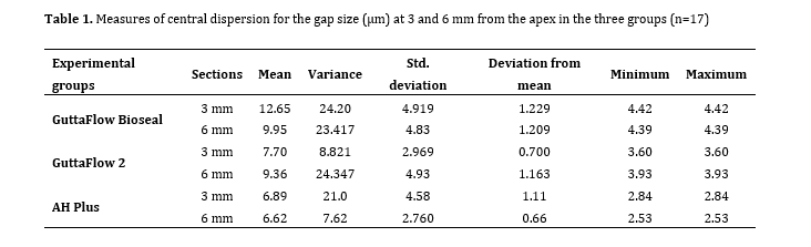
Results: At 3 mm distance from the apex, the mean gap size in the GuttaFlow Bioseal group (12.65±4.919 µm) was significantly larger than that in GuttaFlow (7.7±2.969 µm) and AH Plus (6.89±4.58 µm) groups (P<0.05), but no significant difference was detected at 6 mm from the apex among the three groups (P=0.121).
Conclusion: Under the limitations of this in vitro study, the results indicated that GuttaFlow Bioseal had poorer marginal adaptation than AH Plus and GuttaFlow at 3 mm from the apex; however, marginal adaptation of sealers was not significantly different at 6 mm from the apex.
Keywords: Dental Marginal Adaptation; Epoxy Resin-Based Root Canal Sealer; GuttaFlow; Root Canal Therapy
Introduction
Microleakage refers to the passage of bacteria, liquids, ions, and molecules through the root filling material and root canal wall interface, which may be clinically undetectable, but is among the main factors that compromise the success of endodontic treatment over time [1]. The reason for microleakage is the marginal gaps at the tooth-filling material interface, which allow the leakage of bacteria [2]. It has been well documented that proliferation of the leaked bacteria can result in dental caries, endodontic reinfection, and periapical diseases [3]. The origin of the bacteria responsible for root canal reinfections has not been clearly identified [4]; however, it is widely believed that the bacteria present in the smear layer can proliferate and cause endodontic reinfection through microleakage [5]. Therefore, achieving a hermetic seal is often one of the main goals of root canal therapy. However, the term “hermetic” is often used for an air-tight seal, which is not the case in endodontic treatment; therefore, terms such as fluid-tight, fluid-impervious, and bacteria-tight seals are currently preferred to describe an ideal endodontic seal [6].
An ideal sealer should be biocompatible, nontoxic, and radiopaque. It should have antimicrobial activity, fill the root canal, and have dimensional stability and proper adhesion to the root canal walls [4]. Also, an ideal sealer should be able to provide an excellent seal for a long period of time [7]. Maximum adaptation of root filling material and sealer to root dentin is highly important in this regard. Microscopic gaps at the sealer-dentin and sealer-root filling material interfaces can compromise the success of root canal treatment, and marginal leakage through such gaps is still one of the main causes of endodontic treatment failure [8]. AH Plus (Dentsply-Sirona, DeTrey GmbH, Konstanz, Germany) is an epoxy resin-based sealer that has excellent physicochemical properties and apical sealing ability, making it a popular choice for
comparative studies [9, 10]. GuttaFlow 2 (Coltene Whaledent, Altstatten, Switzerland) is a silicone-based root canal sealer available in Automix syringe form. It contains gutta-percha powder, polydimethylsiloxane, platinum catalyst, zirconium dioxide, and micro-silver [11, 12].
The bioactive ingredients in Guttaflow Bioseal (Coltene/Whaledent AG, Alstatten, Switzerland) are supposed to support tissue regeneration and repair [12]. GuttaFlow Bioseal differs from other GuttaFlow sealers by incorporating bioactive glass, comprising silica, calcium oxide, sodium oxide, and phosphorus oxide [13].
Different obturation techniques and several sealer types are used to provide an optimal seal in the root canal system. Considering the lack of information regarding the comparative marginal adaptation of different sealer types, this study aimed to compare the marginal adaptation of GuttaFlow 2, GuttaFlow Bioseal, and AH Plus in the apical third of single-canal human teeth obturated by the single-cone obturation technique. The null hypothesis was that the marginal adaptation of the above-mentioned three types of sealers would not be significantly different in the single-canal human teeth obturated by the single-cone obturation technique.
Materials and Methods
This in vitro study was conducted on human anterior teeth extracted because of poor periodontal prognosis. The study was approved by the ethics committee of the Dental School at Islamic Azad University of Medical Sciences, Tehran, Iran (IR.IAU.DENTAL.REC.1399.219).
Eligibility criteria
Human extracted single-canal incisors with no fracture, crack, canal calcification, and internal or external root resorption, and 0 to 15-degree root canal curvature as measured by Schneider’s method [14] were included. Open-apex teeth and those with previous endodontic treatment were excluded [15].
Sample size calculation
The sample size was calculated to be 17 in each group according to a study by Shinde et al, [15], assuming a one-unit standard deviation of the mean difference between the groups, alpha=5%, and study power of 80% using 2015 Power and Precision software version 3.
Sample preparation
A total of 51 eligible anterior teeth were collected and subsequently disinfected by immersion in 5.25% NaOCl (Morvabon, Tehran, Iran) for 1 minute. A #10 K-file (Mani, Japan) was introduced into the canal, and a periapical radiograph was obtained to ensure apical patency and desirable degree of curvature. BioRaCe rotary files (FKG Dentaire, La Chaux de Fonds, Switzerland) were used to #40/4% for chemo-mechanical root canal preparation. Patency was ensured with a #10 K-file (Mani, Japan) while a #25 K-file could not pass through the apex. Following the use of each rotary file, the root canal was rinsed with 2 mL of NaOCl (Morvabon, Tehran, Iran). The teeth were then randomly divided into three groups (n=17) for obturation with GuttaFlow 2, GuttaFlow Bioseal, and AH Plus sealers. After rinsing the canals with 17% EDTA (Morvabon, Tehran, Iran) for 1 minute, 5.25% NaOCl (Morvabon, Tehran, Iran) was used for 30 seconds, followed by 2 mL of distilled water for smear layer removal [16]. For single-cone obturation of the canals in each group, the sealer was injected into the canal by an auto-mix tip, and the matched gutta-percha point was inserted into canal, and its excess length was cut 1 mm below the orifice by a hot hand plugger (Mani Inc., Tochigi, Japan).
Finally, a cold plugger was used to vertically pack the gutta-percha at the canal orifice. The obturated teeth were incubated at 37°C and 100% humidity for 7 days in an incubator (Memmert GmbH, Germany). The teeth were then mounted in auto-polymerizing acrylic resin (Acropars; Marlic Medical Industries, Iran) and transversely sectioned using a diamond cutting disc (Buehler, Lake Bluff, USA) mounted on a high precision CNC machine (Delta, Neihu, Taipei) at 3- and 6-mm distances from the apex. Sectioning was performed under water coolant to prevent overheating. The slices were then gold-plated to 10-15 nm thickness and inspected under a scanning electron microscope (SEM; Seron Technologies Inc., South Korea, Gyeonggi) with 15 kV voltage at x25 to x2000 magnifications. The gap at the root filling material-root canal wall interface was measured at 3 points with 120-degree angles relative to each other at 3 and 6 mm from the apex using Image J software, and the mean values were reported (Figures 1-3) [8].
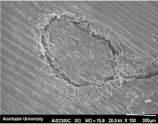
Figure 1. Representative SEM micrographs of the cross-section of obturated teeth using GuttaFlow Bioseal at x150 magnification
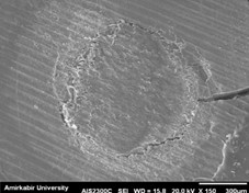
Figure 2. Representative SEM micrographs of the cross-section of obturated teeth using AH Plus at x 150 magnification
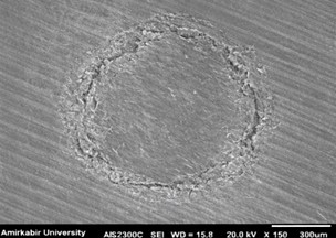
Figure 3. Representative SEM micrographs of the cross-section of obturated teeth using GuttaFlow 2 at x150 magnification
Statistical analysis:
Table 1. Measures of central dispersion for the gap size (µm) at 3 and 6 mm from the apex in the three groups (n=17)
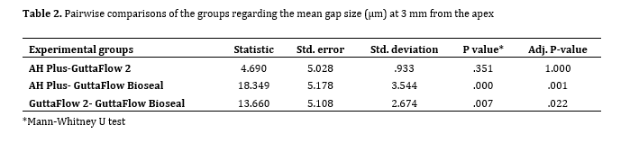
Table 2. Pairwise comparisons of the groups regarding the mean gap size (µm) at 3 mm from the apex
Discussion
This study compared AH Plus, GuttaFlow 2, and GuttaFlow Bioseal regarding marginal adaptation at 3 and 6 mm from the apex in single-canal anterior teeth filled by the single-cone obturation technique. The results showed that GuttaFlow Bioseal had significantly poorer marginal adaptation at 3 mm from the apex compared with the other two sealers; however, the difference among the three sealers was not significant at 6 mm from the apex. Thus, the null hypothesis of the study was rejected at 3 mm, and accepted at 6 mm from the apex.
Marginal adaptation of root canal sealers plays a critical role in preventing microleakage and subsequent reinfection. However, the literature regarding microleakage assessment remains controversial [8]. Several techniques have been employed to evaluate the sealing ability and marginal adaptation of endodontic sealers, including confocal laser scanning microscopy, which reveals sealer penetration into dentinal tubules [17], cone-beam computed tomography [18], bacterial leakage models [19], micro-computed tomography [20], a combination of micro-computed tomography and SEM [21], and dye penetration methods, which are among the oldest methods used for this assessment [22]. Among these methods, SEM has been widely used due to its high resolution, deep field of view, and high magnification, allowing precise evaluation of the core material interface [8, 23]. Accordingly, in the present study, marginal adaptation of sealers was evaluated using SEM, as recommended by several previous investigations [6, 15, 23]. Nevertheless, SEM has certain limitations, including the possibility of mechanical damage to the root filling margins during specimen sectioning and its inherently two-dimensional assessment of the dentin–filling material interface, which should be considered when interpreting the findings.
The single-cone obturation technique was adopted in the present study to simulate a commonly used obturation method in clinical practice and to achieve maximum standardization among the experimental groups, while exhibiting lower complexity compared with three-dimensional condensation techniques [8, 24]. Mandibular premolars [21, 25] and maxillary molars [26] have been commonly used in previous studies for the assessment of marginal adaptation of sealers. In the present study, single-canal anterior teeth were selected to minimize anatomical variations and ensure methodological standardization [6].
A more favorable seal with GuttaFlow Bioseal using the single-cone obturation technique, compared with AH Plus, has been reported by Lee et al. [25]. However, in the present study, GuttaFlow Bioseal exhibited significantly inferior marginal adaptation compared with AH Plus and GuttaFlow 2 at 3 mm from the apex. This discrepancy may be attributed to differences in tooth type (anterior teeth in the present study versus mandibular premolars in their study) as well as variations in gap measurement techniques (SEM in the present study versus liquid flow rate measurement in their study) [25].
Superior adaptation of GuttaFlow in the apical third compared with AH Plus and RealSeal has also been demonstrated by Adhikari and Jain [8]. In contrast, the present study found no significant difference in marginal adaptation between GuttaFlow 2 and AH Plus, which may be explained by the use of GuttaFlow 2, a more recent formulation of GuttaFlow.
No significant difference between GuttaFlow Bioseal and AH Plus regarding penetration into dentinal tubules has been reported by Ackay et al. [17] using confocal laser scanning microscopy. The discrepancy between their findings and the results of the present study may be related to differences in the evaluation method and tooth selection, as single-rooted mandibular premolars were used in their study.
Superior sealing ability of GuttaFlow Bioseal compared with GuttaFlow and bioceramic sealers using the single-cone obturation technique has also been reported by Naji and Al-Gharrawi [27]. In their study, no significant difference was observed between AH Plus and GuttaFlow Bioseal; however, AH Plus showed higher sealability than GuttaFlow. These findings are in contrast with the present results and may be attributed to the use of maxillary first molars and the dye penetration technique for sealability assessment. It has also been reported by Kaul et al. [28] that none of the evaluated sealers could provide a complete apical seal, with no significant difference observed between AH Plus and GuttaFlow, which is consistent with the findings of the present study.
The use of SEM for the assessment of marginal adaptation may be regarded as a limitation, as it provides only a two-dimensional evaluation of a three-dimensional and highly complex root canal system. Therefore, future studies are recommended to employ more advanced three-dimensional assessment methods and to evaluate different tooth types with varying degrees of root curvature.
Conclusion
Within the limitations of this in vitro study, the results showed that sealer type affected marginal adaptation in the apical third when the single-cone obturation technique was used. GuttaFlow Bioseal demonstrated significantly poorer marginal adaptation than AH Plus and GuttaFlow 2 at 3 mm from the apex; whereas, no significant differences were observed among the three sealers at 6 mm from the apex. These findings suggest that, despite its bioactive formulation, GuttaFlow Bioseal may not provide superior apical adaptation under the conditions of this study. Further investigations using three-dimensional evaluation methods and different tooth anatomies are recommended to confirm these results.
Background and Aim: This study compared the marginal adaptation of GuttaFlow 2, GuttaFlow Bioseal, and AH Plus in the apical third of human single-canal teeth obturated by the single-cone technique.
Materials and Methods: This in vitro study evaluated 51 eligible extracted single-canal maxillary incisors. The teeth underwent biomechanical preparation with BioRaCe rotary system to #40/4%, and were then randomly divided into 3 experimental groups (n=17) of GuttaFlow 2, GuttaFlow Bioseal, and AH Plus. The canals were rinsed with 17% EDTA for 1 minute, 5.25% NaOCl for 30 seconds, and 2 mL of distilled water for smear layer removal, and were obturated with the single-cone obturation technique. The roots were then transversely sectioned at 3 and 6 mm from the apex, and the gap at the root filling material-canal wall interface was measured under a scanning electron microscope (SEM). Data were analyzed with the Kruskal-Wallis and Mann-Whitney U tests (alpha=0.05).
Results: At 3 mm distance from the apex, the mean gap size in the GuttaFlow Bioseal group (12.65±4.919 µm) was significantly larger than that in GuttaFlow (7.7±2.969 µm) and AH Plus (6.89±4.58 µm) groups (P<0.05), but no significant difference was detected at 6 mm from the apex among the three groups (P=0.121).
Conclusion: Under the limitations of this in vitro study, the results indicated that GuttaFlow Bioseal had poorer marginal adaptation than AH Plus and GuttaFlow at 3 mm from the apex; however, marginal adaptation of sealers was not significantly different at 6 mm from the apex.
Keywords: Dental Marginal Adaptation; Epoxy Resin-Based Root Canal Sealer; GuttaFlow; Root Canal Therapy
Introduction
Microleakage refers to the passage of bacteria, liquids, ions, and molecules through the root filling material and root canal wall interface, which may be clinically undetectable, but is among the main factors that compromise the success of endodontic treatment over time [1]. The reason for microleakage is the marginal gaps at the tooth-filling material interface, which allow the leakage of bacteria [2]. It has been well documented that proliferation of the leaked bacteria can result in dental caries, endodontic reinfection, and periapical diseases [3]. The origin of the bacteria responsible for root canal reinfections has not been clearly identified [4]; however, it is widely believed that the bacteria present in the smear layer can proliferate and cause endodontic reinfection through microleakage [5]. Therefore, achieving a hermetic seal is often one of the main goals of root canal therapy. However, the term “hermetic” is often used for an air-tight seal, which is not the case in endodontic treatment; therefore, terms such as fluid-tight, fluid-impervious, and bacteria-tight seals are currently preferred to describe an ideal endodontic seal [6].
An ideal sealer should be biocompatible, nontoxic, and radiopaque. It should have antimicrobial activity, fill the root canal, and have dimensional stability and proper adhesion to the root canal walls [4]. Also, an ideal sealer should be able to provide an excellent seal for a long period of time [7]. Maximum adaptation of root filling material and sealer to root dentin is highly important in this regard. Microscopic gaps at the sealer-dentin and sealer-root filling material interfaces can compromise the success of root canal treatment, and marginal leakage through such gaps is still one of the main causes of endodontic treatment failure [8]. AH Plus (Dentsply-Sirona, DeTrey GmbH, Konstanz, Germany) is an epoxy resin-based sealer that has excellent physicochemical properties and apical sealing ability, making it a popular choice for
comparative studies [9, 10]. GuttaFlow 2 (Coltene Whaledent, Altstatten, Switzerland) is a silicone-based root canal sealer available in Automix syringe form. It contains gutta-percha powder, polydimethylsiloxane, platinum catalyst, zirconium dioxide, and micro-silver [11, 12].
The bioactive ingredients in Guttaflow Bioseal (Coltene/Whaledent AG, Alstatten, Switzerland) are supposed to support tissue regeneration and repair [12]. GuttaFlow Bioseal differs from other GuttaFlow sealers by incorporating bioactive glass, comprising silica, calcium oxide, sodium oxide, and phosphorus oxide [13].
Different obturation techniques and several sealer types are used to provide an optimal seal in the root canal system. Considering the lack of information regarding the comparative marginal adaptation of different sealer types, this study aimed to compare the marginal adaptation of GuttaFlow 2, GuttaFlow Bioseal, and AH Plus in the apical third of single-canal human teeth obturated by the single-cone obturation technique. The null hypothesis was that the marginal adaptation of the above-mentioned three types of sealers would not be significantly different in the single-canal human teeth obturated by the single-cone obturation technique.
Materials and Methods
This in vitro study was conducted on human anterior teeth extracted because of poor periodontal prognosis. The study was approved by the ethics committee of the Dental School at Islamic Azad University of Medical Sciences, Tehran, Iran (IR.IAU.DENTAL.REC.1399.219).
Eligibility criteria
Human extracted single-canal incisors with no fracture, crack, canal calcification, and internal or external root resorption, and 0 to 15-degree root canal curvature as measured by Schneider’s method [14] were included. Open-apex teeth and those with previous endodontic treatment were excluded [15].
Sample size calculation
The sample size was calculated to be 17 in each group according to a study by Shinde et al, [15], assuming a one-unit standard deviation of the mean difference between the groups, alpha=5%, and study power of 80% using 2015 Power and Precision software version 3.
Sample preparation
A total of 51 eligible anterior teeth were collected and subsequently disinfected by immersion in 5.25% NaOCl (Morvabon, Tehran, Iran) for 1 minute. A #10 K-file (Mani, Japan) was introduced into the canal, and a periapical radiograph was obtained to ensure apical patency and desirable degree of curvature. BioRaCe rotary files (FKG Dentaire, La Chaux de Fonds, Switzerland) were used to #40/4% for chemo-mechanical root canal preparation. Patency was ensured with a #10 K-file (Mani, Japan) while a #25 K-file could not pass through the apex. Following the use of each rotary file, the root canal was rinsed with 2 mL of NaOCl (Morvabon, Tehran, Iran). The teeth were then randomly divided into three groups (n=17) for obturation with GuttaFlow 2, GuttaFlow Bioseal, and AH Plus sealers. After rinsing the canals with 17% EDTA (Morvabon, Tehran, Iran) for 1 minute, 5.25% NaOCl (Morvabon, Tehran, Iran) was used for 30 seconds, followed by 2 mL of distilled water for smear layer removal [16]. For single-cone obturation of the canals in each group, the sealer was injected into the canal by an auto-mix tip, and the matched gutta-percha point was inserted into canal, and its excess length was cut 1 mm below the orifice by a hot hand plugger (Mani Inc., Tochigi, Japan).
Finally, a cold plugger was used to vertically pack the gutta-percha at the canal orifice. The obturated teeth were incubated at 37°C and 100% humidity for 7 days in an incubator (Memmert GmbH, Germany). The teeth were then mounted in auto-polymerizing acrylic resin (Acropars; Marlic Medical Industries, Iran) and transversely sectioned using a diamond cutting disc (Buehler, Lake Bluff, USA) mounted on a high precision CNC machine (Delta, Neihu, Taipei) at 3- and 6-mm distances from the apex. Sectioning was performed under water coolant to prevent overheating. The slices were then gold-plated to 10-15 nm thickness and inspected under a scanning electron microscope (SEM; Seron Technologies Inc., South Korea, Gyeonggi) with 15 kV voltage at x25 to x2000 magnifications. The gap at the root filling material-root canal wall interface was measured at 3 points with 120-degree angles relative to each other at 3 and 6 mm from the apex using Image J software, and the mean values were reported (Figures 1-3) [8].
Figure 1. Representative SEM micrographs of the cross-section of obturated teeth using GuttaFlow Bioseal at x150 magnification
Figure 2. Representative SEM micrographs of the cross-section of obturated teeth using AH Plus at x 150 magnification
Figure 3. Representative SEM micrographs of the cross-section of obturated teeth using GuttaFlow 2 at x150 magnification
Statistical analysis:
Statistical analysis was performed using SPSS version 24.0 (IBM Corp., Armonk, NY, USA). The normality of data distribution was assessed with the Kolmogorov-Smirnov test. The results indicated that the gap size data at both 3 mm and 6 mm from the apex were not normally distributed (P<0.05). Accordingly, comparisons among the three groups were performed using the Kruskal-Wallis test. When a statistically significant difference was detected, pairwise comparisons were conducted using the Mann-Whitney U test with Bonferroni correction as a non-parametric post-hoc analysis to adjust for multiple comparisons. A corrected P value<0.05 was considered statistically significant.
Results
A total of 102 sections were assessed in all three groups. The measures of central dispersion for the gap size at 3 and 6 mm from the apex in the three groups are presented in Table 1. The Kruskal-Wallis test showed a significant difference in the mean gap size among the three groups (P=0.001). At 3 mm distance from the apex, the mean gap size in the GuttaFlow Bioseal group (12.65±4.919 µm) was significantly larger than that in the GuttaFlow (7.7±2.969 µm) and AH Plus (6.89±4.58 µm) groups (P<0.05). Thus, pairwise comparisons were carried out by the Mann-Whitney U test (Table 2), which showed that the mean gap size in the GuttaFlow Bioseal group was significantly higher than that in the GuttaFlow2 (P=0.022) and AH Plus (P=0.001) groups at 3 mm from the apex. However, no significant difference was detected at 6 mm from the apex among the three groups (P=0.121).
Table 1. Measures of central dispersion for the gap size (µm) at 3 and 6 mm from the apex in the three groups (n=17)
Table 2. Pairwise comparisons of the groups regarding the mean gap size (µm) at 3 mm from the apex
Discussion
This study compared AH Plus, GuttaFlow 2, and GuttaFlow Bioseal regarding marginal adaptation at 3 and 6 mm from the apex in single-canal anterior teeth filled by the single-cone obturation technique. The results showed that GuttaFlow Bioseal had significantly poorer marginal adaptation at 3 mm from the apex compared with the other two sealers; however, the difference among the three sealers was not significant at 6 mm from the apex. Thus, the null hypothesis of the study was rejected at 3 mm, and accepted at 6 mm from the apex.
Marginal adaptation of root canal sealers plays a critical role in preventing microleakage and subsequent reinfection. However, the literature regarding microleakage assessment remains controversial [8]. Several techniques have been employed to evaluate the sealing ability and marginal adaptation of endodontic sealers, including confocal laser scanning microscopy, which reveals sealer penetration into dentinal tubules [17], cone-beam computed tomography [18], bacterial leakage models [19], micro-computed tomography [20], a combination of micro-computed tomography and SEM [21], and dye penetration methods, which are among the oldest methods used for this assessment [22]. Among these methods, SEM has been widely used due to its high resolution, deep field of view, and high magnification, allowing precise evaluation of the core material interface [8, 23]. Accordingly, in the present study, marginal adaptation of sealers was evaluated using SEM, as recommended by several previous investigations [6, 15, 23]. Nevertheless, SEM has certain limitations, including the possibility of mechanical damage to the root filling margins during specimen sectioning and its inherently two-dimensional assessment of the dentin–filling material interface, which should be considered when interpreting the findings.
The single-cone obturation technique was adopted in the present study to simulate a commonly used obturation method in clinical practice and to achieve maximum standardization among the experimental groups, while exhibiting lower complexity compared with three-dimensional condensation techniques [8, 24]. Mandibular premolars [21, 25] and maxillary molars [26] have been commonly used in previous studies for the assessment of marginal adaptation of sealers. In the present study, single-canal anterior teeth were selected to minimize anatomical variations and ensure methodological standardization [6].
A more favorable seal with GuttaFlow Bioseal using the single-cone obturation technique, compared with AH Plus, has been reported by Lee et al. [25]. However, in the present study, GuttaFlow Bioseal exhibited significantly inferior marginal adaptation compared with AH Plus and GuttaFlow 2 at 3 mm from the apex. This discrepancy may be attributed to differences in tooth type (anterior teeth in the present study versus mandibular premolars in their study) as well as variations in gap measurement techniques (SEM in the present study versus liquid flow rate measurement in their study) [25].
Superior adaptation of GuttaFlow in the apical third compared with AH Plus and RealSeal has also been demonstrated by Adhikari and Jain [8]. In contrast, the present study found no significant difference in marginal adaptation between GuttaFlow 2 and AH Plus, which may be explained by the use of GuttaFlow 2, a more recent formulation of GuttaFlow.
No significant difference between GuttaFlow Bioseal and AH Plus regarding penetration into dentinal tubules has been reported by Ackay et al. [17] using confocal laser scanning microscopy. The discrepancy between their findings and the results of the present study may be related to differences in the evaluation method and tooth selection, as single-rooted mandibular premolars were used in their study.
Superior sealing ability of GuttaFlow Bioseal compared with GuttaFlow and bioceramic sealers using the single-cone obturation technique has also been reported by Naji and Al-Gharrawi [27]. In their study, no significant difference was observed between AH Plus and GuttaFlow Bioseal; however, AH Plus showed higher sealability than GuttaFlow. These findings are in contrast with the present results and may be attributed to the use of maxillary first molars and the dye penetration technique for sealability assessment. It has also been reported by Kaul et al. [28] that none of the evaluated sealers could provide a complete apical seal, with no significant difference observed between AH Plus and GuttaFlow, which is consistent with the findings of the present study.
The use of SEM for the assessment of marginal adaptation may be regarded as a limitation, as it provides only a two-dimensional evaluation of a three-dimensional and highly complex root canal system. Therefore, future studies are recommended to employ more advanced three-dimensional assessment methods and to evaluate different tooth types with varying degrees of root curvature.
Conclusion
Within the limitations of this in vitro study, the results showed that sealer type affected marginal adaptation in the apical third when the single-cone obturation technique was used. GuttaFlow Bioseal demonstrated significantly poorer marginal adaptation than AH Plus and GuttaFlow 2 at 3 mm from the apex; whereas, no significant differences were observed among the three sealers at 6 mm from the apex. These findings suggest that, despite its bioactive formulation, GuttaFlow Bioseal may not provide superior apical adaptation under the conditions of this study. Further investigations using three-dimensional evaluation methods and different tooth anatomies are recommended to confirm these results.
Type of Study: Original article |
Subject:
Endodontics
References
1. Muliyar S, Shameem KA, Thankachan RP, Francis PG, Jayapalan CS, Hafiz KA. Microleakage in endodontics. J Int Oral Health. 2014 Nov-Dec;6(6):99-104.
2. Savadkouhi ST, Bakhtiar H, Ardestani SE. In vitro and ex vivo microbial leakage assessment in endodontics: A literature review. J Int Soc Prev Community Dent. 2016 Nov-Dec;6(6):509-16. [DOI:10.4103/2231-0762.195516] [PMID] [PMCID]
3. Ramamurthy P, Rath A, Sidhu P, Fernandes B, Nettem S, Fee PA, et al. Sealants for preventing dental caries in primary teeth. Cochrane Database Syst Rev. 2022 Feb;2(2):Cd012981. [DOI:10.1002/14651858.CD012981.pub2] [PMID] [PMCID]
4. Modaresi J, Mokhtari F, Khodarahmi E. In vitro comparison of the marginal adaptation of cold ceramic sealer with the single-cone obturation technique versus AH-26 sealer with the lateral compaction technique in single-canal teeth. BMC Oral Health. 2025 Jan;25(1):69. [DOI:10.1186/s12903-024-05314-2] [PMID] [PMCID]
5. Jokstad A. Secondary caries and microleakage. Dent Mater. 2016 Nov;32(1):11-25. [DOI:10.1016/j.dental.2015.09.006] [PMID]
6. Upadhyay V, Upadhyay M, Panday RK, Chturvedi TP, Bajpai U. A SEM evaluation of dentinal adaptation of root canal obturation with GuttaFlow and conventional obturating material. Indian J Dent Res. 2011 Nov-Dec;22(6):881. [DOI:10.4103/0970-9290.94696] [PMID]
7. Komabayashi T, Colmenar D, Cvach N, Bhat A, Primus C, Imai Y. Comprehensive review of current endodontic sealers. Dent Mater J. 2020 Sep;39(5):703-20. [DOI:10.4012/dmj.2019-288] [PMID]
8. Adhikari HD, Jain S. Scanning electron microscopic evaluation of marginal adaptation of AH-Plus, GuttaFlow, and RealSeal at apical one-third of root canals - Part II: Core-sealer interface. J Conserv Dent. 2018 Jan-Feb;21(1):90-4.
9. El Sayed M. Comparing sectional and total dentin bond strengths of three endodontic sealers after using the single-cone obturation technique: An in vitro study. J Contemp Dent Pract. 2024 Oct;25(10):976-82. [DOI:10.5005/jp-journals-10024-3765] [PMID]
10. Kunam D, Uppalapati Y, Ponnapalli AT, Krishna C, Bode Y, Yadatha S. Comparative evaluation of wettability of AH plus, Ceraseal and Guttaflow bioseal root canal sealers on root canal dentin: An in vitro study. J Conserv Dent Endod. 2024 Oct;27(10):1021-5. [DOI:10.4103/JCDE.JCDE_484_24] [PMID] [PMCID]
11. Gpv S, Ghosh M, Chatterjee R, Gajpal A, Mustafa M, Almokhatieb AA. Field emission scanning electron microscope analysis of the marginal adaptation of various root canal sealers at the dentin-sealer and sealer-gutta percha interfaces at three root canal levels: An in vitro study. Cureus. 2024 Aug;16(8):e66156. [DOI:10.7759/cureus.66156]
12. Eissa MH, Boghdadi R, Bedier M. Evaluation of the push-out bond strength and penetration of Guttaflow bBoseal, Endoseal MTA and Guttaflow 2 versus AH-Plus root canal sealers into the dentinal tubules in mandibular premolar teeth (a randomized in vitro comparative study). Adv Dent J. 2024 Jan;6(1):181-92. [DOI:10.21608/adjc.2023.191652.1251]
13. Behery K, Elbaz A, Ali SM, Elgendy AY. Comparative histological and immuno-histochemical analysis of the biocompatibility of three root canal sealers: An in vivo study. Egypt Dent J. 2024 Apr;70(2):2019-29. [DOI:10.21608/edj.2024.261680.2874]
14. Schneider SW. A comparison of canal preparations in straight and curved root canals. Oral Surg Oral Med Oral Pathol. 1971 Aug;32(2):271-5. [DOI:10.1016/0030-4220(71)90230-1] [PMID]
15. Shinde A, Kokate S, Hegde V. Comparative assessment of apical sealing ability of three different endodontic sealers: A scanning electron microscopic study. J Pierre Fauchard Acad (India Section). 2014 Sep;28(3):78-82. [DOI:10.1016/j.jpfa.2014.09.002]
16. Rajakumaran A, Ramesh H, Ashok R, Balaji L, Ganesh A. Smear layer removal and microhardness alteration potential of a naturally occurring antioxidant - An in vitro study. Cureus. 2019 Jul;11(7):e5241. [DOI:10.7759/cureus.5241] [PMID] [PMCID]
17. Akcay M, Arslan H, Durmus N, Mese M, Capar ID. Dentinal tubule penetration of AH Plus, iRoot SP, MTA Fillapex, and Guttaflow bioseal root canal sealers after different final irrigation procedures: A confocal microscopic study. Lasers Surg Med. 2016 Jan;48(1):70-6. [DOI:10.1002/lsm.22446] [PMID]
18. Raghuwanshi S, Jain P, Patni PM, Pandey SH, Hiremath H, Baghel S. Dentinal adaptation of warm thermoplastic obturating material and cold thermoplastic obturating material: An in vitro study. Contemp Clin Dent. 2019 Jan-Mar;10(1):64-8. [DOI:10.4103/ccd.ccd_312_18] [PMID] [PMCID]
19. Elias I, Guimarães GO, Caldeira CL, Gavini G, Cai S, Akisue E. Apical sealing ability comparison between GuttaFlow and AH Plus: in vitro bacterial and dye leakage. J Health Sci Inst. 2010 Jan-Mar;28(1):77-9.
20. Zare S, Shen I, Zhu Q, Ahn C, Primus C, Komabayashi T. Micro-computed tomographic evaluation of single-cone obturation with three sealers. Restor Dent Endod. 2021 May;46(2):e25. [DOI:10.5395/rde.2021.46.e25] [PMID] [PMCID]
21. Huang Y, Orhan K, Celikten B, Orhan AI, Tufenkci P, Sevimay S. Evaluation of the sealing ability of different root canal sealers: a combined SEM and micro-CT study. J Appl Oral Sci. 2018 Jan-Feb;26:e20160584. [DOI:10.1590/1678-7757-2016-0584] [PMID] [PMCID]
22. Sadr S, Golmoradizadeh A, Raoof M, Tabanfar MJ. Microleakage of single-cone gutta-percha obturation technique in combination with different types of sealers. Iran Endod J. 2015 Summer;10(3):199-203.
23. Punitha P, Shashikala K. Evaluation of the adaptation of resin based sealers epiphany, AH plus and AH 26 to the root canal dentin by scanning electron microscope. Indian J Stomatol. 2011 Dec;2(4):207.
24. Wu MK, van der Sluis LW, Wesselink PR. A 1-year follow-up study on leakage of single-cone fillings with RoekoRSA sealer. Oral Surg Oral Med Oral Pathol Oral Radiol Endod. 2006 May;101(5):662-7. [DOI:10.1016/j.tripleo.2005.03.013] [PMID]
25. Lee SH, Oh S, Al-Ghamdi AS, Mandorah AO, Kum KY, Chang SW. Sealing ability of AH Plus and GuttaFlow Bioseal. Bioinorg Chem Appl. 2020 Jan;2020:8892561. [DOI:10.1155/2020/8892561] [PMID] [PMCID]
26. Yanpiset K, Banomyong D, Chotvorrarak K, Srisatjaluk RL. Bacterial leakage and micro-computed tomography evaluation in round-shaped canals obturated with bioceramic cone and sealer using matched single cone technique. Restor Dent Endod. 2018 Aug;43(3):e30. [DOI:10.5395/rde.2018.43.e30] [PMID] [PMCID]
27. Naji AN, Al-Gharrawi HA. Comparison of the sealing ability of GuttaFlow Bioseal with different obturation systems (an in vitro study). J Int Dent Med Res. 2020 Dec;13(4):1632-6.
28. Kaul S, Kumar A, Badiyani BK, Sukhtankar L, Madhumitha M, Kumar A. Comparison of sealing ability of bioceramic sealer, AH Plus, and GuttaFlow in conservatively prepared curved root canals obturated with single-cone technique: An in vitro study. J Pharm Bioallied Sci. 2021 Jun;13(Suppl 1):S857-60. [DOI:10.4103/jpbs.jpbs_52_21] [PMID] [PMCID]
Send email to the article author
| Rights and permissions | |
 |
This work is licensed under a Creative Commons Attribution-NonCommercial 4.0 International License. |