Journal of Research in Dental
and Maxillofacial Sciences
Volume 10, Issue 4 (12-2025)
J Res Dent Maxillofac Sci 2025, 10(4): 292-298 |
Back to browse issues page

Ethics code: SRB/SDC-Perio/0167/2024
Download citation:
BibTeX | RIS | EndNote | Medlars | ProCite | Reference Manager | RefWorks
Send citation to:


BibTeX | RIS | EndNote | Medlars | ProCite | Reference Manager | RefWorks
Send citation to:
Chakraborty P, Gurumoorthy K, Suresh N. Preparation and Characterization of Europium-Doped Bioglass for Bone Regeneration: An In Vitro Study. J Res Dent Maxillofac Sci 2025; 10 (4) :292-298
URL: http://jrdms.dentaliau.ac.ir/article-1-970-en.html
URL: http://jrdms.dentaliau.ac.ir/article-1-970-en.html
Preparation and Characterization of Europium-Doped Bioglass for Bone Regeneration: An In Vitro Study


1- Department of Periodontology, Saveetha Dental College and Hospitals, Saveetha Institute of Medical and Technical Sciences, Saveetha University, Chennai, India
Keywords: Anti-Bacterial Agents, Bioglass, Bone Regeneration, Europium, Materials Science, Microscopy, Electron, Scanning
Full-Text [PDF 453 kb]
(234 Downloads)
| Abstract (HTML) (567 Views)
Full-Text: (254 Views)
Abstract
Background and Aim: Europium (Eu) is the most reactive and volatile rare-earth element. This study aimed to develop Eu-doped bioglass for applications in bone regeneration.
Materials and Methods: To synthesize Eu-doped bioglass, 1.3 g of cetyltrimethylammonium bromide was dissolved in 40 mL of ethanol to form a homogeneous surfactant solution. Europium nitrate, tetraethyl orthosilicate, calcium nitrate, and orthophosphoric acid were then sequentially added under continuous stirring. The mixture was stirred for 24 hours at room temperature to allow complete reaction and precursor integration. Next, 50 mL of acetone was added to induce precipitation, followed by centrifugation at 12,000 rpm for 6 minutes. The supernatant was discarded, and the collected precipitate was sintered to obtain the final bioglass. The fabricated Eu-doped bioglass was subsequently characterized using scanning electron microscopy (SEM), attenuated total reflectance–infrared spectroscopy (ATR-IR), and antimicrobial analysis to evaluate its morphological, structural, and biological properties.
Results: The ATR-IR spectra revealed typical silicate bands, and SEM images displayed a hollow-shaped structure. The superior crystallinity of Eu contributed to the bioglass network’s enhanced mechanical properties. Antimicrobial evaluation revealed a notable reduction in the number of colonies for both Escherichia coli (E. coli) and Staphylococcus aureus (S. aureus).
Conclusion: The synthesized Eu-doped bioglass exhibited notable antimicrobial activity and favorable characteristics for bone tissue engineering. Incorporation of Eu ions into the bioglass matrix enhanced biological performance, suggesting its potential applicability in bone regeneration. These findings indicate that Eu-doped bioglass could serve as a promising material for biomedical applications, particularly for promoting bone healing.
Keywords: Anti-Bacterial Agents; Bioglass; Bone Regeneration; Europium; Materials Science; Microscopy, Electron, Scanning
Introduction
Materials science has shown increasing interest in rare-earth elements due to their unique physicochemical and biological properties. Recent studies suggest that incorporating rare-earth elements into biomaterials can significantly enhance their biological performance. Among them, europium (Eu) is particularly notable for its versatility, reactivity, and softness, typically existing in its trivalent state (Eu³⁺) [1]. While widely used in industrial applications, Eu has also gained attention in biomedical research for its fluorescent properties, making it useful in cell imaging, drug release tracking, and quantitative detection [2].
Bioglass is a well-established material in bone tissue engineering, known for its excellent osteoinductive properties. It forms a strong bond with host bone through surface dissolution and subsequent formation of an apatite layer. The release of ions such as silica and calcium further stimulates cellular activities that are critical for bone matrix production [2,3]. Despite its advantages, bioglass suffers from limitations, including low mechanical strength and poor fracture toughness, which restrict its application in load-bearing scenarios [4]. Its high porosity, even greater than that of cancellous bone, can also pose challenges for structural integrity.
Recent studies have explored the incorporation of Eu into the bioglass matrix to enhance its biological functionalities. Eu-doped biomaterials have demonstrated a range of beneficial effects, including promotion of osteogenesis, angiogenesis, nerve regeneration, and notable antimicrobial and anticancer activities [5,6]. In particular, Eu-doped mesoporous bioactive glasses have been shown to stimulate bone formation in vivo and offer concentration-dependent antibacterial properties, paving the way for antibiotic-free, infection-resistant materials [7-9].
Given these promising attributes, the present study aimed to synthesize Eu-doped bioglass and evaluate its morphology and antimicrobial properties. The goal was to assess the potential of Eu-doped bioglass as a multifunctional material for bone regeneration and infection control in bone tissue engineering.
Materials and Methods
This in vitro study was conducted at the research laboratory of Saveetha Dental College and Hospital. The study protocol was reviewed and approved by the Institutional Review and Ethical Committee prior to commencement, under the reference number SRB/SDC/PERIO-2204/23/060.
Fabrication of material
All reagents used in this study were of analytical grade and used without further purification. Cetyltrimethylammonium bromide (1.3 g; SRL Chem, Mumbai, India) was dissolved in 40 mL of ethanol (SS Chemicals, U.P., India) and mixed with 55 mL of distilled water. The solution was stirred continuously for 1 hour using a magnetic stirrer.
Separately, 5.56 g of calcium nitrate tetrahydrate [Ca(NO₃)₂·4H₂O; Loba Chemie Pvt Ltd., Mumbai, India] was dissolved in 10 mL of distilled water and added to the cetyltrimethylammonium bromide-ethanol solution. Then, 0.45 g of europium (III) nitrate hexahydrate [Eu(NO₃)₃·6H₂O; Central Drug House (P) Ltd., New Delhi, India] was dissolved in 10 mL of distilled water and added to the same mixture. Next, 10.25 mL of tetraethyl orthosilicate (Central Drug House (P) Ltd., New Delhi, India), 2.08 g of sodium nitrate (NaNO₃; Central Drug House (P) Ltd., New Delhi, India), and 21.1 g of orthophosphoric acid (H₃PO₄;Kesari Scientific Chemicals, Chennai, India) were sequentially added. The final solution was stirred overnight at room temperature to ensure uniform mixing and precursor interaction.
After 24 hours, 50 mL of acetone (Nutan Chemical, Chakan, India) was added, and the solution was stirred for an additional 10 minutes. The mixture was then centrifuged at 12,000 rpm for 6 minutes. The supernatant was discarded, and the precipitate was collected, dried in a hot-air oven, and subsequently sintered to obtain the Eu-doped bioglass. The fabricated bioglass was then subjected to further analysis [10].
Characterization of Eu-doped bioglass
Scanning electron microscopy (SEM) and energy dispersive X-ray spectroscopy (EDS) analyses:
The morphological and elemental characterization of Eu-doped bioglass was conducted using field emission SEM (FESEM) integrated with EDS. A JEOL JSM-IT800 FESEM system (JEOL Ltd., Tokyo, Japan) was used for this purpose. Powdered samples were mounted on aluminum stubs using conductive carbon tape and then coated with a thin gold layer using a sputter coater to enhance conductivity and reduce charging effects. The imaging was carried out under high-vacuum conditions with an accelerating voltage of 14.0 kV, a spot size of 30, and a working distance of 9.2 mm. SEM images were captured at a magnification of 5,000×, with a field of view of approximately 34.65 µm. EDS analysis was performed at the same regions imaged under SEM to assess the elemental composition of the sample. The system was operated with Tru-Q® software to collect and process the elemental data.
Attenuated total reflectance Fourier-transform infrared (ATR-FTIR) spectroscopy:
ATR-FTIR was performed to characterize the chemical structure and functional groups in Eu-doped bioglass. The analysis was conducted using an ATR-IR spectrometer (Bruker, Billerica, MA, USA). Before analysis, the bioglass samples were finely ground into a uniform powder using an agate mortar and pestle. A small amount of the powder was placed directly onto the diamond ATR crystal, and firm contact was ensured using the built-in pressure arm of the instrument. Spectra were recorded in the range of 4000 cm⁻¹ to 400 cm⁻¹ at a resolution of 4 cm⁻¹, and 32 scans were averaged per sample to enhance spectral clarity. A background spectrum was collected before each sample run, and automatically subtracted to eliminate environmental interference.
Antimicrobial assay
The antimicrobial activity of Eu-doped bioglass was evaluated against two common bacterial strains associated with bone infections: Escherichia coli (E. Coli; ATCC 25922) and Staphylococcus aureus (S. aureus; ATCC 25923). These strains were obtained from a recognized microbial culture collection and maintained under aseptic conditions. Both bacterial strains were cultured in nutrient broth and incubated at 37°C for 18 hours to reach the logarithmic growth phase. After incubation, the bacterial suspensions were adjusted to match 0.5 McFarland standard turbidity, corresponding to approximately 1 × 10⁸ colony-forming units (CFUs)/mL [11].
To assess bactericidal activity, the CFU counting method was employed. The bioglass samples were incubated with standardized bacterial suspensions at 37°C for 24 hours under static conditions. Post-incubation, aliquots of the bacterial suspensions were serially diluted in sterile physiological saline. From each dilution, 100 µL was plated on nutrient agar plates using the spread plate technique. The plates were then incubated at 37°C for 24 hours, after which the number of colonies formed was manually counted. The percentage reduction in CFUs relative to the control groups (without bioglass) was calculated to quantify the antimicrobial effectiveness of Eu-doped bioglass [12].
Results
SEM and EDX analyses
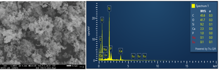
The SEM analysis of Eu-doped bioglass revealed that the material exhibited a well-formed, porous structure, which is crucial for bone regeneration applications. The pores observed in the SEM images were within the ideal range for facilitating cellular infiltration and tissue integration, which is necessary for successful bone healing as illustrated in Figure 1. The EDX analysis of the novel Eu-doped bioglass revealed its surface elemental composition, consisting of 45.6% carbon (C), 41.7% oxygen (O), 9.2% silicon (Si), 2.3% calcium (Ca), 1% phosphorus (P), 0.1% sodium (Na), and 0.1% Eu by weight, shown in Figure 1.
Figure 1. SEM analysis and EDX spectra of Eu-doped bioglass
ATR analysis
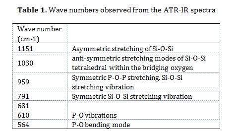
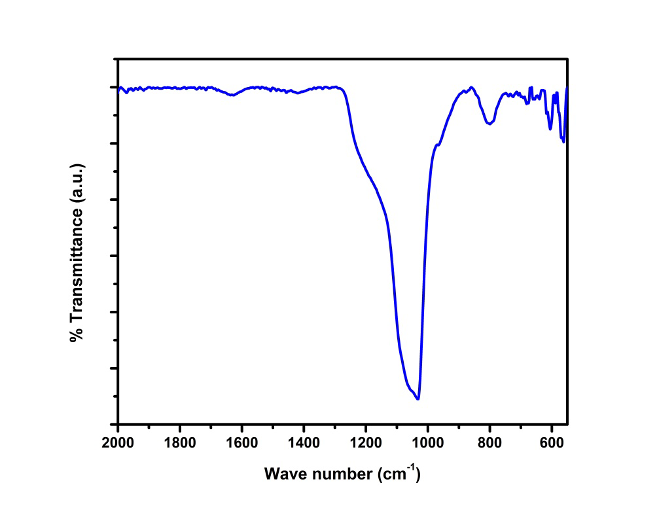
Figure 2 presents the ATR-IR results of Eu-doped bioglass network, which displayed consistent silicate-related bands throughout the spectra. These included vibration peaks at 1151 cm⁻¹ and 791 cm⁻¹, corresponding to the asymmetric and symmetric stretching of Si-O-Si bonds, respectively. Additional notable peaks were detected: a peak at 959 cm⁻¹ associated with P-O-P stretching in the bioglass matrix; a peak at 1030 cm⁻¹ linked to the antisymmetric stretching of Si-O-Si tetrahedra involving bridging oxygen atoms; a smaller peak at 564 cm⁻¹ attributed to P-O bending indicative of crystalline apatite-like calcium phosphate; and a peak at 681 cm⁻¹ related to the symmetric stretching of P-O bonds in crystalline silicate, as detailed in Table 1.
Table 1. Wave numbers observed from the ATR-IR spectra
Antimicrobial analysis
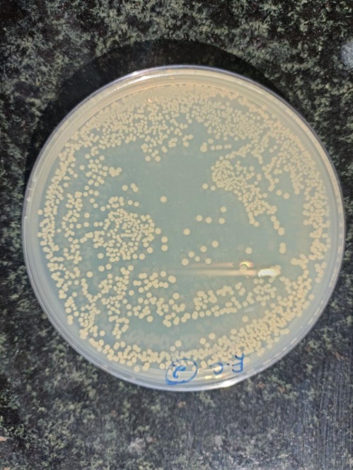
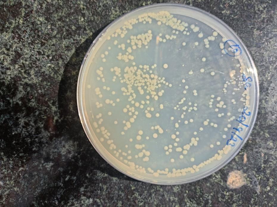
Antimicrobial testing was conducted by placing bacterial cultures of E. coli and S. aureus onto the bioglass samples. The growth inhibition was assessed through colony counting after incubation at 37°C for 24 hours. Initial bacterial counts before treatment were approximately 200,000,000 CFUs. After incubating with Eu-doped bioglass, bacterial growth was reduced by approximately 70-80% for E. coli, and 80-85% for S. aureus, as shown in Figures 3 and 4.
Figure 2. ATR-IR spectrum of Eu-doped bioglass
Figure 3. Antimicrobial analysis against E. coli
Figure 4. Antimicrobial analysis against S. aureus
Discussion
The SEM analysis of Eu-doped bioglass revealed that the material exhibited a well-formed, porous structure, which is crucial for bone regeneration applications. The pores observed in the SEM images were within the ideal range for facilitating cellular infiltration and tissue integration, which is necessary for successful bone healing as illustrated in Figure 1. The EDX analysis of the novel Eu-doped bioglass revealed its surface elemental composition, consisting of 45.6% carbon (C), 41.7% oxygen (O), 9.2% silicon (Si), 2.3% calcium (Ca), 1% phosphorus (P), 0.1% sodium (Na), and 0.1% Eu by weight, shown in Figure 1.
Figure 1. SEM analysis and EDX spectra of Eu-doped bioglass
ATR analysis
Figure 2 presents the ATR-IR results of Eu-doped bioglass network, which displayed consistent silicate-related bands throughout the spectra. These included vibration peaks at 1151 cm⁻¹ and 791 cm⁻¹, corresponding to the asymmetric and symmetric stretching of Si-O-Si bonds, respectively. Additional notable peaks were detected: a peak at 959 cm⁻¹ associated with P-O-P stretching in the bioglass matrix; a peak at 1030 cm⁻¹ linked to the antisymmetric stretching of Si-O-Si tetrahedra involving bridging oxygen atoms; a smaller peak at 564 cm⁻¹ attributed to P-O bending indicative of crystalline apatite-like calcium phosphate; and a peak at 681 cm⁻¹ related to the symmetric stretching of P-O bonds in crystalline silicate, as detailed in Table 1.
Table 1. Wave numbers observed from the ATR-IR spectra
Antimicrobial analysis
Antimicrobial testing was conducted by placing bacterial cultures of E. coli and S. aureus onto the bioglass samples. The growth inhibition was assessed through colony counting after incubation at 37°C for 24 hours. Initial bacterial counts before treatment were approximately 200,000,000 CFUs. After incubating with Eu-doped bioglass, bacterial growth was reduced by approximately 70-80% for E. coli, and 80-85% for S. aureus, as shown in Figures 3 and 4.
Figure 2. ATR-IR spectrum of Eu-doped bioglass
Figure 3. Antimicrobial analysis against E. coli
Figure 4. Antimicrobial analysis against S. aureus
Discussion
The present study aimed to synthesize and characterize Eu-doped bioglass to assess its morphological, chemical, and antimicrobial properties, with a focus on its potential for bone regeneration in periodontal applications.
Periodontal regeneration remains a clinical challenge, particularly due to the need for biomaterials that not only support bone growth but also prevent bacterial colonization. Bioglass has long been recognized for its osteoconductive and osteoinductive effects, with studies showing its ability to form strong chemical bonds with bone via formation of a hydroxycarbonate apatite layer [13-16]. However, conventional bioglass suffers from low fracture toughness, poor mechanical strength, and delayed degradation, which limits its application in load-bearing environments [17]. In a systematic review, Abushahba et al. [18] emphasized the effectiveness of bioglass for treating periodontal bone defects, highlighting its promise for clinical use. Nevertheless, bioglass has drawbacks, including slow degradation, insufficient mechanical strength, and limited fracture resistance, which restrict its broader applications in periodontal treatment. To address these limitations, we developed a Eu-doped bioglass. SEM analysis revealed that the Eu-doped bioglass maintained a highly porous morphology. This is a critical structural feature, as interconnected porosity enhances cell infiltration, vascularization, and nutrient diffusion, all of which are necessary for successful osteointegration [19]. Wu et al. [20] emphasized the importance of such porosity in bioactive glasses for promoting cellular attachment and differentiation. The present findings are consistent with a previous study demonstrating that rare-earth-doped bioactive glasses retained surface roughness and porosity favorable for bone regeneration [21].
In the present study, ATR-IR confirmed the incorporation of Eu into the silicate glass network. The presence of Eu–O vibrational bands, along with shifts in phosphate and silicate peak positions suggest successful integration without disrupting the network structure. These shifts imply a possible modification in the bonding environment around phosphate groups, which may enhance the material's reactivity with physiological fluids and promote faster apatite layer formation. Similar observations were reported in another study, noting that doping with rare-earth ions like Eu³⁺ altered the glass matrix structure and increased bioactivity [22].
One of the most significant outcomes of this study was the antimicrobial behavior exhibited by the Eu-doped bioglass. The material demonstrated a concentration-dependent inhibition of S. aureus and E. coli, both of which are clinically relevant pathogens in bone infections. These findings align with previous reports where Eu³⁺ exhibited antibacterial properties via disruption of microbial cell membranes and interference with ion transport [23]. In another study, it was found that Eu³⁺ ions reduced bacterial viability through mechanisms involving oxidative stress and membrane destabilization, supporting our hypothesis regarding the antimicrobial action of the doped material [24].
The bactericidal effect observed in this study may also be due to the slow and sustained release of Eu ions from the glass matrix, which is advantageous compared to conventional antibiotics, as it minimizes the risk of developing resistance. This is supported by a previous study, showing similar antimicrobial efficacy using silver-doped bioglass, highlighting the potential of ion-releasing bioactive materials for infection control [25].
Taken together, integration of Eu into the bioglass matrix not only maintained its essential physical features (porosity and stability) but also imparted new functional capabilities, such as antibacterial activity. The combined effects of structural integrity, ion release, and antimicrobial potential make Eu-doped bioglass a promising candidate for periodontal regeneration, where both bone healing and infection prevention are essential.
Conclusion
The results of this study showed that Eu-doped bioglass exhibited promising antimicrobial properties and structural characteristics suitable for bone regeneration. SEM and ATR-IR analyses confirmed the successful integration of Eu into the glass matrix, while antimicrobial assays demonstrated effective inhibition of common bone-infecting bacteria. These findings suggest that Eu-doped bioglass has potential for supporting bone healing. Further in vivo studies are recommended to evaluate its long-term biocompatibility and clinical applicability.
Type of Study: Original article |
Subject:
Periodontology
References
1. Zhang Y, Hu M, Wang X, Zhou Z, Liu Y. Design and Evaluation of Europium Containing Mesoporous Bioactive Glass Nanospheres: Doxorubicin Release Kinetics and Inhibitory Effect on Osteosarcoma MG 63 Cells. Nanomaterials (Basel). 2018 Nov 21;8(11):961. [DOI:10.3390/nano8110961] [PMID] []
2. Chen F, Huang P, Zhu YJ, Wu J, Zhang CL, Cui DX. The photoluminescence, drug delivery and imaging properties of multifunctional Eu3+/Gd3+ dual-doped hydroxyapatite nanorods. Biomaterials. 2011 Dec;32(34):9031-9. [DOI:10.1016/j.biomaterials.2011.08.032] [PMID]
3. Hou X, Zhang L, Zhou Z, Luo X, Wang T, Zhao X, et al, Chen F, Zheng L. Calcium Phosphate-Based Biomaterials for Bone Repair. J Funct Biomater. 2022 Oct 14;13(4):187. [DOI:10.3390/jfb13040187] [PMID] []
4. Meskher H, Sharifianjazi F, Tavamaishvili K, Irandoost M, Nejadkoorki D, Makvandi P. Limitations, challenges and prospective solutions for bioactive glasses-based nanocomposites for dental applications: A critical review. J Dent. 2024 Nov;150:105331. [DOI:10.1016/j.jdent.2024.105331] [PMID]
5. Wu L, Yang F, Xue Y, Gu R, Liu H, Xia D, et al. The biological functions of europium-containing biomaterials: A systematic review. Mater Today Bio. 2023 Feb 24;19:100595. [DOI:10.1016/j.mtbio.2023.100595] [PMID] []
6. Shi M, Xia L, Chen Z, Lv F, Zhu H, Wei F, et al. Europium-doped mesoporous silica nanosphere as an immune-modulating osteogenesis/angiogenesis agent. Biomaterials. 2017 Nov;144:176-87. [DOI:10.1016/j.biomaterials.2017.08.027] [PMID]
7. Cui Y, Hong S, Jiang W, Li X, Zhou X, He X, et al. Engineering mesoporous bioactive glasses for emerging stimuli-responsive drug delivery and theranostic applications. Bioact Mater. 2024 Jan 12;34:436-62. [DOI:10.1016/j.bioactmat.2024.01.001] [PMID] []
8. Wu C, Xia L, Han P, Mao L, Wang J, Zhai D, et al. Europium-Containing Mesoporous Bioactive Glass Scaffolds for Stimulating in Vitro and in Vivo Osteogenesis. ACS Appl Mater Interfaces. 2016 May 11;8(18):11342-54. [DOI:10.1021/acsami.6b03100] [PMID]
9. Hussein L, Moaness M, Mabrouk M, Farahat MG, Beherei HH. Advancements in mesoporous bioactive glasses for effective bone cancer therapy: Recent developments and future perspectives. Biomater Biosyst. 2025 Feb 15;17:100108. [DOI:10.1016/j.bbiosy.2025.100108] [PMID] []
10. Chitra S, Bargavi P, Balasubramaniam M, Chandran RR, Balakumar S. Impact of copper on in-vitro biomineralization, drug release efficacy and antimicrobial properties of bioactive glasses. Mater Sci Eng C Mater Biol Appl. 2020 Apr;109:110598. [DOI:10.1016/j.msec.2019.110598] [PMID]
11. Iconaru SL, Groza A, Gaiaschi S, Rokosz K, Raaen S, Ciobanu SC, Chapon P, Predoi D. Antimicrobial properties of samarium doped hydroxyapatite suspensions and coatings. Coatings. 2020 Nov 20;10(11):1124. [DOI:10.3390/coatings10111124]
12. Iconaru SL, Predoi D, Ciobanu CS, Negrila CC, Trusca R, Raaen S, Rokosz K, Ghegoiu L, Badea ML, Cimpeanu C. Novel Antimicrobial Agents Based on Zinc-Doped Hydroxyapatite Loaded with Tetracycline. Antibiotics (Basel). 2024 Aug 25;13(9):803. [DOI:10.3390/antibiotics13090803] [PMID] []
13. Yu F, Lian R, Liu L, Liu T, Bi C, Hong Ket al. Biomimetic Hydroxyapatite Nanorods Promote Bone Regeneration via Accelerating Osteogenesis of BMSCs through T Cell-Derived IL-22. ACS Nano. 2022 Jan 25;16(1):755-70. [DOI:10.1021/acsnano.1c08281] [PMID]
14. Liu DD, Zhang JC, Zhang Q, Wang SX, Yang MS. TGF-β/BMP signaling pathway is involved in cerium-promoted osteogenic differentiation of mesenchymal stem cells. J Cell Biochem. 2013 May;114(5):1105-14. [DOI:10.1002/jcb.24451] [PMID]
15. Marycz K, Smieszek A, Targonska S, Walsh SA, Szustakiewicz K, Wiglusz RJ. Three dimensional (3D) printed polylactic acid with nano-hydroxyapatite doped with europium(III) ions (nHAp/PLLA@Eu3+) composite for osteochondral defect regeneration and theranostics. Mater Sci Eng C Mater Biol Appl. 2020 May;110:110634. [DOI:10.1016/j.msec.2020.110634] [PMID]
16. Velraj MS, Suresh N, Kaarthikeyan G, Shivalingam C. In vitro study on the synthesis and characterization of erbium-doped hydroxyapatite/bioglass-polyvinyl alcohol scaffold for periodontal bone regeneration. J Pharm Bioallied Sci. 2025;21(2):239-49. [DOI:10.58240/1829006X-2025.2-239]
17. Sergi R, Bellucci D, Cannillo V. A comprehensive review of bioactive glass coatings: State of the art, challenges and future perspectives. Coatings. 2020 Aug 3;10(8):757. [DOI:10.3390/coatings10080757]
18. Abushahba F, Algahawi A, Areid N, Hupa L, Närhi TO. Bioactive glasses in periodontal regeneration: a systematic review. Tissue Eng. Part C Methods. 2023 May 1;29(5):183-96. [DOI:10.1089/ten.tec.2023.0036] [PMID]
19. Li F, Wang M, Pi G, Lei B. Europium Doped Monodispersed Bioactive Glass Nanoparticles Regulate the Osteogenic Differentiation of Human Marrow Mesenchymal Stem Cells. J Biomed Nanotechnol. 2018 Apr 1;14(4):756-64. [DOI:10.1166/jbn.2018.2504] [PMID]
20. Wu C, Zhou Y, Xu M, Han P, Chen L, Chang J, Xiao Y. Copper-containing mesoporous bioactive glass scaffolds with multifunctional properties of angiogenesis capacity, osteostimulation and antibacterial activity. Biomaterials. 2013 Jan;34(2):422-33. [DOI:10.1016/j.biomaterials.2012.09.066] [PMID]
21. Suresh N, Kaarthikeyan G. Green Synthesis and the Evaluation of Osteogenic Potential of Novel Europium-Doped-Monetite Calcium Phosphate by Cissus quadrangularis. Cureus. 2024 Apr 28;16(4):e59202. [DOI:10.7759/cureus.59202]
22. Reddy SB, Arumugam P, Kishore OG, K S. Development, Characterization, and Antibacterial Analysis of the Selenium-Doped Bio-Glass-Collagen-Gelatin Composite Scaffold for Guided Bone Regeneration. Cureus. 2023 Nov 15;15(11):e48838. [DOI:10.7759/cureus.48838]
23. Shih KY, Yu SC. Microwave-Assisted Rapid Synthesis of Eu(OH)3/RGO Nanocomposites and Enhancement of Their Antibacterial Activity against Escherichia coli. Materials (Basel). 2021 Dec 22;15(1):43. [DOI:10.3390/ma15010043] [PMID] []
24. Çinar Avar E, Türkmen KE, Erdal E, Loğoğlu E, Katircioğlu H. Biological Activities and Biocompatibility Properties of Eu(OH)3 and Tb(OH)3 Nanorods: Evaluation for Wound Healing Applications. Biol Trace Elem Res. 2023 Apr;201(4):2058-2070. [DOI:10.1007/s12011-022-03264-w] [PMID]
25. Zheng K, Balasubramanian P, Paterson TE, Stein R, MacNeil S, Fiorilli S, et al. Ag modified mesoporous bioactive glass nanoparticles for enhanced antibacterial activity in 3D infected skin model. Mater Sci Eng C Mater Biol Appl. 2019 Oct;103:109764. [DOI:10.1016/j.msec.2019.109764] [PMID]
| Rights and permissions | |
 |
This work is licensed under a Creative Commons Attribution-NonCommercial 4.0 International License. |