Journal of Research in Dental
and Maxillofacial Sciences
Volume 10, Issue 2 (6-2025)
J Res Dent Maxillofac Sci 2025, 10(2): 82-87 |
Back to browse issues page


Ethics code: IR.IAU.DENTAL.REC.1399.299
Download citation:
BibTeX | RIS | EndNote | Medlars | ProCite | Reference Manager | RefWorks
Send citation to:


BibTeX | RIS | EndNote | Medlars | ProCite | Reference Manager | RefWorks
Send citation to:
Fazlyab M, Hosseini Z S, Vatanpour M, Rezaei F. Effect of 660 nm and 808 nm Low-Level Diode Laser Irradiation on Production of Vascular Endothelial Growth Factor by Human Gingival Fibroblasts: An in vitro Study. J Res Dent Maxillofac Sci 2025; 10 (2) :82-87
URL: http://jrdms.dentaliau.ac.ir/article-1-797-en.html
URL: http://jrdms.dentaliau.ac.ir/article-1-797-en.html


1- Department of Endodontics, TeMS.C., Islamic Azad University, Tehran, Iran
2- Private Dentist, Tehran, Iran.
3- Department of Endodontics, TeMS.C., Islamic Azad University, Tehran, Iran ,dr.fatemeh.rezaeii@gmail.com
2- Private Dentist, Tehran, Iran.
3- Department of Endodontics, TeMS.C., Islamic Azad University, Tehran, Iran ,
Full-Text [PDF 340 kb]
(887 Downloads)
| Abstract (HTML) (2341 Views)
Full-Text: (709 Views)
Abstract
Background and Aim: This study aimed to assess the effect of 660 nm and 808 nm low-level diode laser irradiation on the production of vascular endothelial growth factor (VEGF) by human gingival fibroblasts (HGFs).
Materials and Methods: In this in vitro experimental study, HGFs were cultured in McCoy’s 5A modified medium and assigned to three groups of control, 660 nm laser irradiation, and 808 nm laser irradiation. The concentration of VEGF was measured in the three groups at 1, 3, and 7 days after the intervention using ELISA. The three groups were compared in this respect using repeated measures ANOVA. Statistical significance was defined as P<0.05. Results: The concentration of VEGF in 808 nm laser group was significantly higher than that in the control and 660 nm laser groups (P<0.05). Also, the concentration of VEGF in 660 nm laser group was significantly higher than that in the control group (P<0.05). The concentration of VEGF at 7 days was significantly higher than that at 1 and 3 days, and the value at 3 days was significantly higher than that at 1 day in all three groups (P<0.05).
Conclusion: Within the limitations of this study, it appears that irradiation of 660 and 808 nm diode laser can increase the production of VEGF by HGFs.
Introduction
Standard endodontic treatment is often recommended for immature teeth with a necrotic pulp due to caries, trauma, or periodontal problems [1, 2]. Regenerative treatments were recently suggested for management of such teeth considering the presence of open apex and the need for using an apical plug in these teeth, as well as the high risk of fracture of the thin root canal walls in the long-term [3]. Regenerative endodontic therapy aims to regenerate the pulp tissue by using a scaffold, stem cells, and growth factors, and enable continuation of dentin formation in the root canal system. Ideally, regenerative treatments should induce the formation of blood clot to serve as a scaffold and induce the migration of cells. Growth factors should also induce the differentiation of stem cells to fibroblasts, nerve cells, and secondary odontoblasts to form connective tissue, nerve fibers, and vasculature [2, 4]. Fibroblasts play an effective role in pulp regeneration [5]. They produce different growth factors such as the vascular endothelial growth factor (VEGF), fibroblast growth factor, and bone morphogenetic proteins in this process. VEGF has numerous effects on the function of vascular endothelial cells and induces angiogenesis. Thus, it is the most important growth factor for differentiation of the vascular system [6, 7]. Impaired blood circulation in the formed scaffold causes the death of the cells migrated to this area. Thus, presence of VEGF is imperative for the pulp regeneration process to continue [8]. Human gingival fibroblasts (HGFs) are easily accessible and show better immune response compared with other cells differentiated from the stem cells [9]. Thus, they are commonly used for research purposes regarding pulp regeneration.
Advances in laser technology paved the way for the use of low-level laser therapy (LLLT) for reduction of tissue inflammation, enhancement of wound healing, pain relief, and induction of tissue regeneration. Low level lasers with a wavelength range of 660-1100 nm can induce pulp regeneration without causing thermal damage [10]. Evidence shows that LLLT induces cell proliferation and differentiation, expression of angiogenic genes, osteogenesis, and dentinogenesis [10-12]. According to El Nawam et al. [10], low level diode laser at 660 nm wavelength increases the expression of VEGF gene and its receptor. Also, Gasparyan et al. [13] indicated that the efficacy of LLLT was comparable to that of VEGF in angiogenesis. Although HGFs are easily accessible and have high similarity to periodontal fibroblasts (as the origin of the cells migrating into the root canal space in the process of regeneration) [9], the production of VEGF by HGFs and the role of this group of cells in pulp regeneration have been less commonly addressed in scientific literature. However, these cells have an undeniably important role in wound healing, particularly in the periodontium [14]. Considering the gap of information regarding the effect of LLLT with different laser wavelengths on the production of VEGF by HGFs, this study aimed to assess the effect of 660 nm and 808 nm low level diode laser irradiation on production of VEGF by HGFs.
Materials and Methods
This in vitro experimental study evaluated HGFs (HGF3-PI 53) that were purchased from the Pasteur Institute of Iran (code C502) [15]. The minimum sample size was 9 wells in each group according to a study by Pansani et al. [14], assuming α=0.05, β=0.1, and mean cycle threshold of VEGF to be 165±43 in the laser and 100±31 in the control group using two-sample t-test power analysis.
Cell culture:
The HGF3-PI 53 cells obtained in cell flasks were cultured in McCoy’s 5A modified medium supplemented with 2% L-glutamine, HEPES, 15% fetal bovine serum, 0.4% antibiotic, 2% sodium pyruvate, and 0.2% antifungal agent under standard conditions at 37°C, 95% moisture, and 5% CO2. When the cells reached adequate confluence, they were detached by using a mixture of trypsin-EDTA (3 cc) and transferred into a new culture plate (first passage). This process was repeated after 2 weeks (second passage). The second-passage cells were trypsinized, detached from the dish, and centrifuged at 1400 rpm for 10 minutes. The sediment was resuspended in the culture medium, and after cell counting, it was transferred into a 96-well plate in equal amounts. The wells were then randomly divided into three groups of control (no laser irradiation), 660 nm laser irradiation, and 808 nm laser irradiation, with 9 wells in each group [16].
LLLT:
The medium was replaced 48 hours after culture. One group of the cells was irradiated with 660 nm diode laser (ASTAR; Polaris2, Bielsko-Biala, Poland) with 80 mW output power and 1 J/cm2 energy density for 3 seconds. The second group was subjected to 808 nm diode laser irradiation (ASTAR; Polaris2, Bielsko-Biala, Poland) with 80 mW output power, and 1 J/cm2 energy density for 3 seconds. To prevent light scattering and focusing on the respective well, the plate was wrapped with aluminum foil, and the wrap was perforated over the respective well for placement of the laser probe and laser irradiation through the hole.
Measuring the concentration of VEGF:
The concentration of VEGF was measured in all three groups at 1, 3 and 7 days after the intervention using a VEGF measurement kit (ZellBio, Germany) operating based on the sandwich technique by the ELISA [16].
Statistical analysis:
Data were analyzed by repeated measures ANOVA with laser irradiation as the between-subject factor using PASS 11 software. Statistical significance was defined as P<0.05.
Results
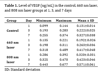
Table 1 presents the concentration of VEGF in the three groups. Two-way ANOVA showed no interaction effect of time and group on the concentration of VEGF (P=0.165). Irrespective of the group, the concentration of VEGF at 3 days was significantly higher than that at 1 day, and its concentration at 7 days was significantly higher than that at 3 days (P=0.001).
Table 1. Level of VEGF (ng/mL) in the control, 660 nm laser, and 808 nm laser groups at 1, 3, and 7 days
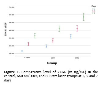
Also, the concentration of VEGF in the control group was significantly lower than that in both laser groups at all time points (P=0.001). Moreover, the concentration of VEGF in 808 nm laser group was significantly higher than that in 660 nm laser group at all time points (P=0.001; Figure 1).
Figure 1. Comparative level of VEGF (in ng/mL) in the control, 660 nm laser, and 808 nm laser groups at 1, 3, and 7 days
Discussion
This study assessed the effect of 660 nm and 808 nm low level diode laser irradiation on production of VEGF by HGFs. The results indicated that the concentration of VEGF in 808 nm laser group was significantly higher than that in the control and 660 nm laser groups. Also, the concentration of VEGF in 660 nm laser group was significantly higher than that in the control group. The concentration of VEGF at 7 days was significantly higher than that at 1 and 3 days, and the value at 3 days was significantly higher than that at 1 day in all three groups.
Pansani et al. [14] evaluated the effect of LLLT on production of VEGF by gingival fibroblasts obtained from young and elderly individuals, and reported an increase in concentration of VEGF in both cell types following irradiation of 780 nm laser with 2 and 3 J/cm2 energy densities, compared with the control group. No significant difference was noted between different energy densities in this respect. They concluded that LLLT of fibroblasts can induce tissue healing and angiogenesis. Although they assessed the effect of a different wavelength of laser (compared with the present study), they confirmed the optimal efficacy of LLLT for upregulation of VEGF gene in fibroblasts, which was in line with the present findings. El Nawam et al. [10] evaluated the effect of 810 nm and 660 nm diode laser irradiation on angiogenesis and formation of dentin-pulp complex and human dentin. They reported higher levels of VEGF in cells irradiated with 660 nm laser with 1 J/cm2 energy density compared with cells subjected to 660 nm laser irradiation with 3 J/cm2 energy density. However, the level of VEGF was not significantly different in cells subjected to irradiation of 810 nm laser with different energy densities, and generally increased in all groups. They concluded that LLLT enhances angiogenesis through the effect of VEGF, and can aid in pulp regeneration and vital pulp therapy. Their methodology was similar to that of the present study and they reported results in line with the present findings; however, they did not assess HGFs. Szezerbaty et al. [17] evaluated the effect of 660 nm laser with 1 and 5 J/cm2 energy densities on production of VEGF. They reported that after 72 hours, irradiation of 660 nm laser with 5 J/cm2 energy density caused a significant increase in level of VEGF compared with 1 J/cm2 energy density. They explained that factors such as energy density of laser can significantly affect the production of VEGF. We did not address this parameter in the current study. However, their results regarding the positive effect of 660 nm laser irradiation on angiogenesis were in agreement with the present findings. Chiarotto et al. [18] evaluated the wound healing effects of 670 nm InGaP and 830 nm GaAlAs lasers on rats, and reported that both laser types significantly increased the expression of VEGF gene, angiogenesis, and proliferation of fibroblasts, compared with the control group. However, 670 nm laser had a higher efficacy for proliferation of fibroblasts. Their results were in agreement with the present findings regarding enhancement of angiogenesis by LLLT. Illescas-Montes et al. [19] evaluated the effect of 940 nm diode laser on production of VEGF. They observed a significant increase in level of VEGF following irradiation of laser on HGFs twice (the second irradiation was performed after 144 hours). They discussed that laser irradiation can be used to increase the level of VEGF and enhance tissue healing. Although the laser wavelength in their study was different from that in the present study, their results regarding the optimal efficacy of LLLT for enhancement of VEGF confirmed the present findings. Szymczyszyn et al. [20] evaluated the effects of LLLT with 808 nm diode laser with 1.6 W/cm2 energy density on endothelial cells from the radial artery of 14 patients. They assessed the blood samples of patients before and 24 hours after laser irradiation, and concluded that laser irradiation had no significant effect on serum level of VEGF. Their results were different from the present findings, which may be due to different methodology, not using ELISA for measurement of the level of VEGF, or laser irradiation through the skin in their study. Cury et al. [21] evaluated the effects of 660 nm and 780 nm laser irradiation on angiogenesis in ischemic skin flaps in rats. They irradiated laser to 24 points of a skin flap and concluded that 660 nm and 780 nm laser induced the production of VEGF and enhanced angiogenesis in ischemic skin flaps in rats. Despite different study designs and methodologies, their results regarding the optimal effects of 660 nm laser irradiation on production of VEGF were in accordance with the present findings. Vitor et al. [22] assessed the effect of 660 nm laser irradiation with 2.5 and 3.7 J/cm2 energy densities on human pulp fibroblasts. They demonstrated that 660 nm laser induced the secretion of angiogenic proteins by human dental pulp fibroblasts, and increased the production of VEGF-A and VEGF-C, and their receptors. No significant difference was noted in the efficacy of different energy densities of laser. The laser wavelength in their study was similar to that in the present study. However, the cell type and laser energy densities were different. Nonetheless, their results confirmed the efficacy of 660 nm laser irradiation for the enhancement of VEGF production, which supports the present results.
The present study evaluated only one energy density of laser, which was a limitation of this study. Future studies should use different energy densities with the same wavelength to assess the effect of energy density on the results. Also, the frequency of laser irradiation cycles should be increased in future studies (e.g., laser irradiation of cells in three consecutive days) to assess the effect of this parameter on the level of VEGF.
Conclusion
Within the limitations of this study, it appears that irradiation of low-level laser, especially 808 nm diode laser with 80 mW output power and 1 J/cm2 energy density for 3 seconds can increase the production of VEGF by HGFs. This finding suggests the potential use of LLLT to promote tissue regeneration through increased angiogenesis.
Background and Aim: This study aimed to assess the effect of 660 nm and 808 nm low-level diode laser irradiation on the production of vascular endothelial growth factor (VEGF) by human gingival fibroblasts (HGFs).
Materials and Methods: In this in vitro experimental study, HGFs were cultured in McCoy’s 5A modified medium and assigned to three groups of control, 660 nm laser irradiation, and 808 nm laser irradiation. The concentration of VEGF was measured in the three groups at 1, 3, and 7 days after the intervention using ELISA. The three groups were compared in this respect using repeated measures ANOVA. Statistical significance was defined as P<0.05. Results: The concentration of VEGF in 808 nm laser group was significantly higher than that in the control and 660 nm laser groups (P<0.05). Also, the concentration of VEGF in 660 nm laser group was significantly higher than that in the control group (P<0.05). The concentration of VEGF at 7 days was significantly higher than that at 1 and 3 days, and the value at 3 days was significantly higher than that at 1 day in all three groups (P<0.05).
Conclusion: Within the limitations of this study, it appears that irradiation of 660 and 808 nm diode laser can increase the production of VEGF by HGFs.
Introduction
Standard endodontic treatment is often recommended for immature teeth with a necrotic pulp due to caries, trauma, or periodontal problems [1, 2]. Regenerative treatments were recently suggested for management of such teeth considering the presence of open apex and the need for using an apical plug in these teeth, as well as the high risk of fracture of the thin root canal walls in the long-term [3]. Regenerative endodontic therapy aims to regenerate the pulp tissue by using a scaffold, stem cells, and growth factors, and enable continuation of dentin formation in the root canal system. Ideally, regenerative treatments should induce the formation of blood clot to serve as a scaffold and induce the migration of cells. Growth factors should also induce the differentiation of stem cells to fibroblasts, nerve cells, and secondary odontoblasts to form connective tissue, nerve fibers, and vasculature [2, 4]. Fibroblasts play an effective role in pulp regeneration [5]. They produce different growth factors such as the vascular endothelial growth factor (VEGF), fibroblast growth factor, and bone morphogenetic proteins in this process. VEGF has numerous effects on the function of vascular endothelial cells and induces angiogenesis. Thus, it is the most important growth factor for differentiation of the vascular system [6, 7]. Impaired blood circulation in the formed scaffold causes the death of the cells migrated to this area. Thus, presence of VEGF is imperative for the pulp regeneration process to continue [8]. Human gingival fibroblasts (HGFs) are easily accessible and show better immune response compared with other cells differentiated from the stem cells [9]. Thus, they are commonly used for research purposes regarding pulp regeneration.
Advances in laser technology paved the way for the use of low-level laser therapy (LLLT) for reduction of tissue inflammation, enhancement of wound healing, pain relief, and induction of tissue regeneration. Low level lasers with a wavelength range of 660-1100 nm can induce pulp regeneration without causing thermal damage [10]. Evidence shows that LLLT induces cell proliferation and differentiation, expression of angiogenic genes, osteogenesis, and dentinogenesis [10-12]. According to El Nawam et al. [10], low level diode laser at 660 nm wavelength increases the expression of VEGF gene and its receptor. Also, Gasparyan et al. [13] indicated that the efficacy of LLLT was comparable to that of VEGF in angiogenesis. Although HGFs are easily accessible and have high similarity to periodontal fibroblasts (as the origin of the cells migrating into the root canal space in the process of regeneration) [9], the production of VEGF by HGFs and the role of this group of cells in pulp regeneration have been less commonly addressed in scientific literature. However, these cells have an undeniably important role in wound healing, particularly in the periodontium [14]. Considering the gap of information regarding the effect of LLLT with different laser wavelengths on the production of VEGF by HGFs, this study aimed to assess the effect of 660 nm and 808 nm low level diode laser irradiation on production of VEGF by HGFs.
Materials and Methods
This in vitro experimental study evaluated HGFs (HGF3-PI 53) that were purchased from the Pasteur Institute of Iran (code C502) [15]. The minimum sample size was 9 wells in each group according to a study by Pansani et al. [14], assuming α=0.05, β=0.1, and mean cycle threshold of VEGF to be 165±43 in the laser and 100±31 in the control group using two-sample t-test power analysis.
Cell culture:
The HGF3-PI 53 cells obtained in cell flasks were cultured in McCoy’s 5A modified medium supplemented with 2% L-glutamine, HEPES, 15% fetal bovine serum, 0.4% antibiotic, 2% sodium pyruvate, and 0.2% antifungal agent under standard conditions at 37°C, 95% moisture, and 5% CO2. When the cells reached adequate confluence, they were detached by using a mixture of trypsin-EDTA (3 cc) and transferred into a new culture plate (first passage). This process was repeated after 2 weeks (second passage). The second-passage cells were trypsinized, detached from the dish, and centrifuged at 1400 rpm for 10 minutes. The sediment was resuspended in the culture medium, and after cell counting, it was transferred into a 96-well plate in equal amounts. The wells were then randomly divided into three groups of control (no laser irradiation), 660 nm laser irradiation, and 808 nm laser irradiation, with 9 wells in each group [16].
LLLT:
The medium was replaced 48 hours after culture. One group of the cells was irradiated with 660 nm diode laser (ASTAR; Polaris2, Bielsko-Biala, Poland) with 80 mW output power and 1 J/cm2 energy density for 3 seconds. The second group was subjected to 808 nm diode laser irradiation (ASTAR; Polaris2, Bielsko-Biala, Poland) with 80 mW output power, and 1 J/cm2 energy density for 3 seconds. To prevent light scattering and focusing on the respective well, the plate was wrapped with aluminum foil, and the wrap was perforated over the respective well for placement of the laser probe and laser irradiation through the hole.
Measuring the concentration of VEGF:
The concentration of VEGF was measured in all three groups at 1, 3 and 7 days after the intervention using a VEGF measurement kit (ZellBio, Germany) operating based on the sandwich technique by the ELISA [16].
Statistical analysis:
Data were analyzed by repeated measures ANOVA with laser irradiation as the between-subject factor using PASS 11 software. Statistical significance was defined as P<0.05.
Results
Table 1 presents the concentration of VEGF in the three groups. Two-way ANOVA showed no interaction effect of time and group on the concentration of VEGF (P=0.165). Irrespective of the group, the concentration of VEGF at 3 days was significantly higher than that at 1 day, and its concentration at 7 days was significantly higher than that at 3 days (P=0.001).
Table 1. Level of VEGF (ng/mL) in the control, 660 nm laser, and 808 nm laser groups at 1, 3, and 7 days
Also, the concentration of VEGF in the control group was significantly lower than that in both laser groups at all time points (P=0.001). Moreover, the concentration of VEGF in 808 nm laser group was significantly higher than that in 660 nm laser group at all time points (P=0.001; Figure 1).
Figure 1. Comparative level of VEGF (in ng/mL) in the control, 660 nm laser, and 808 nm laser groups at 1, 3, and 7 days
Discussion
This study assessed the effect of 660 nm and 808 nm low level diode laser irradiation on production of VEGF by HGFs. The results indicated that the concentration of VEGF in 808 nm laser group was significantly higher than that in the control and 660 nm laser groups. Also, the concentration of VEGF in 660 nm laser group was significantly higher than that in the control group. The concentration of VEGF at 7 days was significantly higher than that at 1 and 3 days, and the value at 3 days was significantly higher than that at 1 day in all three groups.
Pansani et al. [14] evaluated the effect of LLLT on production of VEGF by gingival fibroblasts obtained from young and elderly individuals, and reported an increase in concentration of VEGF in both cell types following irradiation of 780 nm laser with 2 and 3 J/cm2 energy densities, compared with the control group. No significant difference was noted between different energy densities in this respect. They concluded that LLLT of fibroblasts can induce tissue healing and angiogenesis. Although they assessed the effect of a different wavelength of laser (compared with the present study), they confirmed the optimal efficacy of LLLT for upregulation of VEGF gene in fibroblasts, which was in line with the present findings. El Nawam et al. [10] evaluated the effect of 810 nm and 660 nm diode laser irradiation on angiogenesis and formation of dentin-pulp complex and human dentin. They reported higher levels of VEGF in cells irradiated with 660 nm laser with 1 J/cm2 energy density compared with cells subjected to 660 nm laser irradiation with 3 J/cm2 energy density. However, the level of VEGF was not significantly different in cells subjected to irradiation of 810 nm laser with different energy densities, and generally increased in all groups. They concluded that LLLT enhances angiogenesis through the effect of VEGF, and can aid in pulp regeneration and vital pulp therapy. Their methodology was similar to that of the present study and they reported results in line with the present findings; however, they did not assess HGFs. Szezerbaty et al. [17] evaluated the effect of 660 nm laser with 1 and 5 J/cm2 energy densities on production of VEGF. They reported that after 72 hours, irradiation of 660 nm laser with 5 J/cm2 energy density caused a significant increase in level of VEGF compared with 1 J/cm2 energy density. They explained that factors such as energy density of laser can significantly affect the production of VEGF. We did not address this parameter in the current study. However, their results regarding the positive effect of 660 nm laser irradiation on angiogenesis were in agreement with the present findings. Chiarotto et al. [18] evaluated the wound healing effects of 670 nm InGaP and 830 nm GaAlAs lasers on rats, and reported that both laser types significantly increased the expression of VEGF gene, angiogenesis, and proliferation of fibroblasts, compared with the control group. However, 670 nm laser had a higher efficacy for proliferation of fibroblasts. Their results were in agreement with the present findings regarding enhancement of angiogenesis by LLLT. Illescas-Montes et al. [19] evaluated the effect of 940 nm diode laser on production of VEGF. They observed a significant increase in level of VEGF following irradiation of laser on HGFs twice (the second irradiation was performed after 144 hours). They discussed that laser irradiation can be used to increase the level of VEGF and enhance tissue healing. Although the laser wavelength in their study was different from that in the present study, their results regarding the optimal efficacy of LLLT for enhancement of VEGF confirmed the present findings. Szymczyszyn et al. [20] evaluated the effects of LLLT with 808 nm diode laser with 1.6 W/cm2 energy density on endothelial cells from the radial artery of 14 patients. They assessed the blood samples of patients before and 24 hours after laser irradiation, and concluded that laser irradiation had no significant effect on serum level of VEGF. Their results were different from the present findings, which may be due to different methodology, not using ELISA for measurement of the level of VEGF, or laser irradiation through the skin in their study. Cury et al. [21] evaluated the effects of 660 nm and 780 nm laser irradiation on angiogenesis in ischemic skin flaps in rats. They irradiated laser to 24 points of a skin flap and concluded that 660 nm and 780 nm laser induced the production of VEGF and enhanced angiogenesis in ischemic skin flaps in rats. Despite different study designs and methodologies, their results regarding the optimal effects of 660 nm laser irradiation on production of VEGF were in accordance with the present findings. Vitor et al. [22] assessed the effect of 660 nm laser irradiation with 2.5 and 3.7 J/cm2 energy densities on human pulp fibroblasts. They demonstrated that 660 nm laser induced the secretion of angiogenic proteins by human dental pulp fibroblasts, and increased the production of VEGF-A and VEGF-C, and their receptors. No significant difference was noted in the efficacy of different energy densities of laser. The laser wavelength in their study was similar to that in the present study. However, the cell type and laser energy densities were different. Nonetheless, their results confirmed the efficacy of 660 nm laser irradiation for the enhancement of VEGF production, which supports the present results.
The present study evaluated only one energy density of laser, which was a limitation of this study. Future studies should use different energy densities with the same wavelength to assess the effect of energy density on the results. Also, the frequency of laser irradiation cycles should be increased in future studies (e.g., laser irradiation of cells in three consecutive days) to assess the effect of this parameter on the level of VEGF.
Conclusion
Within the limitations of this study, it appears that irradiation of low-level laser, especially 808 nm diode laser with 80 mW output power and 1 J/cm2 energy density for 3 seconds can increase the production of VEGF by HGFs. This finding suggests the potential use of LLLT to promote tissue regeneration through increased angiogenesis.
Type of Study: Original article |
Subject:
Endodontics
References
1. Raedel M, Hartmann A, Bohm S, Walter MH. Three-year outcomes of root canal treatment: Mining an insurance database. J Dent. 2015 Apr;43(4):412-7. [DOI:10.1016/j.jdent.2015.01.013]
2. Bakhtiar H, Mazidi S A, Mohammadi Asl S, Ellini MR, Moshiri A, Nekoofar MH, Dummer PMH. The role of stem cell therapy in regeneration of dentine-pulp complex: a systematic review. Prog Biomater. 2018 Dec;7(4):249-68. [DOI:10.1007/s40204-018-0100-7] [PMID]
3. Soares PV, Santos-Filho PCF, Queiroz EC, Araújo TC, Campos RE, Araújo CA, Soares CJ. Fracture resistance and stress distribution in endodontically treated maxillary premolars restored with composite resin. J Prosthodont. 2008 Feb;17(2):114-9. [DOI:10.1111/j.1532-849X.2007.00258.x] [PMID]
4. Galler KM, D'Souza RN, Federlin M, Cavender AC, Hartgerink JD, Hecker S, Schmalz G. Dentin conditioning codetermines cell fate in regenerative endodontics. J Endod. 2011 Nov;37(11):1536-41. [DOI:10.1016/j.joen.2011.08.027] [PMID]
5. Martignago CC, Oliveira RF, Pires-Oliveira DA, Oliveira PD, Pacheco Soares C, Monzani PS, Poli-Frederico RC. Effect of low-level laser therapy on the gene expression of collagen and vascular endothelial growth factor in a culture of fibroblast cells in mice. Lasers Med Sci. 2015 Jan;30(1):203-8. [DOI:10.1007/s10103-014-1644-y]
6. Saghiri MA, Asatourian A, Sorenson CM, Sheibani N. Role of angiogenesis in endodontics: contributions of stem cells and proangiogenic and antiangiogenic factors to dental pulp regeneration. J Endod. 2015 Jun;41(6):797-803. [DOI:10.1016/j.joen.2014.12.019] [PMID] []
7. Ferrara N. Vascular endothelial growth factor. Eur J Cancer. 1996 Dec;32A(14):2413-22. [DOI:10.1016/S0959-8049(96)00387-5] [PMID]
8. Laschke MW, Harder Y, Amon M, Martin I, Farhadi J, Ring A, et al. Angiogenesis in tissue engineering: breathing life into constructed tissue substitutes. Tissue Eng. 2006 Aug;12(8):2093-104. [DOI:10.1089/ten.2006.12.2093] [PMID]
9. Malhotra N. Induced Pluripotent Stem (iPS) Cells in Dentistry: A Review. Int J Stem Cells. 2016 Nov 30;9(2):176-85. [DOI:10.15283/ijsc16029] [PMID] []
10. El Nawam H, El Backly R, Zaky A, Abdallah A. Low-level laser therapy affects dentinogenesis and angiogenesis of in vitro 3D cultures of dentin-pulp complex. Lasers Med Sci. 2019 Oct;34(8):1689-98. [DOI:10.1007/s10103-019-02804-6] [PMID]
11. Arany PR, Cho A, Hunt TD, Sidhu G, Shin K, Hahm E, et al. Photoactivation of endogenous latent transforming growth factor-β1 directs dental stem cell differentiation for regeneration. Sci Transl Med. 2014 May 28;6(238):238ra69. [DOI:10.1126/scitranslmed.3008234] [PMID]
12. de Oliveira TS, Serra AJ, Manchini MT, Bassaneze V, Krieger JE, de Tarso Camillo de Carvalho Pet al. Effects of low level laser therapy on attachment, proliferation, and gene expression of VEGF and VEGF receptor 2 of adipocyte-derived mesenchymal stem cells cultivated under nutritional deficiency. Lasers Med Sci. 2015 Jan;30(1):217-23. [DOI:10.1007/s10103-014-1646-9] [PMID]
13. Gasparyan L, Brill GE, Makela A. Activation of angiogenesis under influence of red low level laser radiation. Proc SPIE. 2005;5968:45-50. [DOI:10.1117/12.660039]
14. Pansani TN, Basso FG, Turrioni AP, Soares DG, Hebling J, de Souza Costa CA. Effects of low-level laser therapy and epidermal growth factor on the activities of gingival fibroblasts obtained from young or elderly individuals. Lasers Med Sci. 2017 Jan;32(1):45-52. [DOI:10.1007/s10103-016-2081-x] [PMID]
15. Eslami H, Motahari P, Safari E, Seyyedi M. Evaluation effect of low level Helium-Neon laser and Iranian propolis extract on Collagen Type I gene expression by human gingival fibroblasts: an in vitro study. Laser Ther. 2017 Jun 30;26(2):105-12. [DOI:10.5978/islsm.17-OR-8] [PMID] []
16. Bloise N, Ceccarelli G, Minzioni P, Vercellino M, Benedetti L, De Angelis MG, Imbriani M, Visai L. Investigation of low-level laser therapy potentiality on proliferation and differentiation of human osteoblast-like cells in the absence/presence of osteogenic factors. J Biomed Opt. 2013 Dec;18(12):128006. [DOI:10.1117/1.JBO.18.12.128006] [PMID]
17. Szezerbaty SKF, de Oliveira RF, Pires-Oliveira DAA, Soares CP, Sartori D, Poli-Frederico RC. The effect of low-level laser therapy (660 nm) on the gene expression involved in tissue repair. Lasers Med Sci. 2018 Feb;33(2):315-21. [DOI:10.1007/s10103-017-2375-7]
18. Chiarotto GB, Neves LM, Esquisatto MA, do Amaral ME, dos Santos GM, Mendonça FA. Effects of laser irradiation (670-nm InGaP and 830-nm GaAlAs) on burn of second-degree in rats. Lasers Med Sci. 2014 Sep;29(5):1685-93. [DOI:10.1007/s10103-014-1573-9] [PMID]
19. Illescas-Montes R, Melguizo-Rodríguez L, García-Martínez O, de Luna-Bertos E, Manzano-Moreno FJ, Ruiz C, Ramos-Torrecillas J. Human Fibroblast Gene Expression Modulation Using 940 NM Diode Laser. Sci Rep. 2019 Aug 19;9(1):12037. [DOI:10.1038/s41598-019-48595-2] [PMID]
20. Szymczyszyn A, Doroszko A, Szahidewicz-Krupska E, Rola P, Gutherc R, Jasiczek J, Mazur G, Derkacz A. Effect of the transdermal low-level laser therapy on endothelial function. Lasers Med Sci. 2016 Sep;31(7):1301-7. [DOI:10.1007/s10103-016-1971-2] [PMID]
21. Cury V, Moretti AI, Assis L, Bossini P, Crusca Jde S, Neto CB, et al. Low level laser therapy increases angiogenesis in a model of ischemic skin flap in rats mediated by VEGF, HIF-1α and MMP-2. J Photochem Photobiol B. 2013 Aug 5;125:164-70. [DOI:10.1016/j.jphotobiol.2013.06.004] [PMID]
22. Vitor LLR, Bergamo MTOP, Lourenço-Neto N, Sakai VT, Oliveira RC, Cruvinel T, et al. Photobiomodulation effect on angiogenic proteins produced and released by dental pulp cells. Clin Oral Investig. 2020 Dec;24(12):4343-54. [DOI:10.1007/s00784-020-03298-1] [PMID]
Send email to the article author
| Rights and permissions | |
 |
This work is licensed under a Creative Commons Attribution-NonCommercial 4.0 International License. |