Journal of Research in Dental
and Maxillofacial Sciences
Volume 9, Issue 4 (12-2024)
J Res Dent Maxillofac Sci 2024, 9(4): 250-256 |
Back to browse issues page


Ethics code: IR.TBZMED.REC.1399.621
Download citation:
BibTeX | RIS | EndNote | Medlars | ProCite | Reference Manager | RefWorks
Send citation to:


BibTeX | RIS | EndNote | Medlars | ProCite | Reference Manager | RefWorks
Send citation to:
Rezaei Y, Ahmad Pour Zabarjad A, Katebi K, Bagheri Maleki S. Effect of Chlorhexidine Application on Microleakage of Pit and Fissure Sealant Therapy with Self-Etch and Etch-And-Rinse Adhesives: An in Vitro Study. J Res Dent Maxillofac Sci 2024; 9 (4) :250-256
URL: http://jrdms.dentaliau.ac.ir/article-1-592-en.html
URL: http://jrdms.dentaliau.ac.ir/article-1-592-en.html


1- Department of Dental Biomaterials, Faculty of Dentistry, Tabriz University of Medical Sciences, Tabriz, Iran
2- Private Dental Practice, Tabriz, Iran
3- Department of Oral and Maxillofacial Medicine, Faculty of Dentistry, Tabriz University of Medical Sciences, Tabriz, Iran ,k_katebi@yahoo.co.uk
2- Private Dental Practice, Tabriz, Iran
3- Department of Oral and Maxillofacial Medicine, Faculty of Dentistry, Tabriz University of Medical Sciences, Tabriz, Iran ,
Full-Text [PDF 369 kb]
(675 Downloads)
| Abstract (HTML) (2302 Views)
Full-Text: (772 Views)
Abstract
Background and Aim: Application of disinfectants in pits and fissures is a commonly used method to lower the cariogenic bacterial count in teeth. This study compared the effect of applying chlorhexidine on microleakage of pit and fissure sealant therapy using two adhesive systems.
Materials and Methods: In this in vitro study, 60 extracted molar teeth were randomly assigned to four groups (n=15): control group 1 (etch-and-rinse adhesive), control group 2 (self-etch adhesive), group 3 (etch-and-rinse adhesive and 0.2% chlorhexidine), and group 4 (self-etch adhesive and 0.2% chlorhexidine). After applying the sealant and subjecting the teeth to thermocycling, the teeth were immersed in 5% methylene blue solution. The teeth were sectioned into four parts and the dye penetration depth was measured under a stereomicroscope. Data were analyzed by the Mann–Whitney and Kruskal–Wallis tests using SPSS 24 (alpha=0.05).
Results: Microleakage was significantly lower in the self-etch adhesive system compared to the etch-and-rinse system in the control groups (P<0.05). For the etch-and-rinse adhesive, there was no significant difference in microleakage between the two groups with chlorhexidine (1.67±1.11) and without chlorhexidine (1.33±0.81) (P>0.05). Similarly, in the self-etch adhesive, microleakage did not significantly differ between groups with chlorhexidine (1.07±0.45) and without chlorhexidine (1.07±0.70) (P>0.05). The interaction effect of adhesive type and chlorhexidine on microleakage was not significant (P=0.428).
Conclusion: Application of chlorhexidine with both self-etch and etch-and-rinse adhesive systems did not significantly affect the microleakage of fissure sealant therapy. Chlorhexidine may be used under fissure sealants for its antimicrobial effects without increasing microleakage.
Keywords: Chlorhexidine; Dental Leakage; Dentin-Bonding Agents; Pit and Fissure Sealants
Introduction
Occlusal pits and fissures pose a significant risk for caries development, particularly affecting permanent first molars in children up to 12 years of age, even in developed countries [1]. To prevent such caries, sealing the pits and fissures is recommended [2,3]. Resin-based sealants are preferred over other sealants due to their higher retention rate after five years [4].
Polymerization shrinkage of adhesives during light curing is a noticeable factor that can result in gap formation between the enamel and adhesive, thus allowing the bacteria, fluids, and ions to penetrate into the gaps [5,6]. One of the crucial factors resulting in fissure sealant failure is microleakage, which happens through the gap between the enamel and sealant [7] and leads to pain, recurrent caries, and discoloration [4]. Therefore, strong bond strength plays an essential role in longevity of fissure sealant therapy and prevention of gap formation [4,7].
Various antibacterial solutions including chlorhexidine, sodium hypochlorite, hydrogen peroxide, and iodine have been used as cavity disinfectants to eradicate residual bacteria from prepared surfaces [8]. Chlorhexidine exhibits broad-spectrum antimicrobial activity against yeast and bacteria, including both Gram-positive and Gram-negative species. It also acts as a bacteriostatic agent by inhibiting bacterial proliferation [9].
The concentration of chlorhexidine, choice of adhesive system, and timing of chlorhexidine application can influence the bond strength of composite resins to dentin [10]. In fissure sealants, the substrate is enamel which differs extensively from dentin [11]. Previous studies by Agrawal and Shigli [12] and Sobhi and Hatam Zade [13] indicated that chlorhexidine application did not compromise the sealant performance. Additionally, Shanmugaavel et al. [14] demonstrated that chlorhexidine incurpo-ration enhanced the antibacterial activity of sealants without adverse effects on mechanical properties. However, conflicting findings exist; for instance, Memarpour and Shafiei [15] reported increased microleakage with chlorhexidine application in fissure sealant therapy, possibly due to differences in study design and methodology.
Most studies on the application of chlorhexidine have been conducted on dentin [11]. Few studies have been conducted on enamel [12,13] mostly showing contradictory results [15]. Therefore, the objective of this study was to compare the microleakage in fissure sealant therapy using different adhesives (etch-and-rinse and self-etch adhesives) with and without the application of chlorhexidine as an antimicrobial agent.
Materials and Methods
In this in vitro, experimental study, 60 recently extracted maxillary and mandibular first and second molars obtained from various dental clinics were evaluated. The inclusion criteria were teeth free from caries, teeth without any previous restorations, and teeth with deep pits and fissures on their occlusal surface [16]. The present study was ethically approved by the regional Ethical Review Committee (IR.TBZMED.REC.1399.621).
The sample size was determined based on a previous study by Memarpour and Shafiei [15], which reported mean (±standard deviation) microleakage scores of 1.36±0.703 and 0.734±0.66 for etching+ bonding+fissure sealant and chlorhexidine + etching+ bonding+fissure sealant groups, respectively. Considering a type I error of 5% and 80% study power, 13 samples per group were calculated. To enhance study reliability, 10% was added, resulting in 15 samples per group.
Immediately after extraction, the teeth were cleaned using fluoride-free pumice powder (Nikdarman, Iran) under running water to remove organic residues and were then placed in 0.9% sodium chloride solution (Iranian Parenteral and Pharmaceutical Company, Iran) at room temperature. Twenty-four hours before the beginning of the experiment, the teeth were removed from saline solution and immersed in deionized water (Kimia, Tehran Acid, Iran) at 37°C temperature [17].
Sixty prepared teeth were divided into 4 groups by simple randomization (n=15) and treated based on their group as follows:
Group 1: Phosphoric acid etching+etch-and-rinse adhesive+fissure sealant (control group 1).
Group 2: Phosphoric acid etching + self-etch adhesive + fissure sealant (control group 2)
Group 3: 0.2% chlorhexidine+phosphoric acid etching+etch-and-rinse adhesive+fissure sealant
Group 4: 0.2% chlorhexidine+phosphoric acid etching+self-etch adhesive+fissure sealant
The occlusal surfaces were etched for 30 seconds using 37% phosphoric acid gel (Master Dent, Charlotte, USA), rinsed for 30 seconds, and air-dried for 10 seconds. In groups 3 and 4, 0.2% chlorhexidine (Najo, Tehran, Iran) was applied with an applicator, and left to dry by itself for 1 minute before etching.
In groups 1 and 3, Tetric N-Bond (Ivoclar Vivadent, Schaan, Liechtenstein) was applied for 20 seconds, and air thinned for 5 seconds. In groups 2 and 4, Optibond all-in-one (Kerr, Orange, CA, USA) was applied for 20 seconds, and air thinned for 5 seconds. All samples were cured with a LED curing unit (Demetron A2; Kerr, Orange, CA, USA) at 1000 mW/cm2 light intensity for 20 seconds.
The sealing material (Avant Dental Supply, Miami, USA) was applied to the pits and fissures and light-cured for 30 seconds. Table 1 lists the materials used in this study.
To simulate the oral cavity conditions, all specimens were incubated in distilled water at 37°C for 24 hours. Next, the teeth were subjected to accelerated aging by 1000 cycles of thermocycling. The samples were alternatively immersed in 5°C and 55°C baths for 20 seconds [18].
After thermocycling, in order to evaluate the degree of microleakage, 5% methylene blue (Pars Shimi Ebtekar, Iran) was used. The apices were sealed with sticky wax (Chemical Dental Laboratory Oppermann, Schwedler, Germany), and the teeth were covered with 2 layers of nail varnish, leaving a 1 mm uncovered area around the margins of the fissure sealant [16]. The teeth were immersed in 5% methylene blue for 24 hours. Then, the specimens were rinsed for 1 minute to eliminate excess methylene blue [19].
Then, the teeth were cut into approximately 4 equal parts by mounting them in acrylic blocks and using a low-speed diamond saw (Diamond. GmbH, D&Z, Berlin) under continuous water spray [20]. The sectioned teeth were examined under a stereomicroscope (Nikon, SMZ 1000, Tokyo, Japan) at x20 magnification. Methylene blue penetration at the material/tooth interface was scored as 0: no penetration, 1: penetration to the outer half of sealant, 2: penetration to the inner half of sealant, and 3: penetration into the underlying groove [21].
Table 1. Materials and their application instructions
Statistical analysis:
Descriptive statistics, and Kruskal–Wallis, and Mann-Whitney tests were used to analyze microleakage based on the type of adhesive and presence or absence of chlorhexidine. Data analysis was done using SPSS 24. A probability value of less than 5% was considered significant.
Results
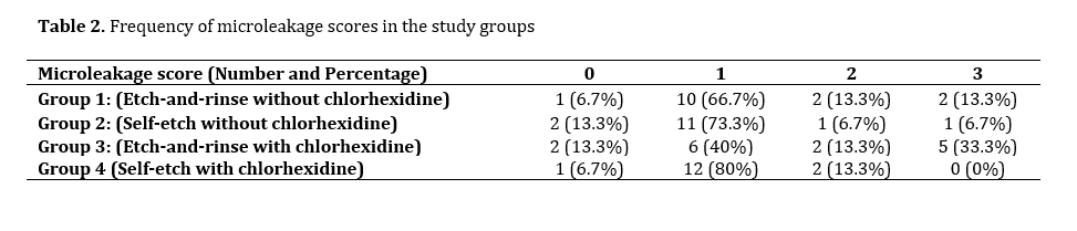
The mean microleakage score was 1.33±0.81 in group 1, 1.07±0.70 in group 2, 1.67±1.11 in group 3 and 1.07±0.45 in group 4. Considering the adhesive system without considering the effect of chlorhexidine, there was a significant difference between the microleakage scores of the two adhesive systems; and the microleakage of the self-etch system was significantly lower than the microleakage of the etch-and-rinse system (P=0.042).
In each adhesive system, applying the chlorhexidine did not have any significant effect on microleakage (P=0.428). Table 2 exhibits the frequency of microleakage scores in the study groups. Score 1 was the most common in all groups (Figure 1).
Statistical analysis:
Descriptive statistics, and Kruskal–Wallis, and Mann-Whitney tests were used to analyze microleakage based on the type of adhesive and presence or absence of chlorhexidine. Data analysis was done using SPSS 24. A probability value of less than 5% was considered significant.
Results
The mean microleakage score was 1.33±0.81 in group 1, 1.07±0.70 in group 2, 1.67±1.11 in group 3 and 1.07±0.45 in group 4. Considering the adhesive system without considering the effect of chlorhexidine, there was a significant difference between the microleakage scores of the two adhesive systems; and the microleakage of the self-etch system was significantly lower than the microleakage of the etch-and-rinse system (P=0.042).
In each adhesive system, applying the chlorhexidine did not have any significant effect on microleakage (P=0.428). Table 2 exhibits the frequency of microleakage scores in the study groups. Score 1 was the most common in all groups (Figure 1).
Figure 1. Stereomicroscopic image showing microleakage score 1 between the enamel surface and the fissure sealant (x50)
The interaction effect of adhesive and chlorhexidine on the microleakage was not significant (P=0.428), indicating that chlorhexidine did not influence the adhesive's effect on microleakage.
Discussion
This study assessed the impact of adhesive type (self-etch and etch-and-rinse) used in fissure sealant therapy and applying chlorhexidine under the fissure sealant layer on microleakage. The results showed that the self-etch adhesive system resulted in a significantly lower amount of microleakage in fissure sealant therapy; however, applying chlorhexidine did not have any significant effect on microleakage.
Microleakage is a major problem in adhesive dentistry, and it is the primary reason for failure of fissure sealant therapy. Methylene blue penetration test is considered as a reliable approach to evaluate microleakage, and has been extensively used for this purpose [22]. One of the problems in using methylene blue for evaluation of microleakage in in vitro studies is that it penetrates more easily than oral bacteria [23].
Self-etch adhesives do not require acid etching of dentin or enamel, but bonding effectiveness is a concern on uncut enamel due to high mineralization [20]. Research indicates that pre-etching of enamel before applying self-etch adhesives significantly enhances the bonding efficacy [24]. For this reason, the enamel was etched before the application of self-etch adhesive in this study.
Table 2. Frequency of microleakage scores in the study groups
Antimicrobial agents can reduce caries following microleakage in enamel margins by decreasing the microflora in the pits and fissures [20]. Chlorhexidine is a well-known antimicrobial agent and has considerable ability to disrupt bacterial cytoplasmic membrane [25]. As chlorhexidine does not have any negative effect on dental materials’ mechanical properties, its application can maximize the success of restorations [26]. Chlorhexidine has an affinity to enamel and increases its free surface energy [27].
Chlorhexidine remnants might interact with the calcium and phosphate ions, and decrease the bond strength; however, some studies have shown that when it is used before sealant application, 0.2% chlorhexidine does not seem to have a negative effect on the sealing ability of dental materials [12,13]. Agrawal and Shigli [12] demonstrated that pit and fissure sealant therapy without using chlorhexidine resulted in greater loss of surface texture and marginal discoloration in comparison to pit and fissure sealant therapy using chlorhexidine after 3, 6, and 12 months; however, the difference was not statistically significant. Also, Sobhi and Hatam Zade [13] found that using 0.2% chlorhexidine prior to sealant placement did not compromise the sealing ability of sealant material. These findings are in accordance with the results of the present study.
Shanmugaavel et al. [14] showed that adding 1% chlorhexidine to conventional glass ionomer sealant and resin-based sealant significantly increased the antibacterial activity of both sealants; in addition, adding chlorhexidine to sealants did not have an adverse effect on their mechanical properties. Their methodology was different from the methodology of the current study because in the present study, chlorhexidine was applied on the tooth surface. However, some studies have shown different results regarding the effect of chlorhexidine on microleakage. In contrast to the present study, Memarpour and Shafiei [15] showed that using chlorhexidine increased microleakage in fissure sealant therapy. In their study, the teeth were kept in water for 6 months and fuchsin was used as dye. However, Haidari Gorji et al. [28] reported that chlorhexidine did not affect the microleakage. Probably the bond to inorganic enamel is more stable than that to dentin; therefore, application of chlorhexidine does not affect the microleakage. This might be due to higher hydroxyapatite inorganic content of the enamel. Due to the presence of controversial results in this regard, more in vitro and clinical studies are needed to reach a definite conclusion.
This study only used methylene blue penetration test; using additional evaluation techniques would allow a more comprehensive analysis. Another limitation of the present study was lack of pH cycling to simulate oral conditions. Long-term clinical trials are warranted to assess the performance of various adhesive systems and chlorhexidine application on maintaining the sealant integrity over extended periods of time.
Conclusion
Application of self-etch adhesive yielded lower microleakage than etch-and-rinse method in fissure sealant therapy. This study also showed that applying chlorhexidine did not have any significant effect on microleakage in using etch-and-rinse or self-etch methods in fissure sealant therapy; thus, it can be used due to its antimicrobial effect in fissure sealant treatments.
Acknowledgement
The authors would like to thank the staff of the Dental and Periodontal Research Center of the Tabriz University of Medical Sciences for their cooperation.
Type of Study: Original article |
Subject:
Restorative Dentistry
References
1. Liu W, Xiong L, Li J, Guo C, Fan W, Huang S. The anticaries effects of pit and fissure sealant in the first permanent molars of school-age children from Guangzhou: a population-based cohort study. BMC Oral Health. 2019 Jul 16;19(1):156. [DOI:10.1186/s12903-019-0846-x] [PMID] []
2. Papageorgiou SN, Dimitraki D, Kotsanos N, Bekes K, van Waes H. Performance of pit and fissure sealants according to tooth characteristics: A systematic review and meta-analysis. J Dent. 2017 Nov;66:8-17. [DOI:10.1016/j.jdent.2017.08.004] [PMID]
3. Wright JT, Crall JJ, Fontana M, Gillette EJ, Nový BB, Dhar V, et al. Evidence-based clinical practice guideline for the use of pit-and-fissure sealants: A report of the American Dental Association and the American Academy of Pediatric Dentistry. J Am Dent Assoc. 2016 Aug;147(8):672-682.e12. [DOI:10.1016/j.adaj.2016.06.001] [PMID]
4. Kühnisch J, Mansmann U, Heinrich-Weltzien R, Hickel R. Longevity of materials for pit and fissure sealing--results from a meta-analysis. Dent Mater. 2012 Mar;28(3):298-303. [DOI:10.1016/j.dental.2011.11.002] [PMID]
5. Paryab M. Effect of chlorhexidine solution on microleakage of pit and fissure sealants in permanent teeth with and without the use of fifth- and sixth-generation adhesive systems. Caspian J Dent Res 2023; 12 (2) :50-6.
6. Buyuk SK, Cantekin K, Demirbuga S, Ali Ozturk M. Are the low-shrinking composites suitable for orthodontic bracket bonding? Eur J Dent. 2013 Jul;7(3):284-8. [DOI:10.4103/1305-7456.115411] [PMID] []
7. Bao Z, Sun H, Fan D, Wang X, Wang Q. Shear bond strength and microleakage of pit and fissure sealants placed after saliva-contaminated etched enamel. Coatings. 2022;12(4):441. [DOI:10.3390/coatings12040441]
8. Esfahanizadeh G, Kooshki R, Karimipoor H, Rastin V. In Vitro Effects of Chlorhexidine and Listerine Mouthwashes on Color Stability of Glazed Bilayer Zirconia and IPS E.Max Ceramics. J Res Dent Maxillofac Sci. 2024;9(1):15-20 [DOI:10.61186/jrdms.9.1.15]
9. Rudolf JL, Moser C, Sculean A, Eick S. In-vitro antibiofilm activity of chlorhexidine digluconate on polylactide-based and collagen-based membranes. BMC Oral Health. 2019 Dec 26;19(1):291. [DOI:10.1186/s12903-019-0979-y] [PMID] []
10. Tavakolinejad Z, razeghi H, Sadeghi M. Effect of Pretreatment with Fluoride, Chlorhexidine, and Mixed Fluoride-Chlorhexidine Gels on Shear Bond Strength of Orthodontic Brackets: An Ex-Vivo Study. J Res Dent Maxillofac Sci 2021;6(1):24-9.
11. Bin-Shuwaish M, AlHussaini A, AlHudaithy L, AlDukhiel S, AlJamhan A, Alrahlah A. Effects of different antibacterial disinfectants on microleakage of bulk-fill composite bonded to different tooth structures. BMC Oral Health. 2021 Jul 16;21(1):348. [DOI:10.1186/s12903-021-01717-7] [PMID] []
12. Agrawal A, Shigli A. Comparison of six different methods of cleaning and preparing occlusal fissure surface before placement of pit and fissure sealant: an in vitro study. J Indian Soc Pedod Prev Dent. 2012 Jan-Mar;30(1):51-5. [DOI:10.4103/0970-4388.95582] [PMID]
13. Sobhi J, Hatam Zade E. Comparison of the Effect of Using Chlorhexidine Solution on Microleakage of Two Pit and Fissure Sealants. Jundishapur Sci Med J. 2015 May-June;14(2):189-98.
14. Shanmugaavel AK, Asokan S, John JB, Priya PG, Devi JG. Effect of One Percent Chlorhexidine Addition on the Antibacterial Activity and Mechanical Properties of Sealants: An in vitro Study. Int J Clin Pediatr Dent. 2015 Sep-Dec;8(3):196-201. [DOI:10.5005/jp-journals-10005-1312] [PMID] []
15. Memarpour M, Shafiei F. The effect of antibacterial agents on fissure sealant microleakage: a 6-month in vitro study. Oral Health Prev Dent. 2014;12(2):149-55.
16. Memarpour M, Abedinzade A, Rafiee A, Hashemian A. Penetration ability and microhardness of infiltrant resin and two pit and fissure sealants in primary teeth with early enamel lesions. Sci Rep. 2022 Mar 17;12(1):4652. [DOI:10.1038/s41598-022-08725-9] [PMID] []
17. Bayrak GD, Gurdogan-Guler EB, Yildirim Y, Ozturk D, Selvi-Kuvvetli S. Assessment of shear bond strength and microleakage of fissure sealant following enamel deproteinization: An in vitro study. J Clin Exp Dent. 2020 Mar 1;12(3):e220-6. [DOI:10.4317/jced.56281] [PMID] []
18. Hu YT, Yu F, Tang XY, Wu WZ, Zhang P, Hu ZH, et al. The antibacterial effect and physical performance of pit and fissure sealants based on an antibacterial core-shell nanocomposite. J Mech Behav Biomed Mater. 2021 May;117:104414. [DOI:10.1016/j.jmbbm.2021.104414] [PMID]
19. Klaisiri A, Vongsang J, Leelaudom T, Krajangta N. Methylene Blue Penetration of Resin Infiltration and Resin Sealant in Artificial White-Spot Lesions. Eur J Dent. 2023 Jul;17(3): 828-33. [DOI:10.1055/s-0042-1756689] [PMID] []
20. Al Khowaiter SS, Al-Bounni RS, Binalrimal S. Comparison of Dentinal Microleakage in Three Interim Dental Restorations: An In Vitro Study. J Int Soc Prev Community Dent. 2022 Dec 30;12(6):590-5. [DOI:10.4103/jispcd.JISPCD_183_21] [PMID] []
21. Shingare P, Chaugule V. An In Vitro Microleakage Study for Comparative Analysis of Two Types of Resin-based Sealants Placed by Using Three Different Types of Techniques of Enamel Preparation. Int J Clin Pediatr Dent. 2021 Jul-Aug;14(4):475-81. [DOI:10.5005/jp-journals-10005-1991] [PMID] []
22. Amend S, Frankenberger R, Boutsiouki C, Scharrelmann V, Winter J, Krämer N. Microleakage of pit and fissure sealings placed after enamel conditioning with phosphoric acid or with self-etching primers/adhesives. Clin Exp Dent Res. 2021 Oct;7(5):763-71. [DOI:10.1002/cre2.420] [PMID] []
23. Erfanparast L, Vatandoust M, Sohrabi A, Mahmoudi L. Clinical Comparison of Fissure Sealants Retention Following Preparation with Seventh Generation Bonding Agent with Prior Etching and Conventional Acid Etch and Bond System (fifth Generation). Dent Hypotheses. 2021 Jan- Mar;12(1):22-7. [DOI:10.4103/denthyp.denthyp_89_20]
24. Van Landuyt KL, Kanumilli P, De Munck J, Peumans M, Lambrechts P, Van Meerbeek B. Bond strength of a mild self-etch adhesive with and without prior acid-etching. J Dent. 2006 Jan;34(1):77-85. [DOI:10.1016/j.jdent.2005.04.001] [PMID]
25. Thangavelu A, Kaspar SS, Kathirvelu RP, Srinivasan B, Srinivasan S, Sundram R. Chlorhexidine: An Elixir for Periodontics. J Pharm Bioallied Sci. 2020 Aug;12(Suppl 1): S57-9. [DOI:10.4103/jpbs.JPBS_162_20] [PMID] []
26. Tavazo Zadeh E, Aref P, Askarizadeh N, Emadi F. In vitro Antimicrobial Effect of Punica granatum Extract versus Chlorhexidine on Streptococcus sobrinus, Streptococcus sanguinis, and Candida albicans. J Res Dent Maxillofac Sci 2023;8(1):18-27. [DOI:10.52547/jrdms.8.1.18]
27. Kapdan A, Öztaş N. Effects of chlorhexidine and gaseous ozone on microleakage and on the bond strength of dentin bonding agents with compomer restoration on primary teeth. J Dent Sci. 2015 March;10(1):46-54. [DOI:10.1016/j.jds.2013.02.026]
28. Haidari Gorji MA, Abolghasemzadeh F, Alaghemand H, Esmaeili B. Effect of 2% Chlorhexidine on the Enamel Microleakage of omposite Restorations Using 5th, 6th, 7th and Universal Generation of Dentine Bonding Agents (In Vitro). J Babol Univ Med Sci.2017;19(12):36-42.
Send email to the article author
| Rights and permissions | |
 |
This work is licensed under a Creative Commons Attribution-NonCommercial 4.0 International License. |