Journal of Research in Dental
and Maxillofacial Sciences
Volume 8, Issue 3 (8-2023)
J Res Dent Maxillofac Sci 2023, 8(3): 203-209 |
Back to browse issues page


Ethics code: IR.MAZUMS.REC.1399.8114.
Download citation:
BibTeX | RIS | EndNote | Medlars | ProCite | Reference Manager | RefWorks
Send citation to:


BibTeX | RIS | EndNote | Medlars | ProCite | Reference Manager | RefWorks
Send citation to:
Hoshyari N, Mesgarani A, Shokrzadeh Lamuki M, Motafeghi F, Mousavi S J, Vaghar Mousavi M. Cytotoxicity of Two Calcium Silicate-Based Sealers and AH Plus Resin-Based Sealer for Human Gingival Fibroblasts. J Res Dent Maxillofac Sci 2023; 8 (3) :203-209
URL: http://jrdms.dentaliau.ac.ir/article-1-464-en.html
URL: http://jrdms.dentaliau.ac.ir/article-1-464-en.html
Narjes Hoshyari *1 

 , Abbas Mesgarani2 , Mohammad Shokrzadeh Lamuki3 , Farzane Motafeghi3 , Seyed Jaber Mousavi4 , Milad Vaghar Mousavi5
, Abbas Mesgarani2 , Mohammad Shokrzadeh Lamuki3 , Farzane Motafeghi3 , Seyed Jaber Mousavi4 , Milad Vaghar Mousavi5
1- Department of Endodontics, School of Dentistry, Mazandaran University of Medical Sciences, Sari, Iran , Narjeshoshyari@rocketmail.com
2- Department of Endodontics, School of Dentistry, Mazandaran University of Medical Sciences, Sari, Iran
3- Department of Pharmacology and Toxicology, School of Pharmacy, Mazandaran University of Medical Sciences, Sari, Iran
4- Department of Community Medicine, School of Medicine, Mazandaran University of Medical Sciences, Sari, Iran
5- Private Dental Practice, Sari, Iran
Full-Text [PDF 666 kb]
(1090 Downloads)
| Abstract (HTML) (2463 Views)
Introduction
The main goal of root canal therapy is three-dimensional obturation of the root canal system. A hermetic seal minimizes coronal, apical, and lateral leakage of fluids and bacteria, prevents the development of apical periodontitis, and eliminates the residual irritants from the root canal system [1].
Different endodontic materials are available for obturation and sealing of the root canal system. Endodontic sealers are imperative to seal the interface of root dentin and root filling materials, and the gaps between them. Sealers should be able to apically and laterally seal the root canal system and fill the gaps and irregularities [2]. Since sealers may contact the periapical tissue, they must be biocompatible and safe for the human body [3].
Biocompatibility of endodontic sealers is highly important for a successful endodontic treatment. A toxic and necrotizing sealer can adversely affect tissue healing and provides a suitable environment for bacterial invasion and leads to treatment failure in the long-term [4].
Different sealer types with various advantages and disadvantages, and different physical and biological properties are available in the market; among which, resin-based sealers, zinc oxide eugenol, calcium hydroxide, glass ionomer, and silicone sealers may be named [5]. Recently, calcium silicate-based sealers were introduced to the market [6].
Resin-based sealers such as AH Plus are optimal sealers, since they are radiopaque, have optimal dimensional stability, good resistance, high flow, and low solubility [7]. On the other hand, MTA Fillapex is a calcium silicate-based sealer that enhances hard tissue mineralization by releasing calcium and creating an alkaline environment [8].
Endoseal MTA is a pozzolan-based sealer in combination with a calcium silicate sealer, which has optimal biocompatibility and results in good obturation quality [9].
The methyl thiazolyl tetrazolium (MTT) assay is a popular tool for estimation of the metabolic activity of viable cells. It is based on enzymatic reduction of tetrazolium salt into purple formazan crystals, which is measured spectrophotometrically [10].
Due to the novelty of calcium-silicate based sealers and the significance of an optimal sealer to maximize the success of endodontic treatment, this study aimed to assess the cytotoxicity of Endoseal MTA and MTA Fillapex by the MTT assay, in comparison with AH Plus resin-based sealer as the gold standard for human gingival fibroblasts (HGFs).
Materials and Methods
This in vitro, experimental study was conducted at Mazandaran University of Medical Sciences and was approved by the ethics committee of the university (IR.MAZUMS.REC.1399.8114). HGFs with normal proliferation and no fungal or bacterial contamination in logarithmic phase of growth were used for this study. Cells with fungal or bacterial contamination were excluded.
Preparation of sealers:
Three commercially available sealers were evaluated in this study including Endoseal MTA (Maruchi; Wonju, Korea), MTA Fillapex (Angelus Solucões Odontológicas, Londrina, PR, Brazil), and AH Plus (Dentsply, De-Trey Konstanz, Germany). AH Plus and MTA Fillapex were prepared as instructed by the manufacturers [11,12]. Endoseal MTA was premixed and injected [13].
Preparation of culture medium:
To prepare the culture medium, 13.48 g of Dulbecco’s modified Eagle’s medium (DMEM; Gibco, USA) and 7.3 g of sodium bicarbonate were dissolved in 1 L of distilled water. The pH of the culture medium was adjusted at 7.4 by using hydrochloric acid and NaOH. The culture medium was then filtered through a filter with 0.2 µm pores under a laminar hood and kept refrigerated in sterile plates. Prior to use, 10% fetal bovine serum (FBS; 1 mL per each 9 mL of the culture medium) and antibiotic (100 U/mL penicillin and 100 µg/mL streptomycin) were added to the culture medium [14].
Cell preparation:
The cells were initially obtained from the Pasteur Institute of Tehran. HGFs were removed from the nitrogen tank and cultured in 75 cm2 flasks (Nunc, Denmark) containing DMEM supplemented with FBS and antibiotics. The cells were incubated at 37°C in presence of 5% CO2. The culture medium was refreshed every 3 days. After reaching confluence, the cells were passaged and transferred to several flasks [14].
Cell passage:
The cell culture flask was rinsed twice with phosphate buffered saline. Next, 2 mm of trypsin (Merck, Germany) was added to the cells in a 75 cm2 flask, and they were incubated at 37°C for 5 minutes. Next, 2 mL of the culture medium containing 10% FBS was added to the flask to stop the activity of trypsin. The cells detached from the bottom of the flask were transferred into a 15 mL sterile tube and centrifuged at 2000 rpm for 5 minutes (Hettich Universal, Germany). The supernatant containing trypsin was discarded, and the cell sediment was transferred to a culture medium containing antibiotic and 10% FBS. The cells were then distributed among three flasks, and the flasks containing cells were incubated at 37°C and 5% CO2 [14].
Cell counting by using trypan blue:
After detachment of cells from the bottom of the flask by using trypsin, the percentage of viable cells was determined by trypan blue. For this purpose, 20 µL of the suspension was transferred into a Neubauer chamber (Tiefe Depth Profondeur, Marienfeld, Germany 0.01 mm). The number of cells was counted in a large square composed of 16 smaller squares, and the following formula was used to calculate the percentage of viable cells:
Number of cells per each 1 mL of the suspension=number of cells in the larger square x 104 [14]
Preparation of sealer extracts:
Endoseal MTA, MTA Fillapex, and AH Plus were prepared as instructed by the manufacturers, and were placed in wells of a 24-well plate (16.2 mm diameter and 2 mm height) prior to setting. Next, 2.5 mL of DMEM containing antibiotic and without FBS was added to the wells. The plate was then incubated at 37°C and 5% CO2 for 48 hours. Next, all three sealers were transferred into test tubes, and 10% FBS was added to them (0.05 mL per each 5 mL of the sealer extract). These test tubes served as sealer extracts with 1:1 concentration. To prepare culture media with lower concentrations of sealer extract, the 1:1 samples were serially diluted using DMEM containing antibiotic and 10% FBS to obtain 1:2, 1:4, and 1:8 concentrations of each sealer [14].
Cytotoxicity:
HGFs were removed from the nitrogen tank and transferred to a 75 cm2 flask containing DMEM enriched with FBS and antibiotics (100 U/mL penicillin and 100 µg/mL streptomycin) and incubated at 37°C and 5% CO2. After cell passage and ensuring normal proliferation of cells, they were detached from the flask using trypsin, and after ensuring their viability by using trypan blue, they were transferred to a 96-well culture plate (8000 cells/0.5 mL/well). The plates were then incubated under standard conditions for 24 hours until cell seeding. They were then incubated along with sealer extracts at the aforementioned concentrations for 48 hours. The percentage of cell viability was then calculated using the MTT assay [14].
MTT assay:
After exposure of cells to the sealer extracts, 20 mL of 5 mg/mL MTT solution was added to each well of the plate. The plates were then incubated for 4 hours. Next, the culture medium was removed, and 100 µL of dimethyl sulfoxide (Merck, Germany) was added to each well to dissolve the formed formazan crystals. The color intensity was then quantified by measuring the optical density of each well at 545 nm wavelength (with 630 nm reference wavelength) using an ELISA Reader. The percentage of cell viability was calculated as follows:
Percentage of viable cells=mean optical density of control wells/optimal density of each well) × 100 [15-17].
All statistical analyses were conducted using PRISM version 3 by two-way ANOVA considering the presence of two factors to be analyzed, followed by post-hoc Tukey test for pairwise comparisons (due to normal data distribution). P<0.05 was considered statistically significant.
Results
The cytotoxicity of Endoseal MTA, MTA Fillapex, and AH Plus in 1:2, 1:4, and 1:8 concentrations was assessed for HGFs in the present study. The negative control group included the culture medium alone which is not cytotoxic.
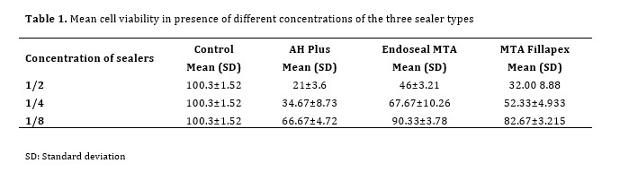
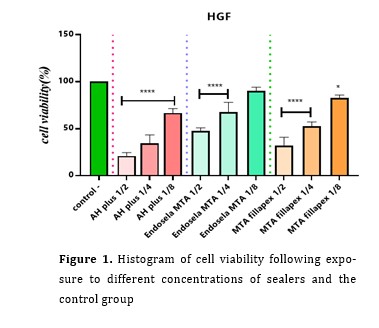
Table 1 shows the mean cell viability in presence of different concentrations of the three sealer types. Table 2 presents the results of pairwise comparisons of different concentrations of the three sealer types regarding cell viability. Figure 1 shows the histogram of cell viability following exposure to different concentrations of sealers and the control group.
Table 1. Mean cell viability in presence of different concentrations of the three sealer types
Table 2. Pairwise comparisons of different concentrations of the three sealer types regarding cell viability
Figure 1. Histogram of cell viability following exposure to different concentrations of sealers and the control group
Discussion
Sealers should have some important characteristics, such as biocompatibility, bacteriostatic properties, and insolubility in oral fluids. Optimal setting time, minimal polymerization shrinkage, not causing tooth discoloration, and opacity are among the criteria for a suitable sealer [18,19].
Extrusion of sealer and gutta-percha into the periapical tissue during root canal obturation can lead to development of a periapical lesion. Apical periodontitis can cause moderate to severe pain.
Immunologically, extrusion of sealer into the periapical tissue elicits a periodontal ligament response characterized by activation of different inflammatory cells, and release of inflammatory mediators such as histamine, cytokines, and prostaglandins [20,21].
Different cell types are used for assessment of cytotoxicity of dental materials and endodontic sealers, such as human periodontal ligament stem cells [3,22], human gingival fibroblasts [23], V79 Hamster fibroblasts [7], and L929 murine fibroblasts.
Also, several methods are available for assessment of cell metabolism, such as assessment of enzymatic activity, membrane permeability, cell adhesion, ATP production, production of coenzymes, and nucleotide excision repair, which can be classified as follows:
(I) Dye removal techniques, such as Trypan blue test, (II) Metabolic activity tests, (III) ATP test, (IV) sulforhodamine B test, (V) protease viability assay, (VI) clonogenic cell survival assay, (VII) DNA synthesis cell proliferation assay, (VIII) and micro-Raman spectroscopy [24].
The MTT assay is fast, quantitative, and accurate, since the results are reported spectrophotometrically. This method is based on enzymatic reduction of tetrazolium salt with a light color to purple-blue formazan crystals [10]. The MTT assay was used in the present study for assessment of cell viability.
In the present study, the percentage of viable cells was lower in AH Plus group than the other two bioceramic sealers. It means that the calcium content of bioceramic sealers increase their biocompatibility while the resin component decreases the biocompatibility. Between the two bioceramic sealers tested in the present study, Endoseal MTA showed higher biocompatibility. MTA Fillapex is a MTA paste-catalyst that includes resin. Thus, it is a resin-based bioceramic sealer. It contains natural resin, salicylate resin, diluted resin, bismuth trioxide, silica nanoparticles, and MTA [25]. On the other hand, Endoseal MTA is a pozzolan-based bioceramic sealer [26]. Pozzolan is a silicon that chemically reacts with calcium hydroxide and forms a cement. The chemical reactions of pozzolan occur between calcium hydroxide and silica, leading to formation of C-S-H hydration reaction as a cement. The pozzolan reaction takes longer than cement formation, and in the setting process, calcium hydroxide is released. Thus, its effects remain longer before setting [27]. Thus, the resin content of MTA Fillapex may decrease biocompatibility, while the pozzolan reaction increases the biocompatibility of Endoseal MTA sealer. Erdogan et al. [28] compared the biocompatibility of AH Plus, MTA Fillapex, and iRoot SP using XTT cytotoxicity measurement test. They reported that AH Plus and MTA Fillapex showed the lowest percentage of viable cells in higher concentrations (1:1, 1:2, 1:4), and iRoot SP resulted in significantly higher number of viable cells in all concentrations compared with the other two sealers. Although their cytotoxicity assessment test was different from that used in the present study, they showed equally high cytotoxicity of AH Plus and MTA Fillapex in 1:2 concentration, which was almost in line with the present findings. Lee et al. [3] compared three calcium silicate-based sealers (Endoseal MTA, nano-ceramic sealer, and WellRoot ST), and two epoxy-resin based sealers (AH Plus and AD Seal) from different aspects such as cytotoxicity for human periodontal ligament stem cells (by the MTT assay), inflammatory response, and osteogenic potential. They showed that calcium silicate-based sealers were more biocompatible and had lower cytotoxicity than epoxy resin-based sealers, which was in agreement with the present findings. Seo et al. [21] compared the cytotoxicity and mineralization activity of three calcium silicate-based sealers (Endoseal MTA, BioRoot RCS, EndoSequence BC) and AH Plus epoxy-resin based sealer using human dental pulp stem cells in monolayer.
Cytotoxicity was evaluated by the MTT assay. They found no significant difference among the four sealers in cell viability. However, in the present study, the percentage of cell viability in Endoseal MTA group in all concentrations was higher than that in AH Plus group, which may be due to using a different cell line, or methodological differences (such as incubation times).
Conclusion
According to the present results, Endoseal MTA in all concentrations showed lower cytotoxicity and higher cell viability than MTA Fillapex and AH Plus.
Full-Text: (947 Views)
| Abstract
Background and Aim: The main goal of root canal treatment is to three-dimensionally seal the root canal system. Since sealer may contact the periapical tissue, it should be biocompatible and safe for the body. This study aimed to assess the cytotoxicity of MTA Fillapex and Endoseal MTA calcium silicate-based sealers and AH Plus resin-based sealer for human gingival fibroblasts (HGFs). Materials and Methods: In this in vitro study, extracts of AH Plus, Endoseal MTA, and MTA Fillapex were obtained, serially diluted 1:2, 1:4, and 1:8, and were exposed to HGFs. Cytotoxicity was assessed by the methyl thiazolyl tetrazolium (MTT) assay. Data were analyzed by ANOVA and Tukey’s test (alpha=0.05). Results: No significant difference existed between AH Plus 1:2 and MTA Fillapex 1:2 concentrations regarding the cell viability percentage (P>0.05). However, the difference between these two sealers in other concentrations was significant (P<0.05). No significant difference existed in AH Pus 1:4, Endoseal MTA 1:2, and MTA Fillapex 1:2 in cell viability (P>0.05); however, the difference among other concentrations of the three sealers was significant (P<0.05). The difference among AH Plus 1:8, Endoseal MTA 1:4, and MTA Fillapex 1:4 and 1:8 concentrations was not significant (P>0.05) but the difference among other concentrations was significant (P<0.05). Endoseal MTA 1:8 showed the highest and AH Plus 1:2 showed the lowest cell viability. Conclusion: Endoseal MTA in all concentrations had lower cytotoxicity than MTA Fillapex and AH Plus and resulted in higher viability of HGFs. Key Words: Root Canal Obturation; Calcium Compounds; Epoxy Resins; Dental Cements; Fibroblast |
Introduction
The main goal of root canal therapy is three-dimensional obturation of the root canal system. A hermetic seal minimizes coronal, apical, and lateral leakage of fluids and bacteria, prevents the development of apical periodontitis, and eliminates the residual irritants from the root canal system [1].
Different endodontic materials are available for obturation and sealing of the root canal system. Endodontic sealers are imperative to seal the interface of root dentin and root filling materials, and the gaps between them. Sealers should be able to apically and laterally seal the root canal system and fill the gaps and irregularities [2]. Since sealers may contact the periapical tissue, they must be biocompatible and safe for the human body [3].
Biocompatibility of endodontic sealers is highly important for a successful endodontic treatment. A toxic and necrotizing sealer can adversely affect tissue healing and provides a suitable environment for bacterial invasion and leads to treatment failure in the long-term [4].
Different sealer types with various advantages and disadvantages, and different physical and biological properties are available in the market; among which, resin-based sealers, zinc oxide eugenol, calcium hydroxide, glass ionomer, and silicone sealers may be named [5]. Recently, calcium silicate-based sealers were introduced to the market [6].
Resin-based sealers such as AH Plus are optimal sealers, since they are radiopaque, have optimal dimensional stability, good resistance, high flow, and low solubility [7]. On the other hand, MTA Fillapex is a calcium silicate-based sealer that enhances hard tissue mineralization by releasing calcium and creating an alkaline environment [8].
Endoseal MTA is a pozzolan-based sealer in combination with a calcium silicate sealer, which has optimal biocompatibility and results in good obturation quality [9].
The methyl thiazolyl tetrazolium (MTT) assay is a popular tool for estimation of the metabolic activity of viable cells. It is based on enzymatic reduction of tetrazolium salt into purple formazan crystals, which is measured spectrophotometrically [10].
Due to the novelty of calcium-silicate based sealers and the significance of an optimal sealer to maximize the success of endodontic treatment, this study aimed to assess the cytotoxicity of Endoseal MTA and MTA Fillapex by the MTT assay, in comparison with AH Plus resin-based sealer as the gold standard for human gingival fibroblasts (HGFs).
Materials and Methods
This in vitro, experimental study was conducted at Mazandaran University of Medical Sciences and was approved by the ethics committee of the university (IR.MAZUMS.REC.1399.8114). HGFs with normal proliferation and no fungal or bacterial contamination in logarithmic phase of growth were used for this study. Cells with fungal or bacterial contamination were excluded.
Preparation of sealers:
Three commercially available sealers were evaluated in this study including Endoseal MTA (Maruchi; Wonju, Korea), MTA Fillapex (Angelus Solucões Odontológicas, Londrina, PR, Brazil), and AH Plus (Dentsply, De-Trey Konstanz, Germany). AH Plus and MTA Fillapex were prepared as instructed by the manufacturers [11,12]. Endoseal MTA was premixed and injected [13].
Preparation of culture medium:
To prepare the culture medium, 13.48 g of Dulbecco’s modified Eagle’s medium (DMEM; Gibco, USA) and 7.3 g of sodium bicarbonate were dissolved in 1 L of distilled water. The pH of the culture medium was adjusted at 7.4 by using hydrochloric acid and NaOH. The culture medium was then filtered through a filter with 0.2 µm pores under a laminar hood and kept refrigerated in sterile plates. Prior to use, 10% fetal bovine serum (FBS; 1 mL per each 9 mL of the culture medium) and antibiotic (100 U/mL penicillin and 100 µg/mL streptomycin) were added to the culture medium [14].
Cell preparation:
The cells were initially obtained from the Pasteur Institute of Tehran. HGFs were removed from the nitrogen tank and cultured in 75 cm2 flasks (Nunc, Denmark) containing DMEM supplemented with FBS and antibiotics. The cells were incubated at 37°C in presence of 5% CO2. The culture medium was refreshed every 3 days. After reaching confluence, the cells were passaged and transferred to several flasks [14].
Cell passage:
The cell culture flask was rinsed twice with phosphate buffered saline. Next, 2 mm of trypsin (Merck, Germany) was added to the cells in a 75 cm2 flask, and they were incubated at 37°C for 5 minutes. Next, 2 mL of the culture medium containing 10% FBS was added to the flask to stop the activity of trypsin. The cells detached from the bottom of the flask were transferred into a 15 mL sterile tube and centrifuged at 2000 rpm for 5 minutes (Hettich Universal, Germany). The supernatant containing trypsin was discarded, and the cell sediment was transferred to a culture medium containing antibiotic and 10% FBS. The cells were then distributed among three flasks, and the flasks containing cells were incubated at 37°C and 5% CO2 [14].
Cell counting by using trypan blue:
After detachment of cells from the bottom of the flask by using trypsin, the percentage of viable cells was determined by trypan blue. For this purpose, 20 µL of the suspension was transferred into a Neubauer chamber (Tiefe Depth Profondeur, Marienfeld, Germany 0.01 mm). The number of cells was counted in a large square composed of 16 smaller squares, and the following formula was used to calculate the percentage of viable cells:
Number of cells per each 1 mL of the suspension=number of cells in the larger square x 104 [14]
Preparation of sealer extracts:
Endoseal MTA, MTA Fillapex, and AH Plus were prepared as instructed by the manufacturers, and were placed in wells of a 24-well plate (16.2 mm diameter and 2 mm height) prior to setting. Next, 2.5 mL of DMEM containing antibiotic and without FBS was added to the wells. The plate was then incubated at 37°C and 5% CO2 for 48 hours. Next, all three sealers were transferred into test tubes, and 10% FBS was added to them (0.05 mL per each 5 mL of the sealer extract). These test tubes served as sealer extracts with 1:1 concentration. To prepare culture media with lower concentrations of sealer extract, the 1:1 samples were serially diluted using DMEM containing antibiotic and 10% FBS to obtain 1:2, 1:4, and 1:8 concentrations of each sealer [14].
Cytotoxicity:
HGFs were removed from the nitrogen tank and transferred to a 75 cm2 flask containing DMEM enriched with FBS and antibiotics (100 U/mL penicillin and 100 µg/mL streptomycin) and incubated at 37°C and 5% CO2. After cell passage and ensuring normal proliferation of cells, they were detached from the flask using trypsin, and after ensuring their viability by using trypan blue, they were transferred to a 96-well culture plate (8000 cells/0.5 mL/well). The plates were then incubated under standard conditions for 24 hours until cell seeding. They were then incubated along with sealer extracts at the aforementioned concentrations for 48 hours. The percentage of cell viability was then calculated using the MTT assay [14].
MTT assay:
After exposure of cells to the sealer extracts, 20 mL of 5 mg/mL MTT solution was added to each well of the plate. The plates were then incubated for 4 hours. Next, the culture medium was removed, and 100 µL of dimethyl sulfoxide (Merck, Germany) was added to each well to dissolve the formed formazan crystals. The color intensity was then quantified by measuring the optical density of each well at 545 nm wavelength (with 630 nm reference wavelength) using an ELISA Reader. The percentage of cell viability was calculated as follows:
Percentage of viable cells=mean optical density of control wells/optimal density of each well) × 100 [15-17].
All statistical analyses were conducted using PRISM version 3 by two-way ANOVA considering the presence of two factors to be analyzed, followed by post-hoc Tukey test for pairwise comparisons (due to normal data distribution). P<0.05 was considered statistically significant.
Results
The cytotoxicity of Endoseal MTA, MTA Fillapex, and AH Plus in 1:2, 1:4, and 1:8 concentrations was assessed for HGFs in the present study. The negative control group included the culture medium alone which is not cytotoxic.
Table 1 shows the mean cell viability in presence of different concentrations of the three sealer types. Table 2 presents the results of pairwise comparisons of different concentrations of the three sealer types regarding cell viability. Figure 1 shows the histogram of cell viability following exposure to different concentrations of sealers and the control group.
Table 1. Mean cell viability in presence of different concentrations of the three sealer types
Table 2. Pairwise comparisons of different concentrations of the three sealer types regarding cell viability
Figure 1. Histogram of cell viability following exposure to different concentrations of sealers and the control group
Discussion
Sealers should have some important characteristics, such as biocompatibility, bacteriostatic properties, and insolubility in oral fluids. Optimal setting time, minimal polymerization shrinkage, not causing tooth discoloration, and opacity are among the criteria for a suitable sealer [18,19].
Extrusion of sealer and gutta-percha into the periapical tissue during root canal obturation can lead to development of a periapical lesion. Apical periodontitis can cause moderate to severe pain.
Immunologically, extrusion of sealer into the periapical tissue elicits a periodontal ligament response characterized by activation of different inflammatory cells, and release of inflammatory mediators such as histamine, cytokines, and prostaglandins [20,21].
Different cell types are used for assessment of cytotoxicity of dental materials and endodontic sealers, such as human periodontal ligament stem cells [3,22], human gingival fibroblasts [23], V79 Hamster fibroblasts [7], and L929 murine fibroblasts.
Also, several methods are available for assessment of cell metabolism, such as assessment of enzymatic activity, membrane permeability, cell adhesion, ATP production, production of coenzymes, and nucleotide excision repair, which can be classified as follows:
(I) Dye removal techniques, such as Trypan blue test, (II) Metabolic activity tests, (III) ATP test, (IV) sulforhodamine B test, (V) protease viability assay, (VI) clonogenic cell survival assay, (VII) DNA synthesis cell proliferation assay, (VIII) and micro-Raman spectroscopy [24].
The MTT assay is fast, quantitative, and accurate, since the results are reported spectrophotometrically. This method is based on enzymatic reduction of tetrazolium salt with a light color to purple-blue formazan crystals [10]. The MTT assay was used in the present study for assessment of cell viability.
In the present study, the percentage of viable cells was lower in AH Plus group than the other two bioceramic sealers. It means that the calcium content of bioceramic sealers increase their biocompatibility while the resin component decreases the biocompatibility. Between the two bioceramic sealers tested in the present study, Endoseal MTA showed higher biocompatibility. MTA Fillapex is a MTA paste-catalyst that includes resin. Thus, it is a resin-based bioceramic sealer. It contains natural resin, salicylate resin, diluted resin, bismuth trioxide, silica nanoparticles, and MTA [25]. On the other hand, Endoseal MTA is a pozzolan-based bioceramic sealer [26]. Pozzolan is a silicon that chemically reacts with calcium hydroxide and forms a cement. The chemical reactions of pozzolan occur between calcium hydroxide and silica, leading to formation of C-S-H hydration reaction as a cement. The pozzolan reaction takes longer than cement formation, and in the setting process, calcium hydroxide is released. Thus, its effects remain longer before setting [27]. Thus, the resin content of MTA Fillapex may decrease biocompatibility, while the pozzolan reaction increases the biocompatibility of Endoseal MTA sealer. Erdogan et al. [28] compared the biocompatibility of AH Plus, MTA Fillapex, and iRoot SP using XTT cytotoxicity measurement test. They reported that AH Plus and MTA Fillapex showed the lowest percentage of viable cells in higher concentrations (1:1, 1:2, 1:4), and iRoot SP resulted in significantly higher number of viable cells in all concentrations compared with the other two sealers. Although their cytotoxicity assessment test was different from that used in the present study, they showed equally high cytotoxicity of AH Plus and MTA Fillapex in 1:2 concentration, which was almost in line with the present findings. Lee et al. [3] compared three calcium silicate-based sealers (Endoseal MTA, nano-ceramic sealer, and WellRoot ST), and two epoxy-resin based sealers (AH Plus and AD Seal) from different aspects such as cytotoxicity for human periodontal ligament stem cells (by the MTT assay), inflammatory response, and osteogenic potential. They showed that calcium silicate-based sealers were more biocompatible and had lower cytotoxicity than epoxy resin-based sealers, which was in agreement with the present findings. Seo et al. [21] compared the cytotoxicity and mineralization activity of three calcium silicate-based sealers (Endoseal MTA, BioRoot RCS, EndoSequence BC) and AH Plus epoxy-resin based sealer using human dental pulp stem cells in monolayer.
Cytotoxicity was evaluated by the MTT assay. They found no significant difference among the four sealers in cell viability. However, in the present study, the percentage of cell viability in Endoseal MTA group in all concentrations was higher than that in AH Plus group, which may be due to using a different cell line, or methodological differences (such as incubation times).
Conclusion
According to the present results, Endoseal MTA in all concentrations showed lower cytotoxicity and higher cell viability than MTA Fillapex and AH Plus.
Type of Study: Original article |
Subject:
Endodontics
References
1. Tavares PB, Bonte E, Boukpessi T, Siqueira JF Jr, Lasfargues JJ. Prevalence of apical periodontitis in root canal-treated teeth from an urban French population: influence of the quality of root canal fillings and coronal restora-tions. J Endod. 2009 Jun;35(6):810-3.. [DOI:10.1016/j.joen.2009.03.048] [PMID]
2. Williamson AE, Marker KL, Drake DR, Dawson DV, Walton RE. Resin-based versus gutta-percha-based root canal obturation: influence on bacterial leakage in an in vitro model system. Oral Surg Oral Med Oral Pathol Oral Radiol Endod. 2009 Aug;108(2):292-6.. [DOI:10.1016/j.tripleo.2009.01.043] [PMID]
3. Lee JK, Kim S, Lee S, Kim HC, Kim E. In Vitro Comparison of Biocompatibility of Calcium Silicate-Based Root Canal Sealers. Materials (Basel). 2019 Jul 29;12(15):2411 [DOI:10.3390/ma12152411] [PMID] [PMCID]
4. Barbosa SV, Araki K, Spångberg LS. Cytotoxicity of some modi-fied root canal sealers and their leachable components. Oral Surg Oral Med Oral Pathol. 1993 Mar;75(3):357-61. [DOI:10.1016/0030-4220(93)90151-S] [PMID]
5. Sevimay S, Dalat D. Evaluation of penetration and adaptation of three different sealers: a SEM study. J Oral Rehabil. 2003 Sep;30(9):951-5. [DOI:10.1046/j.1365-2842.2003.01084.x] [PMID]
6. Kuga MC, Duarte MA, Sant'anna-Júnior A, Keine KC, Faria G, Dantas AA, Guiotti FA. Effects of calcium hydroxide addition on the physical and chemical properties of a calcium silicate-based sealer. J Appl Oral Sci. 2014 Jun; 22 (3):180-4. [DOI:10.1590/1678-775720130032] [PMID] [PMCID]
7. Bin CV, Valera MC, Camargo SE, Rabelo SB, Silva GO, Balducci I, Camargo CH. Cytotoxicity and genotoxicity of root canal sealers based on mineral trioxide aggregate. J Endod. 2012 Apr;38(4):495-500. [DOI:10.1016/j.joen.2011.11.003] [PMID]
8. Yoshino P, Nishiyama CK, Modena KC, Santos CF, Sipert CR. In vitro cytotoxicity of white MTA, MTA Fillapex® and Portland cement on human periodontal ligament fibroblasts. Braz Dent J. 2013;24(2):111-6. [DOI:10.1590/0103-6440201302115] [PMID]
9. Kim JA, Hwang YC, Rosa V, Yu MK, Lee KW, Min KS. Root Canal Filling Quality of a Premixed Calcium Silicate Endodontic Sealer Applied Using Gutta-percha Cone-mediated Ultrasonic Activation. J Endod. 2018 Jan;44 (1):133-8. [DOI:10.1016/j.joen.2017.07.023] [PMID]
10. Grela E, Kozłowska J, Grabowiecka A. Current methodology of MTT assay in bacteria - A review. Acta Histochem. 2018 May;120(4):303-11. [DOI:10.1016/j.acthis.2018.03.007] [PMID]
11. Yang R, Tian J, Huang X, Lei S, Cai Y, Xu Z, Wei X. A comparative study of dentinal tubule penetration and the re-treatability of EndoSequence BC Sealer HiFlow, iRoot SP, and AH Plus with different obturation techniques. Clin Oral Investig. 2021 Jun;25(6):4163-73. [DOI:10.1007/s00784-020-03747-x] [PMID] [PMCID]
12. Elsayed MA, Hassanien EE, Elgendy AAE. Ageing of TotalFill BC Sealer and MTA Fillapex in Simulated Body Fluid. Eur Endod J. 2021 Apr 14;6(2):183-8.
13. Dastorani M, Shourvarzi B, Nojoumi F, Ajami M. Comparison of Bacterial Microleakage of Endoseal MTA Sealer and Pro-Root MTA in Root Perforation. J Dent (Shiraz). 2021 Jun;22(2):96-101.
14. Yan P, Yuan Z, Jiang H, Peng B, Bian Z. Effect of bioaggregate on differentiation of human periodontal ligament fibroblasts. Int Endod J. 2010 Dec;43(12):1116-21. [DOI:10.1111/j.1365-2591.2010.01786.x] [PMID]
15. Shokrzadeh M, Mortazavi P, Moghadami A, Khayambashi B, Motafeghi F. Synergistic Antiproliferative and Anticancer Activi-ty of Carotenoid Lutein or Coenzyme Q10 in Combination with Doxorubicin on the MCF7 Cell Line. Applied In Vitro Toxicology. 2021;7(4):167-74. [DOI:10.1089/aivt.2021.0008]
16. Motafeghi F, Mortazavi P, Ghassemi-Barghi N, Zahedi M, Shokrzadeh M. Dexamethasone as an anti-cancer or hepatotoxic. Toxicol Mech Methods. 2022 Aug 8:1-11.
17. Ghorbani A, Sadeghnia HR, Asadpour E. Mechanism of pro-tective effect of lettuce against glucose/serum deprivation-induced neurotoxicity. Nutr Neurosci. 2015 Apr; 18(3):103-9. [DOI:10.1179/1476830513Y.0000000107] [PMID]
18. Tomson RM, Polycarpou N, Tomson PL. Contemporary obturation of the root canal system. Br Dent J. 2014 Mar; 216(6):315-22 [DOI:10.1038/sj.bdj.2014.205] [PMID]
19. GROSSMAN LI. An improved root canal cement. J Am Dent Assoc. 1958 Mar;56(3):381-5. [DOI:10.14219/jada.archive.1958.0055] [PMID]
20. Caviedes-Bucheli J, Castellanos F, Vasquez N, Ulate E, Munoz HR. The influence of two reciprocating single-file and two rota-ry-file systems on the apical extrusion of debris and its biologi-cal relationship with symptomatic apical periodontitis. A systematic review and meta-analysis. Int Endod J. 2016 Mar;49(3):255-70. [DOI:10.1111/iej.12452] [PMID]
21. Seo DG, Lee D, Kim YM, Song D, Kim SY. Biocompatibility and Mineralization Activity of Three Calcium Silicate-Based Root Canal Sealers Compared to Conventional Resin-Based Sealer in Human Dental Pulp Stem Cells. Materials (Basel). 2019 Aug 5;12(15):2482. [DOI:10.3390/ma12152482] [PMID] [PMCID]
22. Derakhshan S, Adl A, Parirokh M, Mashadiabbas F, Haghdoost AA. Comparing subcutaneous tissue responses to freshly mixed and set root canal sealers. Iran Endod J. 2009 Fall;4(4):152-7.
23. Rodríguez-Lozano FJ, García-Bernal D, Oñate-Sánchez RE, Ortolani-Seltenerich PS, Forner L, Moraleda JM. Evaluation of cytocompatibility of calcium silicate-based endo-dontic sealers and their effects on the biological responses of mesenchymal dental stem cells. Int Endod J. 2017 Jan;50(1):67-76. [DOI:10.1111/iej.12596] [PMID]
24. Adan A, Kiraz Y, Baran Y. Cell Proliferation and Cytotoxicity Assays. Curr Pharm Biotechnol. 2016;17(14): 1213-21. [DOI:10.2174/1389201017666160808160513] [PMID]
25. Jafari F, Jafari S. Composition and physicochemical properties of calcium silicate based sealers: A review article. J Clin Exp Dent. 2017 Oct 1;9(10):e1249-e55. [DOI:10.4317/jced.54103] [PMID] [PMCID]
26. Yoo YJ, Baek SH, Kum KY, Shon WJ, Woo KM, Lee W. Dynamic intratubular biomineralization following root canal obturation with pozzolan-based mineral trioxide aggregate sealer cement. Scanning. 2016 Jan-Feb;38(1):50-6. [DOI:10.1002/sca.21240] [PMID] [PMCID]
27. Kim MA, Rosa V, Neelakantan P, Hwang YC, Min KS. Characterization, Antimicrobial Effects, and Cytocompatibility of a Root Canal Sealer Produced by Pozzolan Reaction between Calcium Hydroxide and Silica. Materials (Basel). 2021 May 27;14(11):2863. [DOI:10.3390/ma14112863] [PMID] [PMCID]
28. Erdogan H, Yildirim S, Cobankara FK. Cytotoxicity and geno-toxicity of salicylate‐and calcium silicate‐based root canal seal-ers on primer human periodontal ligament fibroblasts. Aust Endod J. 2021 Dec;47(3):645-53. [DOI:10.1111/aej.12537] [PMID]
Send email to the article author
| Rights and permissions | |
 |
This work is licensed under a Creative Commons Attribution-NonCommercial 4.0 International License. |