


BibTeX | RIS | EndNote | Medlars | ProCite | Reference Manager | RefWorks
Send citation to:
URL: http://jrdms.dentaliau.ac.ir/article-1-257-en.html

 , M Shariati 2, MH Salari3 , SH Soltanzade4 , AR Banifatemeh5 , E Hashemi5 , M Mohammadi5
2- Postgraduate Student, Prosthodontics Dept, Faculty of Dentistry, Tehran Medical Sciences, Islamic Azad University, Tehran, Iran , m_shariati67@yahoo.com
3- Associate Professor, Prosthodontics Dept, Faculty of Dentistry, Tehran Medical Sciences
4- Dentist
5- Postgraduate Student, Prosthodontics Dept, Faculty of Dentistry, Tehran Medical Sciences
Abstract
Background and Aim: This study aimed to assess the effect of G-Bond and Z-Prime Plus on fracture resistance of prefabricated zirconia posts bonded to root canal walls.
Materials and Methods: This in-vitro experimental study evaluated 22 mandibular premolars with equal diameter and length. The teeth were cut at the cementoenamel junction (CEJ), underwent root canal treatment, and were randomly divided into two groups (n=11). One tooth from each group served as a control. Post space was prepared in the remaining teeth with a 10-mm length. Intracanal dentin was then etched, rinsed, and dried. Panavia F2 resin cement was applied to the canal. Z-Prime Plus and G-Bond were applied to the surfaces of zirconia posts in groups 1 and 2, respectively, and the posts were then cemented into the canals. The cores were built-up using Photo Core resin composite. The teeth underwent a compressive force applied to the central fossa of the core along their longitudinal axes at a crosshead speed of 0.5 mm/minute. The load at fracture was recorded. Data were analyzed using t-test considering their normal distribution.
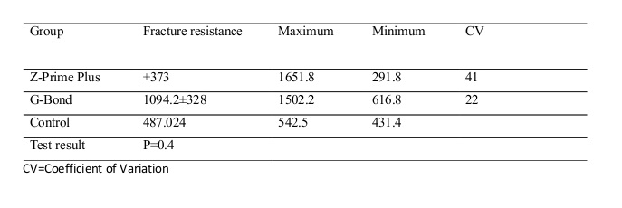
Results: The mean fracture resistance was 1094.2±328.0 N with G-Bond and 912.6±373.0 N with Z-Prime Plus; the difference was not significant (P=0.4).
Conclusion: G-Bond and Z-Prime Plus were not significantly different in fracture resistance of zirconia posts bonded to root canal walls. However, G-Bond is recommended for this purpose since it had a lower coefficient of variation (CV) and slightly higher fracture resistance.
Keywords: Endodontics, Fracture Strength, G-Bond, Post and Core Technique, Zirconia, Z-Prime Plus
Introduction
Fracture resistance of dental roots is an important factor to consider in the rehabilitation of endodontically treated teeth that have lost a great portion of their coronal structure. Intracanal posts are commonly used in endodontically treated teeth to provide more uniform load distribution. (1) Endodontically treated teeth have often lost a large portion of their structure and are at a high risk of fracture. Post space preparation in such teeth may further weaken the tooth structure and is associated with the risk of microfracture and perforation. (2) The bond strength of non-metallic posts to root canal walls is one of the most important factors in choosing the best technique for the restoration of endodontically treated teeth that have lost most of their coronal structure. (3) Zirconia posts regularly need to be reshaped and may be airborne particle abraded to enhance adhesion. Consequently, the surface of zirconia ceramics may be malformed; this influences the mechanical properties of zirconia. (4) Teeth with fiber posts reportedly undergo fracture at much higher loads compared to teeth with zirconia posts. (5) A study on fracture resistance of different intracanal posts bonded using new generations of bonding agents concluded that silane might be necessary to reinforce the bond of fiber posts cemented with resin cements. (6) Another study assessed the effect of primers on tensile bond strength of zirconia ceramic to composite resins after surface treatment with different primers and found no significant difference in this respect. (7)
G-Bond is a 7th-generation bonding agent comprising a strong resin, urethane dimethacrylate (UDMA), dimethacrylates, acidic resins, 4-Methacryloxyethyl trimellitic anhydride (4-MET), and phosphoric acid ester monomers. It has shown promising results in the bonding of restorations. (8) It is supplied in one bottle and does not require separate etching. It creates a thin void-free layer that covers the entire surface and is even suitable for shallow cavities. It is not visible after polymerization. The new generation of G-Bond does not contain 2-Hydroxyethyl methacrylate (HEMA); therefore, water sorption and discoloration do not occur. It is biocompatible and durable in the oral environment. It is used for surface treatment of zirconia posts since it is easy to use, provides a strong bond, is single-component, has low technical sensitivity, does not require a separate etching step, and saves time (30 seconds). (8)
Z-Prime Plus is a single-component primer manufactured by Bisco. (9) It is used to enhance the bonding between indirect restorative materials and composite resin cements. Due to its unique chemical formulation, it can be used for surface treatment of zirconia, alumina, metal, and composite posts. This primer has been exclusively manufactured to create a strong bond, irrespective of the method of curing. (9) Its advantages include strong chemical bonding to zirconia, compatibility with self-cure and light-cure resin cements, high durability, and easy application. (10)
Because zirconia posts are not cemented with the use of resin cements, their surface treatment is imperative to achieve a strong bond. Considering the gap of information regarding the application of G-Bond and Z-Prime Plus for bonding of zirconia posts, this study aimed to assess and compare the effect of G-Bond and Z-Prime Plus on fracture resistance of prefabricated zirconia posts bonded to root canal walls.
Materials and Methods
This in-vitro experimental study evaluated 22 extracted mandibular premolars. The teeth had no carious lesions, root cracks or internal and external root resorption and were of the same size and length. The Ethics Committee of the Dental Faculty of Islamic Azad University of Medical Sciences, Tehran, Iran, has approved this study. The teeth had been extracted for orthodontic reasons, and informed consent was obtained from the subjects. The teeth were thrown away afterward. The teeth were without names and could not be assigned to any individual.
The teeth were selected using convenience sampling. After extraction, they were immersed in 0.5% chloramine-T solution (Chloramine T trihydrate, Merck KGaA, Darmstadt, Germany) for disinfection, and were stored in saline at 37°C until the experiment One tooth from each group served as the control and did not undergo any intervention (no post surface treatment).
In the remaining teeth, the crown was cut using a metal disc with a 0.2-mm thickness mounted on a high-speed handpiece under copious water irrigation such that the remaining root length was 14 mm. The working length (WL) was radiographically determined using a #35 K-file (Dentsply/Maillefer, Ballaigues, Switzerland). The canals were rinsed with a 5.25% sodium hypochlorite (NaOCl) solution. A #45 master apical file (MAF) was selected for all the canals, which were then flared up to file #60 using the step-back technique (25 mm file length). The canals were dried using paper points. Next, A #35 master cone and #15 accessory cones were used for root canal filling. The root canals were filled using AH-26 sealer (Dentsply/Maillefer, Ballaigues, Switzerland) and gutta-percha via the lateral compaction technique. A #25 finger spreader (Dentsply/Maillefer, Ballaigues, Switzerland) was also used one mm shorter than the WL.
After obturation, the teeth were randomly divided into two groups of 10 samples each for the application of G-Bond (GC, Tokyo, Japan) or Z-Prime Plus (Bisco Inc., Irving Park, France). Two teeth were considered as the control samples as follows: (1,8,10-13)
Group 1 (n=10): Post space was prepared using peeso reamers #1, #2, and #3 (Dentsply/Maillefer, Ballaigues, Switzerland) in an orderly fashion and then using the universal drill available in the kit (DT Universal Drill, Munich, Germany). Post space was prepared in the canals with a 10-mm length and at a 3- to 4-mm distance from the apex. (9,12) Then, the #3 finishing drill was used for final preparation. A rubber-stop was used as a guide for the preparation length according to the radiograph. The prepared post space was cleaned using oil-free water and air spray. The posts (Ice light, Danville, USA) were tried in the canals to ensure their easy placement and passive fit. (13-15) The coronal part of all posts was cut using a 008 fissure diamond bur for all posts to have an equal length of 14 mm. (12) The post space was then cleaned with paper points and dried. Next, 37% phosphoric acid (Ivoclar Vivadent, Schaan, Liechtenstein) was applied to the canals for 15 seconds, rinsed, and dried for 3 seconds such that the radicular dentin remained slightly moist. (13,14) Panavia F2 resin cement (Kuraray, Tokyo, Japan) was prepared according to the manufacturer’s instructions and was applied to the canal using a microbrush. It was gently air-dried after 30 seconds. The excess cement was removed using a paper point. Next, Z-Prime Plus was prepared according to the manufacturer’s instructions, applied to the post surface, and cured. Next, A and B pastes (Panavia F2, Kuraray, Japan) were mixed at a 1:1 ratio and applied uniformly to the surface of the post. Zirconia posts were then inserted into the canals and compressed with finger pressure. The excess cement was removed using a microbrush. The cement was light-cured for 60 seconds using a light-curing unit (Woodpecker Light Cure - LED-D, China). Oxyguard gel (Kuraray, Osaka, Japan) was applied around the orifice for 3 minutes to ensure the complete set of the cement. (15) All fiber-reinforced composite (FRC) posts with zirconia coating (size 2; Ice light, Danville, USA) had a 14-mm length; 10 mm of their length was in the canal space, and 4 mm remained outside the canal for core retention. Next, the core was built-up using Photo Core composite resin (CLEARFIL™. PHOTO CORE, Kuraray, Osaka, Japan). Prefabricated polyester crowns were filled with Photo Core composite resin and placed on the coronal part of the posts. The cores were then cured for 40 seconds at 750mW/cm2 from four directions such that the core thickness was the same for all teeth.
Group 2 (n=10): Post space was prepared, and the canal was etched as explained for group 1. G-Bond was applied to the canal according to the manufacturer’s instructions using a microbrush. After 30 seconds, it was gently air-dried with air spray, and the excess bonding agent was removed using paper points. A and B pastes (Panavia F2, Kuraray, Japan) were then mixed and applied to the post surface. The post was placed in the canal and compressed with finger pressure for 5 to 10 seconds. (16) The excess cement was removed using a microbrush, and light-curing was performed for 60 seconds. Oxyguard was applied for 3 minutes to ensure the complete set of the cement. (15) The next steps were the same as those in group 1 (Figure 1).
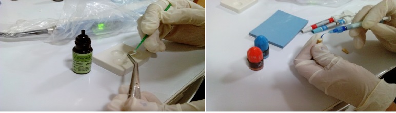
Figure 1. Z-Prime Plus was prepared according to the manufacturer’s instructions and was applied to the post surface using a microbrush.
The control group (n=2): The control teeth did not receive any surface treatment. Post space was prepared, and the canal was etched as explained for group 1. A and B pastes (Panavia F2, Kuraray, Japan) were then mixed and applied to the post surface. The post was placed in the canal and compressed with finger pressure for 5 to 10 seconds. (16) The excess cement was removed using a microbrush, and light-curing was performed for 60 seconds. Oxyguard was applied for 3 minutes to ensure the complete set of the cement. (15) The next steps were the same as those in group 1.
All teeth were then subjected to a compressive load applied to the central fossa of the core at a 90° angle relative to their longitudinal axes at a crosshead speed of 0.5 mm/minute in the Instron machine (Z050, Zwick/Roell, Ulm, Germany; Figure 2).
Figure 2. Compressive load application to the central fossa of the core at a 90° angle relative to the longitudinal axis of the tooth at a crosshead speed of 0.5 mm/minute using the Instron machine (Z050, Zwick/Roell, Ulm, Germany)
The load at fracture was recorded. The Kolmogorov-Smirnov test confirmed the normal distribution of data. Thus, data were compared using t-test via SPSS 22 software (SPSS Inc., Chicago, IL, USA).
Results
This study was carried out on 22 premolars: 10 prepared with G-Bond, 10 with Z-Prime Plus, and two control teeth with no surface treatment.
The mean fracture resistance of the control samples was 487.02 N.
Table 1 presents the mean fracture resistance (N) and coefficient of variation (CV) in the two groups. Accordingly, the difference between the two groups regarding fracture resistance was not significant (P=0.4).
Table 1. Fracture resistance (N) of the two groups
Discussion
Fracture resistance of dental roots should be taken into account when selecting the best method for reconstruction of endodontically treated teeth that have lost a large portion of their coronal structure. Intracanal posts have long been used for uniform stress distribution in endodontically treated teeth. (1)
Factors that affect the fracture resistance of endodontically treated teeth include the diameter, length, design, and adaptability of the post, the remaining dentin, the type of cement, the method of cementation, the core material and design, the design of the crown, and biocompatibility of the post. (17)
This study assessed the effect of G-Bond and Z-Prime Plus on fracture resistance of prefabricated zirconia posts bonded to root canal walls. The results showed that G-Bond and Z-Prime Plus were not significantly different in terms of the fracture resistance of zirconia posts bonded to canal walls. They both were suitable for bonding of zirconia posts to dentin although the fracture resistance provided by G-Bond was slightly (but not significantly) higher. Thus, they can both be successfully used in clinical settings for the bonding of zirconia posts to the radicular dentin. An important finding of our study was that although G-Bond provided slightly higher retention, it had a smaller standard deviation (SD) and a lower CV. Thus, G-Bond is expected to show a more predictable clinical behavior than Z-Prime Plus. Therefore, it may be preferred over Z-Prime Plus in clinical settings.
Kivanc and Gorgul evaluated the fracture resistance of teeth restored with different posts and new bonding agents and concluded that silane is imperative to reinforce the bond of fiber posts cemented with resin cements. (6) Their result was in line with our findings. Sanohkan et al evaluated the effect of different primers on the tensile bond strength of zirconia ceramic to composites. (7) They concluded that surface treatment with primers caused no significant change in bond strength, whereas G-Bond and Z-Prime Plus significantly increased the fracture resistance in our study.
Maleki pour et al evaluated the effect of different surface treatments on the flexural strength and modulus of elasticity of FRC posts and concluded that surface treatment of glass fiber and quartz fiber posts with laser and 10% hydrogen peroxide (H2O2) had no significant effect on flexural strength and modulus of elasticity. (18) However, in our study, the application of G-Bond and Z-Prime Plus as surface treatment increased the fracture resistance of zirconia posts. Habibzadeh et al (19) evaluated the fracture resistance of zirconia, cast nickel-chromium (Ni-Cr), and FRC post systems under all-ceramic crowns and reported significantly lower fracture resistance (435.34 N) and non-restorable fractures for zirconia posts, which is consistent with the results of the present study. In a recent study, prefabricated zirconia posts demonstrated higher fracture resistance than carbon fiber posts. (20,21)
This study had some limitations. Finding teeth with the same length and thickness was problematic and led to our small sample size. Future studies are required to assess a higher number of teeth with the same size and age to increase the accuracy of the results.
Conclusion
Within the limitations of this in-vitro study, there was no significant difference between G-Bond and Z-Prime Plus in the bonding of zirconia posts to root canal walls of endodontically treated teeth. However, since G-Bond provided slightly higher fracture resistance and lower CV, its use may be preferred for bonding of zirconia posts in clinical settings.
| Rights and permissions | |
 |
This work is licensed under a Creative Commons Attribution-NonCommercial 4.0 International License. |
