


BibTeX | RIS | EndNote | Medlars | ProCite | Reference Manager | RefWorks
Send citation to:
URL: http://jrdms.dentaliau.ac.ir/article-1-345-en.html


2- Oral Medicine Dept, Faculty of Dentistry,Tehran Medical Sciences,
3- Endodontics Dept, Faculty of Dentistry, Tehran Medical Sciences
4- Endodontics Dept, Faculty of Dentistry, Tehran Medical Sciences,Islamic Azad University, Tehran, Iran , s_savadkouhi@yahoo.com
Abstract
Background and Aim: Methylene blue and curcumin are effective photosensitizers for inactivation of bacteria. This study assessed the penetration depth of methylene blue and curcumin in presence/absence of smear layer into dentinal tubules.
Materials and Methods: Thirty-two human central and lateral incisors were included in this experimental study. The initially prepared specimens were randomly allocated to 4 experimental groups: Group 1: methylene blue with smear layer, group 2: methylene blue without smear layer, group 3: curcumin with smear layer, group 4: curcumin without smear layer. Root specimens were sectioned by a diamond disc at 4 and 8 mm from the apex to obtain apical, middle, and coronal sections. The mean penetration depth was measured at the buccal, mesial, distal and palatal areas on cross sections. ANOVA was used to assess the effect of photosensitizer type, smear layer, and root level on penetration depth. Pairwise comparisons were performed by the Student’s t-test.
Results: The maximum penetration depth was in the apical third in group 2 (0.98±0.25 mm) and the minimum penetration depth was in the coronal third in group 1 (0.21±0.15 mm); this difference was significant (P=0.001). Smear layer removal from the apical and middle thirds was correlated with higher photosensitizer penetration depth (P=0.000) but this difference was not significant in the coronal third (P=0.6). Curcumin had significantly greater penetration depth in presence of smear layer in all three parts compared with methylene blue (P<0.05).
Conclusion: Curcumin can penetrate more into dentinal tubules than methylene blue in presence of smear layer.
Key words
Curcumin; Dentin; Methylene Blue; Photosensitizing Agents; Smear Layer
Introduction
There has been a growing interest in use of photodynamic therapy (PDT) as an adjunct for treatment of endodontic infections. PDT requires a light source to activate a non-toxic photosensitizing agent to create singlet oxygen and free radicals, which can induce the death of microorganisms. The half-life of singlet oxygen and free radicals is very short (1). PDT was first developed as a treatment option for malignancies and was proven to be a highly promising alternative against microorganisms for treatment of localized infections. The advantages of PDT over more traditional treatments (chemical irrigants, ultrasonic or high power lasers) in disinfecting the root canal system include minimal toxicity, minimal effect on the surrounding tissues, and disruption of biofilm structure instead of directly targeting the microorganisms (2).The penetration depth of photosensitizers into dentinal tubules will determine the killing effect of PDT against microorganisms. It has been shown that methylene blue and curcumin are effective photosensitizing agents for inactivation of endodontic bacteria (3-5). Curcumin is a natural photosensitizer, which is extracted from the Curcuma longa. In addition to antibacterial properties when activated by blue light, curcumin has great bioactive properties such as anti-inflammatory and anti-tumor effects (6-8). Methylene blue is a synthetic photosensitizer, which is easily dissolved in water and penetrates into the bacterial cell wall. Red-wavelength lasers can activate methylene blue (9-11).
Dentin permeability is partly determined by the presence or absence of smear layer on root canal walls. Smear layer is characterized by inorganic and organic substances which may be contaminated by microorganisms. It has been shown that antibacterial properties of irrigants and intracanal medicaments diminishes in presence of smear layer. However, smear layer removal remains a controversial topic in endodontic therapy and restorative dentistry (12, 13). This study aimed to determine the penetration depth of methylene blue and curcumin in presence and absence of smear layer into human root dentin.
Materials and Methods
Thirty-two human central and lateral incisors were included in this experimental ex-vivo study from the dental bank of Dental School, Islamic Azad University, Tehran, Iran and stored in sterile saline until the experiment. The proposal of this study was approved and registered in the ethic committee of this university (IR.IAU.DENTAL.REC.1399.065).
The sample size of 8 for each group was calculated based on a study by Kosarieh et al, (14) using one-way ANOVA power analysis, α=0.05, β=0.2, effect size=0.67, and standard deviation=0.9.
Periapical radiographs were obtained from each tooth. The inclusion criteria were human single-rooted and single-canal central and lateral incisors with minimal curvature (<15º) and mature apex (#10 K-file could pass throw the apical foramen while #20 K-file could not). The exclusion criteria were root caries, root resorptions, calcifications and cracks.
Sample preparation:
The included teeth were decoronated to obtain 12 mm equal working length. The root canals were prepared by BioRace rotary files (FKG, Switzerland) to 40/0.04, and irrigation was done with 2 mL of 2.5% sodium hypochlorite after each instrumentation.
The external root surface was covered with two layers of nail polish; then, the specimens were subjected to autoclave-sterilization (121oC, 15 psi for 30 minutes).
The initially prepared specimens were randomly allocated to 4 experimental groups:
Group 1: methylene blue with smear layer
Group 2: methylene blue without smear layer
Group 3: curcumin with smear layer
Group 4: curcumin without smear layer
Samples in groups 1 and 3 underwent final irrigation with 2 mL of sterile saline but groups 2 and 4 were irrigated with 2 mL of 17% EDTA for 1 minute followed by 2 mL of 5.25% sodium hypochlorite for 30 seconds (15). Finally, photosensitizers (100 µg/mL methylene blue and 300 µg/mL curcumin) were poured into the respective root canals until filled, and they were incubated for 10 minutes. Next, they were removed from the root canals by using paper points.
Stereomicroscopic analysis:
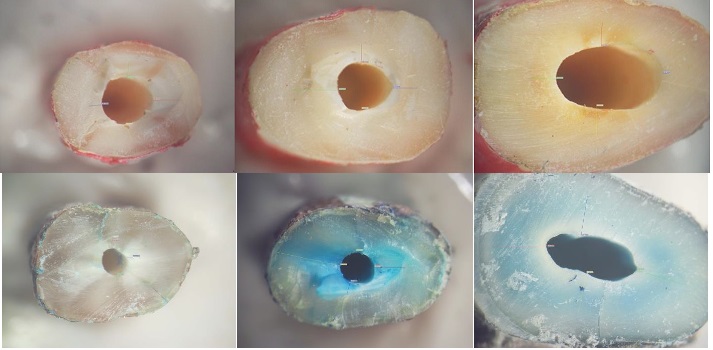
Thirty-two prepared specimens in 4 experimental groups were subjected to stereomicroscopic analysis (Nikon SMZ 1500) under ×20 magnification to determine the photosensitizer penetration into root dentinal tubules. Root specimens were sectioned by a diamond disc at 4 and 8 mm from the apex to obtain specimens from the apical, middle and coronal thirds. The mean penetration depth was calculated at the buccal, mesial, distal and palatal surfaces to measure the level of photosensitizer penetration into root dentin cross-sections. The measurements were made by an equipped software connected to the stereomicroscope (Figure 1).
Statistical analysis:
Data analysis was done using SPSS version 16. Multivariate ANOVA was used to analyze the effect of photosensitizer type (methylene blue or curcumin), smear layer (presence/absence) and root level (apical, middle or coronal third) on penetration depth. Pairwise comparisons were done by the Student’s t-test.
Figure 1. steriomicroscopic images of methylene blue and curcumin at different root levels
Results
A total of 96 samples were evaluated. Data analysis showed the maximum penetration depth in the apical third in group 2 (methylene blue without smear layer; 0.98±0.25 mm) and the minimum penetration depth in the coronal third in group 1 (methylene blue with smear layer; 0.21±0.15 mm); this difference was significant (P=0.001; Table 1).
Table 1: Minimum, maximum, mean and standard deviation (mm) of photosensitizer penetration depth in different experimental groups at different root levels (n=8)
ANOVA showed a significant difference among the experimental groups in terms of photosensitizer penetration depth into dentinal tubules (P=0.001). In the apical third, presence of smear layer decreased the penetration depth of curcumin and methylene blue (0.354 mm and 0.656 mm, respectively; P=0.009 and P=0.000, respectively). In the middle third, presence of smear layer decreased the penetration depth of curcumin and methylene blue (0.2 mm and 0.56 mm, respectively; P=0.04 and P=0.000, respectively). In the coronal third, presence of smear layer decreased the penetration depth of curcumin and methylene blue (0.045 mm and 0.34 mm, respectively; P=0.6 and P=0.000, respectively). In the apical third, when smear layer was present, curcumin had 0.24 mm more tubular penetration depth than methylene blue (P=0.001) but when smear layer was removed, the difference was not significant (P=0.6). In the middle and coronal thirds, when smear layer was present, curcumin had greater tubular penetration depth than methylene blue (P=0.001 and P=0.000, respectively; Figure 2).
Discussion
This study assessed the penetration depth of curcumin and methylene blue into dentin tubules of single-canal human teeth. Curcumin in presence of smear layer penetrated deeper into dentinal tubules than methylene blue. But when the smear layer was removed, the penetration depth of the two photosensitizers was close to each other, and was not significantly different. Also, both photosensitizers in the apical region had a greater penetration depth in absence of smear layer.
There are many factors that affect the penetration depth of photosensitizers into dentinal tubules, including the size of the molecules, solubility in water, time allowed for penetration of photosensitizer, presence or absence of smear layer, surface tension, and osmotic and hydrostatic pressure (3).
According to a study by Pashley and Livingston (16), increasing the radius of the penetrant molecules by 19 times decreased permeability by 100 times. Researchers also examined the penetration depth of chlorhexidine and fluoride, regardless of their molecular weight and size. Low penetration could also be related to the bonding of molecules to dentin. Therefore, in addition to the size and weight of the molecules, dentin bond strength is also another determining factor in penetration depth of photosensitizers.
Therefore, the study of physical and chemical properties of materials can indicate their penetrability into dentinal tubules.
Curcumin is a yellow compound derived from a plant with the scientific name Curcuma longa. This substance belongs to the curcuminoid family. It has been used in traditional medicine for centuries, and no toxic effect has been reported for it so far. This material is stable in acidic physiological pH but degrades rapidly in alkaline environments. Other biological properties of this substance are due to the complex molecular structures that curcumin makes when combined with other molecules. In addition to these properties, the antimicrobial properties of curcumin are also important (7, 8).
The minimum inhibitory concentration (MIC) of curcumin has been determined in several studies (17-19). We used 300 µg/mL curcumin to meet the MIC for maximum bacterial coverage. The MIC of methylene blue to meet maximum bacterial coverage is 100 µg/mL (20-22). The present study showed that the difference in concentration of the two photosensitizers had no significant effect on their penetration depth into dentin.
Methylene blue is a highly soluble substance in water that can easily penetrate into dentinal tubules in presence of water and does not damage the dental matrix or hydroxyapatite. It also has bactericidal properties (11). In the present study, it was observed that in spite of the lower molecular weight of methylene blue (319.85 g/mol) compared with curcumin (368.38 g/mol), curcumin had a greater tubular penetration depth in presence of smear layer, possibly due to its acidic nature (23). Smear layer showed less ability to prevent tubular penetration of curcumin compared with methylene blue. Accordingly, Kosarieh et al. (14) found that despite the lower molecular weight of tolonium chloride in comparison with indocyanine green, the mean penetration depth of indocyanine green was significantly higher in all root areas of the tooth.
As reported in the present study, after removal of the smear layer, methylene blue, which has a higher solubility in water than curcumin, had a greater penetration depth probably because dentinal tubules are hydrophilic; thus, water solubility can also affect the penetrability of the substance (11).
In this study, in the groups where smear layer was removed, the highest penetration depth was observed in the apical region, which was in contrast to the study by Kosarieh et al, (14) who concluded that the penetration depth of tolonium chloride and indocyanine green decreased from the coronal towards the apical region. They attributed this finding to the increase in the number and diameter of dentinal tubules from the apical towards the coronal part of the root canal. Paque et al, (24) also reported that the permeability of dentinal tubules decreased from the coronal towards the apical part of the root canal. As mentioned in the present study, both materials had significantly greater penetration depth in absence of smear layer. This indicates that the removal of the smear layer increased the penetration depth of both curcumin and methylene blue. This has been reported in many other studies (13, 25, 26).
| Rights and permissions | |
 |
This work is licensed under a Creative Commons Attribution-NonCommercial 4.0 International License. |
