Journal of Research in Dental
and Maxillofacial Sciences
Volume 3, Issue 3 (8-2018)
J Res Dent Maxillofac Sci 2018, 3(3): 19-26 |
Back to browse issues page


Download citation:
BibTeX | RIS | EndNote | Medlars | ProCite | Reference Manager | RefWorks
Send citation to:


BibTeX | RIS | EndNote | Medlars | ProCite | Reference Manager | RefWorks
Send citation to:
kaviani R, Mackinejad A, Pourgholi Takromi Z, Mirmehdi S. Evaluating the Symmetry of Permanent Teeth Dimensions on Two Sides of Dental Arches with Different Occlusions and Related Factors. J Res Dent Maxillofac Sci 2018; 3 (3) :19-26
URL: http://jrdms.dentaliau.ac.ir/article-1-210-en.html
URL: http://jrdms.dentaliau.ac.ir/article-1-210-en.html


1- Assistant Professor,Faculty Member and Head of Department, Anatomy and Morphology Department, , rakavira@yahoo.com
2- Assistant professor, Faculty Member and Head of Department, Anatomy and Morphology Department,
3- Dentist
2- Assistant professor, Faculty Member and Head of Department, Anatomy and Morphology Department,
3- Dentist
Full-Text [PDF 318 kb]
(2102 Downloads)
| Abstract (HTML) (5675 Views)
Abstract
Background and Aim: Considering the importance of teeth dimensions in their adequacies and aesthetics, their significant racial differences, and various statistics regarding the dimensions of symmetrical teeth in previous articles, we aimed to evaluate the dimensions of symmetrical permanent teeth on two sides of the maxillary and mandibular dental arches and related factors in different occlusions of students and patients of Islamic Azad Dental University of Tehran.
Materials and Methods: This cross-sectional study was conducted on 210 people. A dental cast was made for each individual. The mesiodistal, labiolingual, and occlusogingival dimensions of the clinical crowns were measured using a caliper with the accuracy of 0.1 mm along with their adaptation and symmetry on two sides of the maxillary and mandibular dental arches and related factors. Data were analyzed using Chi-square test.
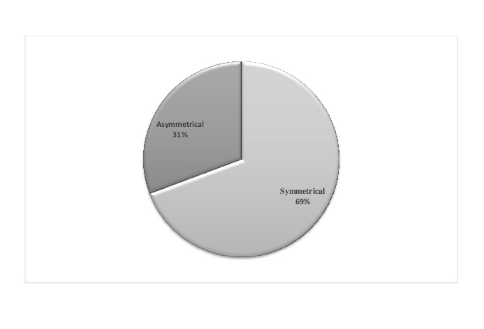
Results: 67.1% of the patients were females and 32.9% were males. There were 69.3% symmetric and 30.7% asymmetric cases; there were no significant differences in terms of the dimensions, gender, and jaws (P<0.8). Regarding teeth types and variations in maxillary and mandibular central incisors, symmetrical values were 10.3%, while non-symmetrical values were 31.3%. Asymmetrical values in central incisors of both dental arches were significantly greater than symmetrical values.
Conclusion: It seems that on the left and right sides of both dental arches of permanent teeth in both genders, symmetrical values are greater than asymmetrical values. Asymmetrical values in central incisors of both arches were significantly greater than symmetrical values.
Keywords: Permanent Dentition, Dental Arch, Dental Occlusion, Odontometry
Introduction
Facial asymmetry is a common phenomenon that was first noticed by Greek sculptors, which was then called “Normal Facial Asymmetry”. (1) This phenomenon causes the face to be more attractive. In 1931, Woo studied the ancient skulls of the 26th to 30th generations in Africans and found out that there were asymmetrical values in the skulls as their right sides (frontal, temporal, and parietal bones) were slightly bigger than their left sides due to more growth of the right hemisphere of the brain, but in their faces, it was vice versa as their zygomatic and maxillary bones were bigger on the left side than on the right side. (1) Lundstrom studied 319 thirteen-year-old children in 1955 and reported changes in the width ratio of the teeth in the two dental arches. (2) Black provided the average teeth size ratio tables for teeth dimensions in 1902. (3) In 1944, Ballard established that 90% of the same teeth on the left and right sides are different from each other by about 0.25 mm or more in their width or mesiodistal dimensions. (3) In 1958, Bolton measured the width of teeth mesiodistally in 55 cases who had ideal occlusions and introduced some ratios known as the anterior and overall teeth ratios. (4) Gillen et al in 1994 discovered that in both Black and White populations, the maxillary anterior teeth in males are wider and longer than those of females. (5) In 1999, Sterrett et al also came to the conclusion that the width and the length of clinical crowns of the maxillary anterior teeth in the Caucasian race are greater in males than in females. (6) According to the brief asymmetries of bipartite proportions of the body and also the presence of slight asymmetries on the left and right sides of the facial region, their research was conducted on asymmetrical values of permanent teeth dimensions on two sides of the dental arches which are a part of the hard tissue in the craniofacial region. (6) The present research aimed to evaluate the symmetry of permanent teeth dimensions on two sides of the dental arches in different occlusions of different individuals to reveal whether there are any symmetrical values related to permanent teeth and what are the differences between those teeth that are symmetric relative to each other and those that are not.
Materials and Methods
This cross-sectional study was conducted on 210 people who were patients and students of Islamic Azad Dental University of Tehran (141 females and 69 males aged between 20-25 years) using the census sampling method. The teeth of these individuals were free from decays, proximal restorations, traumatic injuries, clinical crown fractures, abrasions, and veneers. There were acceptable slight spaces between the teeth (diastema), malocclusion, and teeth rotation in the individuals who participated in this research as they all had normal occlusions. (3,5) Then, by using premade dental impression trays, impressions were taken from the maxillary and mandibular dental arches of each individual using an alginate impression material (Zhermack Co., Germany). All the impressions were wrapped in moist gauze bandages after being disinfected and were then immediately cast using dental stone (GC Fuji Rock; GC. Corporation, Tokyo, Japan), and finally, dental casts were attained from each individual. Then, using a caliper with the accuracy of 0.1 mm, the mesiodistal (the maximum width between the mesial and distal surfaces of a tooth), (7) labiolingual (the maximum width from the height of contour of the labial surface to the lingual surface), (8) and occlusogingival (the clinical crown) dimensions were measured along with their adaptation and symmetry on two sides of the maxillary and mandibular dental arches and related factors, which were then recorded in datasheets. Next, data were analyzed and compared statistically using Chi-square test.
Results
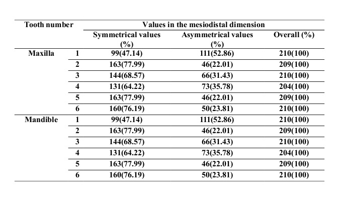
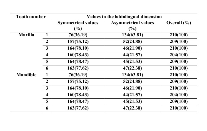
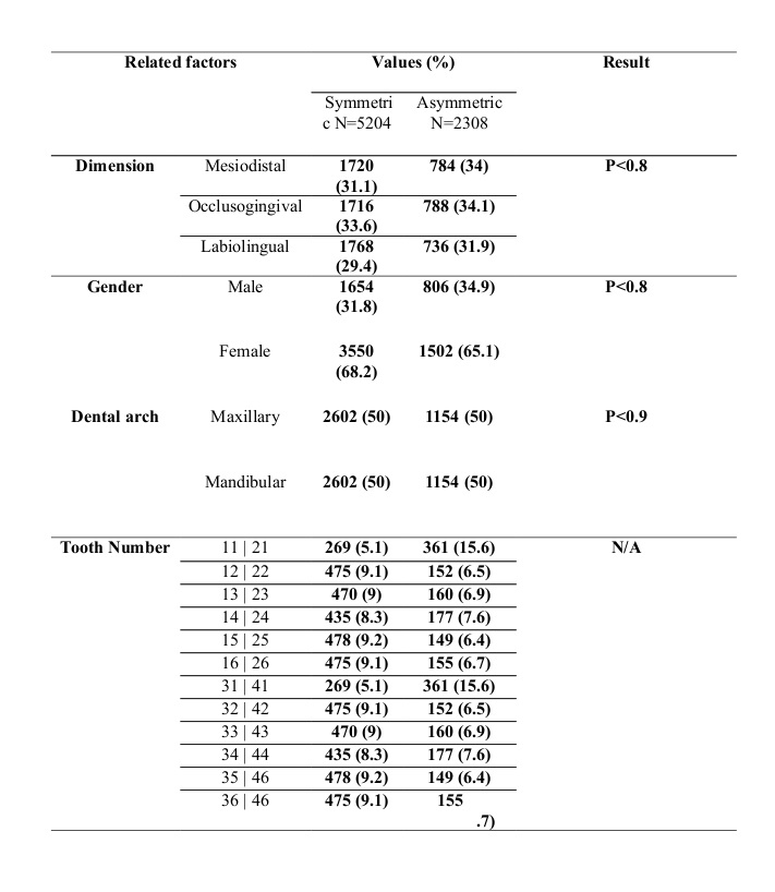
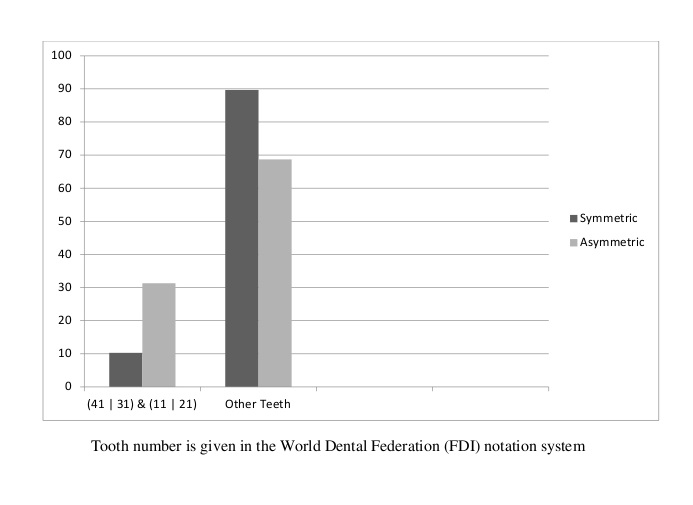
The symmetrical values are respectively presented in Tables 1, 2, and 3, categorized by the type of teeth in the three dimensions (mesiodistal, occlusogingival, and labiolingual). The distribution of the individuals taking part in this research showed that 69.3% had symmetric values and 30.7% did not, who were placed in the asymmetric group of individuals as it is shown in Figure 1. According to the prevalence of symmetrical values among all the participants, the real asymmetrical values with a 95% confidence interval are calculated from the minimum of 24.5% to the maximum of 36.5%. The distribution of the studied individuals in terms of symmetrical values and related factors is summarized in Table 4 which shows no significant differences in terms of the dimensions (mesiodistal, labiolingual, and occlusogingival), gender, and jaws (P<0.8). The distribution of the studied individuals in terms of the tooth number and tooth type is presented in Figure 2 which shows 10.3% symmetrical and 31.3% asymmetrical values in maxillary and mandibular central incisors. By comparing them to other teeth, we could acknowledge that asymmetrical values were significantly greater than symmetrical values.
Table 1. Symmetrical values of the mesiodistal dimension by tooth type
Table 2. Symmetrical values of the occlusogingival dimension by tooth type.
Table 3. Symmetrical values of the labiolingual dimension by tooth type.
Table 4. Distribution of the studied individuals in terms of symmetrical values and related factors.
Figure 1. Distribution of 210 individuals based on symmetrical and asymmetrical values
Figure 2. Distribution of the studied individuals in terms of the tooth number and tooth type
Discussion
This study showed that there were 69.3% symmetrical and 30.7% asymmetrical values in the permanent teeth of both dental arches, and also, individuals who lacked symmetrical values showed no significant differences in terms of the dimensions (mesiodistal, labiolingual, and occlusogingival), gender, and jaws. Correspondingly, in terms of the tooth types, this study showed 10.3% symmetrical and 31.3% asymmetrical values in maxillary and mandibular central incisors, and by comparing them to other teeth, we could acknowledge that asymmetrical values were significantly greater than symmetrical values.
By reviewing previous studies, no statements have been made regarding the measurements of symmetrical values; therefore, no similarity could be established; nevertheless, the width of teeth is not the only factor related to symmetry. In Mosby’s medical dictionary, the definition of the word “symmetry” is the correspondence in the size ratio, form, and arrangement of parts on opposite sides of a plane or around an axis. (9) Clinically, the word “symmetry” is defined as a phenomenon which is balanced correctly. (9) In our study, three dental dimensions were considered, and their balance was evaluated on both poles (sides) of the body, while in previous studies, the only dimension that has been evaluated is the width of the mesiodistal dimension. In 2004, Zarringhalam conducted a research aiming to evaluate the symmetry of teeth and found out that there were no significant differences in the overall size ratio of teeth in females and males. (10) Similarly, in our study, the overall symmetrical value was 69.3%, which was more than asymmetrical values and showed a slight difference in the teeth dimensions in both dental arches. On the other hand, in the mentioned research, the number of individuals was limited although the number of females and males was equal. In a study by Jalali and Pousti in 2004, the outcome revealed that the dimensions of the maxillary and mandibular teeth in the group with Class I malocclusion were significantly greater than that in the group with normal occlusions. (11) Obviously, the cited study was conducted on crowded teeth, but in our study, Class I malocclusion and perceptible crowded teeth were not observed in the studied individuals. In 2006, Başaran et al established that the results of the measurements in the mesiodistal dimension were not significantly different in different malocclusions (Class I, Class II, and Class III). (7) In the mentioned research, an electronic digital caliper was used which is a very accurate device. In 2005, Eslamian et al concluded that in the mesiodistal dimension, there was a significant difference between the proportions of the anterior teeth compared to the overall size ratio of other teeth in females and males, and also, the comparison of the teeth revealed that the maxillary second premolars, mandibular second premolars, mandibular central incisors, and maxillary lateral incisors show the highest diversity in size ratio, respectively. (12) A study by Kachoei et al in Tabriz in 2011 showed no significant difference between the teeth measurements on two sides of the dental arches (left and right). (13) In our study, symmetrical values were greater than asymmetrical values, which shows the slight difference in the dimensions of the teeth size ratios. In this study, a digital caliper with 0.1-mm precision was used, which is an accurate device. In 2004, Zarringhalam conducted another research to evaluate the overall symmetry of the teeth and found out that the overall maxillary and mandibular teeth size ratios in males were significantly greater than that in females. (14) A study by Scanavini et al in Brazil in 2012 revealed that asymmetrical values were observed in the dental arches of individuals with normal occlusion and Class II and Class III malocclusions. (15) The level of asymmetry in the mandibular arch was greater than that in the maxillary arch, but in our study, symmetrical values were evaluated regarding the teeth size ratios. In 2011, Malkoç et al in Turkey discovered that there was a significant difference in the mesiodistal dimensions of the teeth between males and females. (16) In our study, no differences could be established in terms of gender. Hussein et al stated that statistically significant differences in tooth sizes are not always accompanied by significant differences in arch width, length or perimeter. (17) A study by Uysal and Sari in 2005 in Turkey showed that variations in the mesiodistal dimensions in the maxillary arch were greater than those in the mandibular arch, and the highest variations were related to the first molars. (8) In our study, the highest diversity of the tooth types was associated with the central incisors, and these differences could be linked to the studied races and ethnicities. In previous studies, the mesiodistal widths have been compared. It would not be rational to only measure the mesiodistal width, and then, base all the results of symmetrical values and comparisons on the similarity of the obtained numbers or that factor alone. In our study, we used the Delphi method to ask several morphology experts based on their scientific experiences and expertise regarding tree-dimensional symmetrical values of teeth, the number of teeth, and the side of the jaws (left and right), and also, how several millimeters of difference in size ratio would not be affecting the diagnostic, therapeutic, and preventive outcomes to be considered symmetric.
Finally, it was concluded that those teeth that are adjacent to each other will be considered as symmetric if the difference in the size ratio is not greater than 0.1 mm, and for the rest of the teeth, not greater than 0.3 mm.
One of the weaknesses of this study is that the research was only conducted on the patients referred to the Islamic Azad Dental University of Tehran, and the results may vary in other societies.
Conclusion
Generally, in the permanent teeth on the left and right sides of both dental arches in both males and females, symmetrical values were greater than asymmetrical values. The central incisors on both dental arches showed more significant differences in terms of asymmetry compared to other teeth.
Full-Text: (3204 Views)
Abstract
Background and Aim: Considering the importance of teeth dimensions in their adequacies and aesthetics, their significant racial differences, and various statistics regarding the dimensions of symmetrical teeth in previous articles, we aimed to evaluate the dimensions of symmetrical permanent teeth on two sides of the maxillary and mandibular dental arches and related factors in different occlusions of students and patients of Islamic Azad Dental University of Tehran.
Materials and Methods: This cross-sectional study was conducted on 210 people. A dental cast was made for each individual. The mesiodistal, labiolingual, and occlusogingival dimensions of the clinical crowns were measured using a caliper with the accuracy of 0.1 mm along with their adaptation and symmetry on two sides of the maxillary and mandibular dental arches and related factors. Data were analyzed using Chi-square test.
Results: 67.1% of the patients were females and 32.9% were males. There were 69.3% symmetric and 30.7% asymmetric cases; there were no significant differences in terms of the dimensions, gender, and jaws (P<0.8). Regarding teeth types and variations in maxillary and mandibular central incisors, symmetrical values were 10.3%, while non-symmetrical values were 31.3%. Asymmetrical values in central incisors of both dental arches were significantly greater than symmetrical values.
Conclusion: It seems that on the left and right sides of both dental arches of permanent teeth in both genders, symmetrical values are greater than asymmetrical values. Asymmetrical values in central incisors of both arches were significantly greater than symmetrical values.
Keywords: Permanent Dentition, Dental Arch, Dental Occlusion, Odontometry
Introduction
Facial asymmetry is a common phenomenon that was first noticed by Greek sculptors, which was then called “Normal Facial Asymmetry”. (1) This phenomenon causes the face to be more attractive. In 1931, Woo studied the ancient skulls of the 26th to 30th generations in Africans and found out that there were asymmetrical values in the skulls as their right sides (frontal, temporal, and parietal bones) were slightly bigger than their left sides due to more growth of the right hemisphere of the brain, but in their faces, it was vice versa as their zygomatic and maxillary bones were bigger on the left side than on the right side. (1) Lundstrom studied 319 thirteen-year-old children in 1955 and reported changes in the width ratio of the teeth in the two dental arches. (2) Black provided the average teeth size ratio tables for teeth dimensions in 1902. (3) In 1944, Ballard established that 90% of the same teeth on the left and right sides are different from each other by about 0.25 mm or more in their width or mesiodistal dimensions. (3) In 1958, Bolton measured the width of teeth mesiodistally in 55 cases who had ideal occlusions and introduced some ratios known as the anterior and overall teeth ratios. (4) Gillen et al in 1994 discovered that in both Black and White populations, the maxillary anterior teeth in males are wider and longer than those of females. (5) In 1999, Sterrett et al also came to the conclusion that the width and the length of clinical crowns of the maxillary anterior teeth in the Caucasian race are greater in males than in females. (6) According to the brief asymmetries of bipartite proportions of the body and also the presence of slight asymmetries on the left and right sides of the facial region, their research was conducted on asymmetrical values of permanent teeth dimensions on two sides of the dental arches which are a part of the hard tissue in the craniofacial region. (6) The present research aimed to evaluate the symmetry of permanent teeth dimensions on two sides of the dental arches in different occlusions of different individuals to reveal whether there are any symmetrical values related to permanent teeth and what are the differences between those teeth that are symmetric relative to each other and those that are not.
Materials and Methods
This cross-sectional study was conducted on 210 people who were patients and students of Islamic Azad Dental University of Tehran (141 females and 69 males aged between 20-25 years) using the census sampling method. The teeth of these individuals were free from decays, proximal restorations, traumatic injuries, clinical crown fractures, abrasions, and veneers. There were acceptable slight spaces between the teeth (diastema), malocclusion, and teeth rotation in the individuals who participated in this research as they all had normal occlusions. (3,5) Then, by using premade dental impression trays, impressions were taken from the maxillary and mandibular dental arches of each individual using an alginate impression material (Zhermack Co., Germany). All the impressions were wrapped in moist gauze bandages after being disinfected and were then immediately cast using dental stone (GC Fuji Rock; GC. Corporation, Tokyo, Japan), and finally, dental casts were attained from each individual. Then, using a caliper with the accuracy of 0.1 mm, the mesiodistal (the maximum width between the mesial and distal surfaces of a tooth), (7) labiolingual (the maximum width from the height of contour of the labial surface to the lingual surface), (8) and occlusogingival (the clinical crown) dimensions were measured along with their adaptation and symmetry on two sides of the maxillary and mandibular dental arches and related factors, which were then recorded in datasheets. Next, data were analyzed and compared statistically using Chi-square test.
Results
The symmetrical values are respectively presented in Tables 1, 2, and 3, categorized by the type of teeth in the three dimensions (mesiodistal, occlusogingival, and labiolingual). The distribution of the individuals taking part in this research showed that 69.3% had symmetric values and 30.7% did not, who were placed in the asymmetric group of individuals as it is shown in Figure 1. According to the prevalence of symmetrical values among all the participants, the real asymmetrical values with a 95% confidence interval are calculated from the minimum of 24.5% to the maximum of 36.5%. The distribution of the studied individuals in terms of symmetrical values and related factors is summarized in Table 4 which shows no significant differences in terms of the dimensions (mesiodistal, labiolingual, and occlusogingival), gender, and jaws (P<0.8). The distribution of the studied individuals in terms of the tooth number and tooth type is presented in Figure 2 which shows 10.3% symmetrical and 31.3% asymmetrical values in maxillary and mandibular central incisors. By comparing them to other teeth, we could acknowledge that asymmetrical values were significantly greater than symmetrical values.
Table 1. Symmetrical values of the mesiodistal dimension by tooth type
Table 2. Symmetrical values of the occlusogingival dimension by tooth type.
Table 3. Symmetrical values of the labiolingual dimension by tooth type.
Table 4. Distribution of the studied individuals in terms of symmetrical values and related factors.
Figure 1. Distribution of 210 individuals based on symmetrical and asymmetrical values
Figure 2. Distribution of the studied individuals in terms of the tooth number and tooth type
Discussion
This study showed that there were 69.3% symmetrical and 30.7% asymmetrical values in the permanent teeth of both dental arches, and also, individuals who lacked symmetrical values showed no significant differences in terms of the dimensions (mesiodistal, labiolingual, and occlusogingival), gender, and jaws. Correspondingly, in terms of the tooth types, this study showed 10.3% symmetrical and 31.3% asymmetrical values in maxillary and mandibular central incisors, and by comparing them to other teeth, we could acknowledge that asymmetrical values were significantly greater than symmetrical values.
By reviewing previous studies, no statements have been made regarding the measurements of symmetrical values; therefore, no similarity could be established; nevertheless, the width of teeth is not the only factor related to symmetry. In Mosby’s medical dictionary, the definition of the word “symmetry” is the correspondence in the size ratio, form, and arrangement of parts on opposite sides of a plane or around an axis. (9) Clinically, the word “symmetry” is defined as a phenomenon which is balanced correctly. (9) In our study, three dental dimensions were considered, and their balance was evaluated on both poles (sides) of the body, while in previous studies, the only dimension that has been evaluated is the width of the mesiodistal dimension. In 2004, Zarringhalam conducted a research aiming to evaluate the symmetry of teeth and found out that there were no significant differences in the overall size ratio of teeth in females and males. (10) Similarly, in our study, the overall symmetrical value was 69.3%, which was more than asymmetrical values and showed a slight difference in the teeth dimensions in both dental arches. On the other hand, in the mentioned research, the number of individuals was limited although the number of females and males was equal. In a study by Jalali and Pousti in 2004, the outcome revealed that the dimensions of the maxillary and mandibular teeth in the group with Class I malocclusion were significantly greater than that in the group with normal occlusions. (11) Obviously, the cited study was conducted on crowded teeth, but in our study, Class I malocclusion and perceptible crowded teeth were not observed in the studied individuals. In 2006, Başaran et al established that the results of the measurements in the mesiodistal dimension were not significantly different in different malocclusions (Class I, Class II, and Class III). (7) In the mentioned research, an electronic digital caliper was used which is a very accurate device. In 2005, Eslamian et al concluded that in the mesiodistal dimension, there was a significant difference between the proportions of the anterior teeth compared to the overall size ratio of other teeth in females and males, and also, the comparison of the teeth revealed that the maxillary second premolars, mandibular second premolars, mandibular central incisors, and maxillary lateral incisors show the highest diversity in size ratio, respectively. (12) A study by Kachoei et al in Tabriz in 2011 showed no significant difference between the teeth measurements on two sides of the dental arches (left and right). (13) In our study, symmetrical values were greater than asymmetrical values, which shows the slight difference in the dimensions of the teeth size ratios. In this study, a digital caliper with 0.1-mm precision was used, which is an accurate device. In 2004, Zarringhalam conducted another research to evaluate the overall symmetry of the teeth and found out that the overall maxillary and mandibular teeth size ratios in males were significantly greater than that in females. (14) A study by Scanavini et al in Brazil in 2012 revealed that asymmetrical values were observed in the dental arches of individuals with normal occlusion and Class II and Class III malocclusions. (15) The level of asymmetry in the mandibular arch was greater than that in the maxillary arch, but in our study, symmetrical values were evaluated regarding the teeth size ratios. In 2011, Malkoç et al in Turkey discovered that there was a significant difference in the mesiodistal dimensions of the teeth between males and females. (16) In our study, no differences could be established in terms of gender. Hussein et al stated that statistically significant differences in tooth sizes are not always accompanied by significant differences in arch width, length or perimeter. (17) A study by Uysal and Sari in 2005 in Turkey showed that variations in the mesiodistal dimensions in the maxillary arch were greater than those in the mandibular arch, and the highest variations were related to the first molars. (8) In our study, the highest diversity of the tooth types was associated with the central incisors, and these differences could be linked to the studied races and ethnicities. In previous studies, the mesiodistal widths have been compared. It would not be rational to only measure the mesiodistal width, and then, base all the results of symmetrical values and comparisons on the similarity of the obtained numbers or that factor alone. In our study, we used the Delphi method to ask several morphology experts based on their scientific experiences and expertise regarding tree-dimensional symmetrical values of teeth, the number of teeth, and the side of the jaws (left and right), and also, how several millimeters of difference in size ratio would not be affecting the diagnostic, therapeutic, and preventive outcomes to be considered symmetric.
Finally, it was concluded that those teeth that are adjacent to each other will be considered as symmetric if the difference in the size ratio is not greater than 0.1 mm, and for the rest of the teeth, not greater than 0.3 mm.
One of the weaknesses of this study is that the research was only conducted on the patients referred to the Islamic Azad Dental University of Tehran, and the results may vary in other societies.
Conclusion
Generally, in the permanent teeth on the left and right sides of both dental arches in both males and females, symmetrical values were greater than asymmetrical values. The central incisors on both dental arches showed more significant differences in terms of asymmetry compared to other teeth.
Type of Study: Original article |
Subject:
Oral & maxillofacial surgery
References
1. Bishara SE, Burkey PS, Kharouf JG. Dental and facial asymmetries: a review. Angle Orthod. 1994;64(2):89-98.
2. LUNDSTROM A. Intermaxillary tooth width ratio and tooth alignment and occlusion. Acta Odontol Scand. 1955 Feb;12(3-4):265-92. [DOI:10.3109/00016355509028167] [PMID]
3. Ballard ML. Asymmetry in Tooth Size: A Factor in the Etiology, Diagnosis and Treatment of Malocclusion. Angle Orthod. 1944 Jul;14(3):67-70.
4. Bolton WA. Disharmony in tooth size and its relation to the analysis and treatment of malocclusion. Angle Orthod. 1958 Jul;28(3):113-30.
5. Gillen RJ, Schwartz RS, Hilton TJ, Evans DB. An analysis of selected normative tooth proportions. Int J Prosthodont. 1994 Sep-Oct;7(5):410-7.
6. Sterrett JD, Oliver T, Robinson F, Fortson W, Knaak B, Russel CM. Width/length ratios of normal clinical crowns of the maxillary anterior dentition in man. J Clin Periodontol. 1999 Mar;26(3):153-7. [DOI:10.1034/j.1600-051X.1999.260304.x] [PMID]
7. Basaran G, Selek M, Hamamci O, Akkuş Z. Intermaxillary Bolton Tooth Size Discrepancies Among Different Malocclusion Groups. Angle Orthod. 2006 Jan;76(1):26-30.
8. Uysal T, Sari Z. Intermaxillary tooth size discrepancy and mesiodistal crown dimensions for a Turkish population. Am J Orthod Dentofacial Orthop. 2005 Aug;128(2):226-30. [DOI:10.1016/j.ajodo.2004.04.029] [PMID]
9. Sheppard JE, Weidner LC, Zakai S, Fountain-Polley S, Williams J. Ambiguous abbreviations: an audit of abbreviations in paediatric note keeping. Arch Dis Child. 2008 Mar;93(3):204-6. [DOI:10.1136/adc.2007.128132] [PMID]
10. Zarringhalam M. A comparison on the mesiodistal width of right and left side teeth in people with normal occlusion. JDM. 2004;17(3):5-11.
11. Jalali T, Pousti M. Comparison of tooth size and arch dimensions between normal occlusion group and class I malocclusion group. J Mashhad Dent Sch. 2004 Autumn;28 (3,4):151-8.
12. Eslamian L, Ghadirian H, Seifi M. Prevalence of tooth size discrepancies (Bolton analysis) in sagittal malocclusions. Beheshti Univ Dent J. 2005;22(4):547-56.
13. Kachoei M, Ahangar-Atashi MH, Pourkhamneh S. Bolton's intermaxillary tooth size ratios among Iranian schoolchildren. Med Oral Patol Oral Cir Bucal. 2011 Jul 1;16(4):e568-72. [DOI:10.4317/medoral.16.e568] [PMID]
14. Zarringhalam M. An investigation on the symmetry of total teeth size in the right and left sides of dental arches among people with normal occlusion. JIDA. 2004;16(4):19-27.
15. Scanavini, PE, Paranhos LR, Torres FC, Vasconcelos MHF, Jóias RP, Scanavini MA. Evaluation of the dental arch asymmetry in natural normal occlusion and Class II malocclusion individuals. Dental Press J Orthod. 2012 Jan-Feb;17(1):125-37. [DOI:10.1590/S2176-94512012000100016]
16. Malkoç S1, Basçiftçi FA, Nur M, Catalbas B. Maxillary and mandibular mesiodistal tooth sizes among different malocclusions in a sample of the Turkish population. Eur J Orthod. 2011 Oct;33(5):592-6. [DOI:10.1093/ejo/cjq111] [PMID]
17. Hussein KW, Rajion ZA, Hassan R, Noor SN. Variations in tooth size and arch dimensions in Malay schoolchildren. Aust Orthod J. 2009 Nov;25(2):163-8.
Send email to the article author
| Rights and permissions | |
 |
This work is licensed under a Creative Commons Attribution-NonCommercial 4.0 International License. |