

Volume 2, Issue 4 (10-2017)
J Res Dent Maxillofac Sci 2017, 2(4): 50-54 |
Back to browse issues page


Download citation:
BibTeX | RIS | EndNote | Medlars | ProCite | Reference Manager | RefWorks
Send citation to:


BibTeX | RIS | EndNote | Medlars | ProCite | Reference Manager | RefWorks
Send citation to:
Hoshyari N, Haddadi A. Endodontic treatment of a mandibular first premolar with three
root canals: a case report. J Res Dent Maxillofac Sci 2017; 2 (4) :50-54
URL: http://jrdms.dentaliau.ac.ir/article-1-167-en.html
URL: http://jrdms.dentaliau.ac.ir/article-1-167-en.html


1- Assistant professor, Endodontics Dept ,School of Dentistry, , narjeshoshyari@rocketmail.com
2- Assistant professor, Endodontics Dept, School of Dentistry,
2- Assistant professor, Endodontics Dept, School of Dentistry,
Full-Text [PDF 306 kb]
(2077 Downloads)
| Abstract (HTML) (4503 Views)
Abstract
Background: A correct diagnosis of the morphology of the root canal system is crucial in order to ensure a successful root canal treatment. The incidence of mandibular first premolars with three root canals has been reported to be low. Case presentation: This clinical case report describes a case of a successful nonsurgical endodontic management of a mandibular first premolar with three root canals. A 27-year-old man presented to our clinic with a pain in the left mandibular premolar. The diagnosis was irreversible pulpitis based on the clinical and radiographic examinations. Therefore, root canal treatment was recommended. After preparing the access cavity, the orifices were found by removing the dentinal shelves. The root canals were treated by using rotary files and were obturated by using the cold lateral compaction technique. Conclusion: Mandibular premolars can present with an extremely complex root canal system morphology, which if not considered during the treatment, it can lead to post-treatment complications, failure or flare-ups.
Keywords: Premolar, pulp canals, Mandible, Root Canal Therapy
Introduction
The main objective of endodontic therapy is mechanical and chemical debridement of the entire root canal followed by a three-dimensional obturation with an inert filling material and coronal restoration. (1) However, the presence of an untreated or missed root canal may lead to endodontic treatment failure. (2) Sometimes, root canals are left untreated because the clinicians fail to identify their presence particularly in teeth that have anatomical variations. (3) It has been reported that mandibular premolars are probably the most difficult teeth to treat endodontically due to the wide variations of the root canal morphology. (4) Vertucci reported that 0.5% of first premolars have three root canals. (3) Thus, practitioners must have a thorough understanding of the internal anatomical configurations of teeth before undertaking endodontic therapy. (5)
Case presentation
A 27-year-old man, with no significant medical history, was referred to the endodontics department of Mazandaran University of Medical Sciences, with a chief complaint of a moderate pain in the right lower jaw. There was an intermittent localized moderate dull pain that was initiated by eating sweet and hot foods. On the clinical examination, the patient’s level of oral hygiene was found to be moderate. A deep carious lesion was observed in tooth #28, but there were no swellings or sinus tracts. Pulp vitality tests showed sensitivity to hot and cold stimuli and to the electric pulp test. The diagnostic radiograph showed a carious coronal lesion and a sudden narrowing of the root canal according to the "fast break" guideline (Fig. 1). Based on these findings, the pulp was diagnosed with irreversible pulpitis with normal periapical tissues.
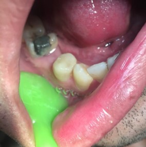
An access cavity was prepared after administration of local anesthesia (2% lidocaine with 1:80,000 adrenaline) under rubber dam isolation. To gain sufficient access to the root canals, the conventional access opening was modified into one that was wider mesiodistally. During the search for root canals, we encountered the lingual root canal first; however, finding the second and third root canals' orifices was difficult as the canals were narrow and calcified, and the orifices were hidden under a dentinal shelf, so we had to extend the access opening distally (Fig. 2). The working length (WL) was determined by the use of an electronic apex locator and was confirmed by radiography (Fig. 3).
Debridement and shaping of the root canals were carried out by the RaCe™ rotary file system (FKG, La Chaux-de-Fonds, Switzerland) with copious irrigation by 5% sodium hypochlorite solution. After the completion of the cleaning and shaping, the root canal system was obturated by using the cold lateral compaction technique with gutta-percha cones and a resin-based sealer (Fig. 4). A post-obturation radiograph was obtained, and the coronal access cavity was restored by composite build-up (Fig. 5 and 6).
Figure 1. Panoramic radiograph
Figure 2. Root canal orifices
Figure 3. Working length (WL) radiography
Figure 4. Obturation radiography
Figure 5. Radiograph of the tooth with coronal restoration
Figure 6. Coronal restoration
Discussion
Successful endodontic treatment of mandibular premolars is often a challenging task. (6) This can be related to the internal morphology of the pulp cavity, the number of root canals, and the presence of apical deltas and lateral root canals. (6) These anatomical variations have been well-documented in anatomic studies and case reports. (7-9) High-quality preoperative radiographs with different horizontal angulations (20° and 40°) should be used to identify the anatomical variations of the root canal system. (10,11) During the radiographic study, examination of the periodontal ligament (PDL) space can help identify the root shape, root position, relative root outline and presence of extra roots. (10) The sudden radiographic disappearance (fast break) of a root canal on parallel radiographs may be an evidence of a dividing root canal or an additional root canal. (12)
In the present case, we noticed a sudden change in the radiolucency of the canal space. A small access cavity in such teeth restricts the visualization of the area; hence, a wider endodontic access is necessary to locate the additional root canals. (13) In order to better visualize the pulp chamber, the access cavity should have divergent walls towards the occlusal aspect, allowing identification of the anatomic map of the pulp chamber floor with its canal orifices. (6) In this case study, we used an Endo-Z drill (Dentsply/Maillefer, Ballaigues, Switzerland) positioned parallel to the long axis of the tooth to shape the access cavity.
Previous studies have shown the presence of one orifice at the lingual aspect and two orifices at the buccal aspect of the pulp chamber floor. (14) In the present case, one canal orifice was located at the lingual aspect, and another canal orifice was found at the buccal aspect, while the third orifice was located distal to the mentioned orifices (Fig. 2). In such cases, tactile sensibility and the direction of the instruments during exploration of the root canal system can be helpful. (6) The sense of touch and radiographic images during treatment will take on further importance, especially in cases in which division of the root canals occurs in the apical half of the root trunk. (11,15)
In the present case, some opening resistance was felt during the initial negotiation of the canals with #08 and #10 k-files, which indicated the presence of extra root canals. In addition, the WL file appeared off-center on the radiograph. Although the root canal morphology was complex, it was clearly determined on the preoperative radiograph. After identification, proper cleaning and shaping were followed by a complete obturation of all the root canals, leading to a predictable long-term endodontic prognosis. (6)
Although it is rare for a mandibular first premolar to have three root canals, each case should be carefully examined radiographically and clinically to locate all the root canals.
Conclusion
Mandibular premolars can present with an extremely complex root canal system morphology, which if not considered during the treatment, it can lead to post-treatment complications, failure or flare-ups.
Conflict of interests
No potential conflict of interests relevant to this article was reported.
Full-Text: (1215 Views)
Abstract
Background: A correct diagnosis of the morphology of the root canal system is crucial in order to ensure a successful root canal treatment. The incidence of mandibular first premolars with three root canals has been reported to be low. Case presentation: This clinical case report describes a case of a successful nonsurgical endodontic management of a mandibular first premolar with three root canals. A 27-year-old man presented to our clinic with a pain in the left mandibular premolar. The diagnosis was irreversible pulpitis based on the clinical and radiographic examinations. Therefore, root canal treatment was recommended. After preparing the access cavity, the orifices were found by removing the dentinal shelves. The root canals were treated by using rotary files and were obturated by using the cold lateral compaction technique. Conclusion: Mandibular premolars can present with an extremely complex root canal system morphology, which if not considered during the treatment, it can lead to post-treatment complications, failure or flare-ups.
Keywords: Premolar, pulp canals, Mandible, Root Canal Therapy
Introduction
The main objective of endodontic therapy is mechanical and chemical debridement of the entire root canal followed by a three-dimensional obturation with an inert filling material and coronal restoration. (1) However, the presence of an untreated or missed root canal may lead to endodontic treatment failure. (2) Sometimes, root canals are left untreated because the clinicians fail to identify their presence particularly in teeth that have anatomical variations. (3) It has been reported that mandibular premolars are probably the most difficult teeth to treat endodontically due to the wide variations of the root canal morphology. (4) Vertucci reported that 0.5% of first premolars have three root canals. (3) Thus, practitioners must have a thorough understanding of the internal anatomical configurations of teeth before undertaking endodontic therapy. (5)
Case presentation
A 27-year-old man, with no significant medical history, was referred to the endodontics department of Mazandaran University of Medical Sciences, with a chief complaint of a moderate pain in the right lower jaw. There was an intermittent localized moderate dull pain that was initiated by eating sweet and hot foods. On the clinical examination, the patient’s level of oral hygiene was found to be moderate. A deep carious lesion was observed in tooth #28, but there were no swellings or sinus tracts. Pulp vitality tests showed sensitivity to hot and cold stimuli and to the electric pulp test. The diagnostic radiograph showed a carious coronal lesion and a sudden narrowing of the root canal according to the "fast break" guideline (Fig. 1). Based on these findings, the pulp was diagnosed with irreversible pulpitis with normal periapical tissues.
An access cavity was prepared after administration of local anesthesia (2% lidocaine with 1:80,000 adrenaline) under rubber dam isolation. To gain sufficient access to the root canals, the conventional access opening was modified into one that was wider mesiodistally. During the search for root canals, we encountered the lingual root canal first; however, finding the second and third root canals' orifices was difficult as the canals were narrow and calcified, and the orifices were hidden under a dentinal shelf, so we had to extend the access opening distally (Fig. 2). The working length (WL) was determined by the use of an electronic apex locator and was confirmed by radiography (Fig. 3).
Debridement and shaping of the root canals were carried out by the RaCe™ rotary file system (FKG, La Chaux-de-Fonds, Switzerland) with copious irrigation by 5% sodium hypochlorite solution. After the completion of the cleaning and shaping, the root canal system was obturated by using the cold lateral compaction technique with gutta-percha cones and a resin-based sealer (Fig. 4). A post-obturation radiograph was obtained, and the coronal access cavity was restored by composite build-up (Fig. 5 and 6).
Figure 1. Panoramic radiograph
Figure 2. Root canal orifices
Figure 3. Working length (WL) radiography
Figure 4. Obturation radiography
Figure 5. Radiograph of the tooth with coronal restoration
Figure 6. Coronal restoration
Discussion
Successful endodontic treatment of mandibular premolars is often a challenging task. (6) This can be related to the internal morphology of the pulp cavity, the number of root canals, and the presence of apical deltas and lateral root canals. (6) These anatomical variations have been well-documented in anatomic studies and case reports. (7-9) High-quality preoperative radiographs with different horizontal angulations (20° and 40°) should be used to identify the anatomical variations of the root canal system. (10,11) During the radiographic study, examination of the periodontal ligament (PDL) space can help identify the root shape, root position, relative root outline and presence of extra roots. (10) The sudden radiographic disappearance (fast break) of a root canal on parallel radiographs may be an evidence of a dividing root canal or an additional root canal. (12)
In the present case, we noticed a sudden change in the radiolucency of the canal space. A small access cavity in such teeth restricts the visualization of the area; hence, a wider endodontic access is necessary to locate the additional root canals. (13) In order to better visualize the pulp chamber, the access cavity should have divergent walls towards the occlusal aspect, allowing identification of the anatomic map of the pulp chamber floor with its canal orifices. (6) In this case study, we used an Endo-Z drill (Dentsply/Maillefer, Ballaigues, Switzerland) positioned parallel to the long axis of the tooth to shape the access cavity.
Previous studies have shown the presence of one orifice at the lingual aspect and two orifices at the buccal aspect of the pulp chamber floor. (14) In the present case, one canal orifice was located at the lingual aspect, and another canal orifice was found at the buccal aspect, while the third orifice was located distal to the mentioned orifices (Fig. 2). In such cases, tactile sensibility and the direction of the instruments during exploration of the root canal system can be helpful. (6) The sense of touch and radiographic images during treatment will take on further importance, especially in cases in which division of the root canals occurs in the apical half of the root trunk. (11,15)
In the present case, some opening resistance was felt during the initial negotiation of the canals with #08 and #10 k-files, which indicated the presence of extra root canals. In addition, the WL file appeared off-center on the radiograph. Although the root canal morphology was complex, it was clearly determined on the preoperative radiograph. After identification, proper cleaning and shaping were followed by a complete obturation of all the root canals, leading to a predictable long-term endodontic prognosis. (6)
Although it is rare for a mandibular first premolar to have three root canals, each case should be carefully examined radiographically and clinically to locate all the root canals.
Conclusion
Mandibular premolars can present with an extremely complex root canal system morphology, which if not considered during the treatment, it can lead to post-treatment complications, failure or flare-ups.
Conflict of interests
No potential conflict of interests relevant to this article was reported.
Type of Study: Case report |
Subject:
Radiology
References
1. Idris M, Sakkir N, Kj N, Kini A. Endodontic Retreatment of a Mandibular Second Molar with four Separate Roots: A Case Report. J Clin Diagn Res 2014;8(3):280-2.
2. Tabassum S, Khan FR. Failure of endodontic treatment: The usual suspects. Eur J Dent. 2016;10(1):144-7.
3. Al-Fouzan KS. The microscopic diagnosis and treatment of a mandibular second premolar with four canals Int Endod J. 2001;34(5):406-10.
4. Mittal S, Kumar T, Mittal S, Sharma J. Mandibular premolars with aberrant canal morphology: An endodontic challengeJ Conserv Dent 2014;17(5):491-4.
5. Diwan A, Sreedevi CR, Nagaraj T, Raghava V, Sinha P, Moushmi CB. Determination of internal anatomy of a permanent dentition: A review. Int J Contemp Dent Med Rev. 2015;2015.
6. Paul B, Dube K. Endodontic Treatment of a Mandibular Second Premolar with Three Roots and Three Canals. Case Rep Dent. 2014; 2014: 973410.
7. Bansal D, Mahajan M. Morphological variations in the root canal system of mandibular premolars in the population of Uttarakhand . IJADS 2016;2(4):68-71.
8. Hariharavel VP, Kumar AA, Ganesh C, Annamalai S, Ramar K, Aravindhan R. Root canal treatment of mandibular second premolar with three separate roots and canals using spiral computed tomographic. Case Rep Dent 2014;2014:816576.
9. Kumar T, Mittal S, Kaur K. Endodontic treatment of Mandibular Second Premolar with three Root Canals- A Case Report. J Adv Med Dent Scie Res 2016;4(3):72-4.
10. Pallavi V, Kumar JR, Mandava RB, Rao SH. Complicated canal morphology of mandibular first premolar. J Pharm Bioallied Sci 2015;7(Suppl 2):S809-11.
11. Moayedi S, Lata D. Mandibular first premolar with three canals. Endodontology. 2004;16:26-9.
12. Daneshvar F, Baziar H, Karkehabadi H, Jafarzadeh H, Bhandi S, Patil S. Mandibular First Premolars with One Root and Three Canals: A Case Series. J Contemp Dent Pract 2015 1;16(6):519-22.
13. Fathi Z, Brahimi S, Tavakoli R, Amini M. A three-rooted mandibular second premolar: A Case Report. J Dent Res Dent Clin Dent Prospects 2014;8(3):184-6.
14. Chauhan R, Singh S. Management of a 3-canal mandibular premolar in a patient with unusual root canal anatomy in all mandibular premolars. Gen Dent 2013;61(6):16-8.
15. Borna Z, Rahimi S, Shahi S, Zand V. Mandibular second premolars with three root canals: a review and 3 case reports Iran Endod J 2011;6(4):179-82.
16. Kakkar P, Singh A. Mandibular first premolar with three roots: a case report. Iran Endod J. 2012;7(4):207-10.
Send email to the article author
| Rights and permissions | |
 |
This work is licensed under a Creative Commons Attribution-NonCommercial 4.0 International License. |
