
Journal of Research in Dental
and Maxillofacial Sciences

Volume 11, Issue 3 (9-2026)
J Res Dent Maxillofac Sci 2026, 11(3): 232-241 |
Back to browse issues page


Ethics code: IR.IAU.DENTAL.REC.1404.009


Download citation:
BibTeX | RIS | EndNote | Medlars | ProCite | Reference Manager | RefWorks
Send citation to:


BibTeX | RIS | EndNote | Medlars | ProCite | Reference Manager | RefWorks
Send citation to:
Farahzadfard M, Alamdari Mahd M. Effect of Commonly Used Pediatric Medications on Primary Enamel Microhardness: An In Vitro Study. J Res Dent Maxillofac Sci 2026; 11 (3) :232-241
URL: http://jrdms.dentaliau.ac.ir/article-1-1626-en.html
URL: http://jrdms.dentaliau.ac.ir/article-1-1626-en.html


1- School of Dentistry, TeMS.c., Islamic Azad University, Tehran, Iran.
2- Department of Pediatric Dentistry, School of Dentistry, TeMS.c., Islamic Azad University, Tehran, Iran. ,mandanaalamdari@iau.ac.ir
2- Department of Pediatric Dentistry, School of Dentistry, TeMS.c., Islamic Azad University, Tehran, Iran. ,
Full-Text [PDF 535 kb]
(118 Downloads)
| Abstract (HTML) (940 Views)
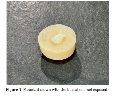
Figure 1. Mounted crown with the buccal enamel exposed
Group A: Acetaminophen syrup (120 mg/5 mL; Velotyl; Kharazmi Co., Iran)
Group B: Amoxicillin syrup (125 mg/5 mL; Susp Amoxicillin, Farabi Co., Iran)
Group C: Iron drops (14 mg/mL; Lipiferr Forte, Kimia Kala Razi Co., Iran)
Group D (Control): Artificial saliva (manufactured by Tehran University of Medical Sciences) containing 1.5 mmol/L Ca, 0.9 mmol/L P, 150 mmol/L KCl, and 0.05 mg/mL F in 0.1 mol/L Tris buffer, with a pH of 7.0.
The pH of the medications was also measured with a pH meter (PT-15; Sartorius, Germany). The pH of each medication was measured 3 times, and the mean value was calculated and recorded.
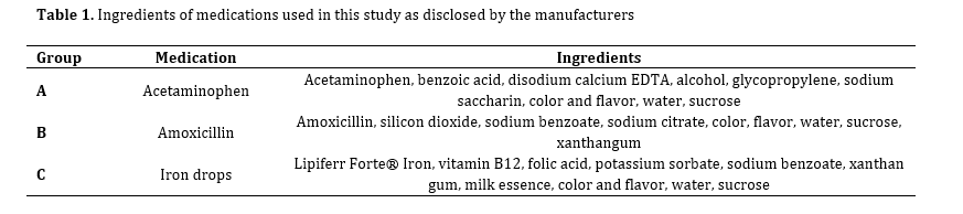
Table 1 shows the ingredients of the medications used in this study as disclosed by the manufacturers.
Baseline microhardness assessment:
Before immersing the teeth in the above-mentioned solutions, the hardness tester (Bareiss profgeratebau GmbH, D-89610 Oberdischingen, Germany) was calibrated against a certified standard reference block in accordance with ISO 6507 before testing. The baseline microhardness of the teeth was then measured at 3 points with a force of 50 g for 10 seconds applied to the specimens by a hardness tester. The mean of the 3 obtained numbers was reported as the microhardness of the respective specimen as the Vickers hardness number (VHN), which was calculated by the device according to the following formula:
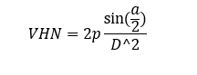
Where D is the mean diameter of the indentation in millimeters, P is the applied force in kilogram force (Kgf), and a is the angle formed between the opposing surfaces of the indenter.
Intervention:
The immersion protocol in groups A and B was as follows: 10 specimens from each group were immersed in 5 mL of each drug for 3 minutes, 3 times a day (with one hour between immersion cycles) for 5 days to simulate the usual number of drug intakes by patients [22]. In group C, 10 teeth were immersed in 5 mL of the drug for 3 minutes once a day for 5 days to match the immersion duration in groups A and B [22]. In group D (control), the specimens were stored in artificial saliva for 5 days and the solution was changed daily. After each immersion cycle, the specimens were first washed with distilled water for 10 seconds, and then stored in 10 mL of artificial saliva with a pH of 7 at 37°C until the next immersion. All immersion and storage cycles were carried out in an incubator at 37°C to simulate the intraoral environment.
Secondary microhardness assessment:
The secondary microhardness was measured after immersion as explained for assessment of baseline microhardness. All microhardness measurements (baseline and after the intervention) were performed by a single trained examiner to ensure intra-examiner consistency. Finally, the change in microhardness was quantified by subtracting the secondary microhardness from the baseline microhardness value.
SEM assessment:
SEM assessment was performed on one random tooth from each group on the last day of the intervention. The specimens were first immersed in distilled water for 10 minutes, and then dried at room temperature for 24 hours. The specimens were then placed on an aluminum stub and fixed with carbon glue. Then, due to the lack of conductivity of the teeth, they were coated with a thin layer of gold and underwent SEM assessment (VEGA3; TESCAN, Czech Republic) at 15 kV with a magnification of x1500. Erosion was classified based on the Primary Enamel Surface (PES) scale as follows [23]:
1: Sporadic rod ends visible
2: Etched prism pattern
3: Crater formation
Statistical analysis:
Normal distribution of data was confirmed by the Shapiro-Wilk test. Thus, the four groups were compared regarding microhardness using repeated measures and one-way ANOVA, followed by pairwise comparisons with the Tamhane’s test. Paired t-test was used to compare baseline and secondary microhardness values within each group. The level of statistical significance was set at 0.05.
Results
pH:
The mean pH was 5.3±0.01 for acetaminophen, 5.89±0.01 for amoxicillin, and 3.41±0.02 for iron drops.
Table 2 presents descriptive statistics for the microhardness in the four groups.
Microhardness change:
The four groups had a significant difference in microhardness change (P<0.001).
Pairwise comparisons of the groups regarding microhardness change (Table 3) showed that the microhardness change was significantly greater in the iron drops than the acetaminophen (P<0.001), amoxicillin (P<0.001) and control (P<0.001) groups. The microhardness change was also significantly greater in the acetaminophen than the amoxicillin group (P<0.001). No other significant differences were found.
Within-group assessment of microhardness change after the intervention compared with baseline (Table 4) showed a significant reduction in acetaminophen (P<0.001) and iron drops (P<0.001) groups, whereas the reduction was not statistically significant in the amoxicillin (P=0.06) and control (P=0.18) groups.
SEM results:
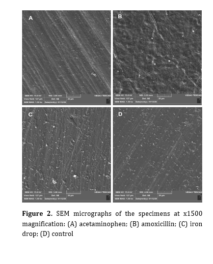
Figure 2 shows SEM micrographs of the specimens at x1500 magnification. The iron drops group showed sporadic rod ends visible, the acetaminophen group showed etched prism pattern, and the amoxicillin group showed crater formation, while the control group showed a sound and intact enamel surface with no discernible erosive changes.
As SEM assessments were limited to one specimen per group, these observations were considered qualitative and illustrative of surface changes.
Table 2. Measures of central dispersion for the microhardness (VHN) in the four groups (n=30)
Table 3. Pairwise comparisons of the groups regarding microhardness change
Table 4. Within-group comparison of microhardness
Figure 2. SEM micrographs of the specimens at x1500 magnification: (A) acetaminophen; (B) amoxicillin; (C) iron drop; (D) control
Discussion
This study assessed the effect of commonly used pediatric medications on primary enamel microhardness. The results showed that the microhardness change was significantly greater in the iron drops than in the acetaminophen, amoxicillin, and control groups. The microhardness change was also significantly greater in the acetaminophen group than in the amoxicillin group. The reduction in microhardness was only significant in the acetaminophen and iron drops groups. Thus, the null hypothesis of the study was rejected for acetaminophen and iron drops, but not for amoxicillin.
Rocha et al. [11] evaluated the effect of four analgesics (ibuprofen, Magnopyrol, paracetamol, Tylenol) on primary enamel erosion. They immersed primary molars in the respective medications 4 times a day for 3 days. Titratable acidity was also measured, and microhardness was quantified by the Knoop hardness test under a force of 35 g for 5 seconds. The results showed that only Magnopyrol resulted in a significant reduction in microhardness compared to the control group, and the change in microhardness of the other study groups was not significant; however, in the present study, acetaminophen syrup (Velotyl) resulted in a significant reduction in microhardness compared to the control group. The reason for the difference in the results is the difference in methodology and study groups. In the present study, the titratable acidity of acetaminophen syrup was not measured and it had a higher pH (pH=5.3) than paracetamol (pH=4.4) and Tylenol (pH=4.3). Also, the Vickers test was used to calculate microhardness, which is different from the Knoop hardness test. According to Jung and Jun [19], the higher the buffering capacity of a drug, the greater the rate of erosion, and the slower the drug is neutralized by salivary remineralizing agents, causing more damage. The higher erosion caused by acetaminophen syrup despite its higher pH compared to paracetamol and Tylenol may plausibly be related to its higher buffering capacity, although buffering capacity was not directly measured in the present study, and this explanation remains speculative. Alqarni [20] assessed primary enamel erosion caused by multivitamin syrup, amoxicillin-clavulanic acid, ibuprofen, and acetaminophen. They used a profilometer to assess surface roughness. They showed that the studied drugs significantly increased the enamel surface roughness of primary teeth, with different intensities compared to the control group. Despite the differences in the study groups and the method of assessing dental erosion, their results are somewhat consistent with the present findings, indicating that liquid pediatric medications with acidic pH may lead to a significant increase in surface roughness, and increase the caries incidence. Similarly, in the present study, both analgesics and antibiotics with low pH led to a decrease in microhardness. Singana and Suma [24] evaluated the cariogenic and erosive potential of liquid pediatric medications (including paracetamol, amoxicillin clavulanate, and various antitussives, antihistamines, anticonvulsants, and nutritional supplements) on primary teeth. They immersed the teeth in the respective medications for 1 minute, 10 minutes, and 8 hours. They showed that, except for paracetamol, the studied drugs increased calcium dissolution to different extents, and there was no significant relationship between the pH of the drug and the rate of calcium dissolution. However, in the present study, acetaminophen syrup resulted in a significant reduction in microhardness. In their study, the pH of paracetamol and amoxicillin syrups was reported to be 6.07 and 4.54, respectively. In the present study, the pH of acetaminophen and amoxicillin syrups was reported to be 5.3 and 5.89, respectively. Differences in immersion time and study methodology, as well as pH below the critical threshold (approximately 5.5) [25] for the initiation of enamel demineralization, are the reasons for the difference in the results. This level of acidity (pH < 5.5) can cause enamel erosion in the absence of bacterial activity, but the composition of the acid, buffering capacity, presence of calcium ions, and duration of contact with tooth are more important than pH [19]. This finding is consistent with a SEM-based study by Mahmoud and Omar [26], which similarly found that all tested pediatric liquid medications produced observable erosive damage to primary enamel regardless of pH; notably, antibiotic syrups with pH values above the commonly cited critical threshold of 5.5 (5.68 and 6.22) still showed an irregular, pitted, and rough enamel surface with numerous pores under SEM, comparable in severity to more acidic formulations. Tupalli and colleagues [23] assessed the erosion potential of various liquid pediatric medications including analgesics, antibiotics, antiepileptics, multivitamins, and cough suppressants. Primary teeth were immersed once for 1 minute, 10 minutes, or 8 hours. Changes in PES were assessed by SEM. The results showed that within 1 minute, an etched prism pattern was observed in the paracetamol and amoxicillin syrup groups, which was consistent with the findings of the present study; however, after 10 minutes and 8 hours of immersion, a different pattern in the form of crater formation was observed. In the present study, acetaminophen syrup had a lower pH (5.3) than paracetamol (6.4) reported in the aforementioned study. The difference in the results can be attributed to the differences in experimental design, especially in terms of immersion time, as well as the different pH of the drugs. Chelating agents present in some liquid pediatric medications, such as sodium salts of various amino acids (alanine, aspartame, glutamate) and lactate, at neutral or near-neutral pH, can remove calcium from enamel, resulting in erosive patterns observed under SEM, and this process is independent of pH [23].
According to the findings reported by Sahebnazar et al. [27], iron drops in general can reduce enamel microhardness. Among them, the Irofant product with lower pH and higher titratable acidity showed the most detrimental effect. In contrast, Sideral iron drops had the least negative effect on enamel microhardness of primary teeth. Lipiferr Forte iron drops used in the present study also have the same type of liposomal formulation and are structurally similar to the aforementioned product. In the present study, Lipiferr Forte caused a significant reduction in primary enamel microhardness. However, the extent of this reduction was smaller compared to the iron drops reported in the study by Sahebnazar et al. [27]. In line with the present study, Elkhodary et al. [4] found that Feromin iron syrup resulted in a significant decrease in enamel microhardness of primary teeth.
Taken together, these findings indicate that the type of drug product, excipients, pH, buffering capacity, titratable acidity, and chemical properties may also plausibly contribute to the severity of changes in enamel microhardness; however, as some of these factors were not directly assessed in the present study, this interpretation remains speculative and warrants confirmation in future studies that measure them directly. Various studies have also pointed to a possible association between dental caries and frequent use of oral liquid medications [14,28,29]. The presence of sucrose in medications leads to a decrease in dental plaque pH and also acts as a substrate for the proliferation of oral bacteria [14] which may contribute to an increased risk of dental caries; however, caries development was not directly assessed in the present study.
Knowledge about the erosive and cariogenic potential of commonly used liquid medications in children is of great importance, especially in Iran where prescription of these medications is highly common. The results of such studies can play an important role in increasing the knowledge level of dentists and pediatricians and help them in a more informed choice of medication and provide preventive recommendations. Also, the provision of appropriate information to parents about the possible effects of these medications on tooth enamel, including reduction of microhardness, increased risk of caries, and discoloration, can be effective in promoting children's oral health. Given the effect of iron drops on tooth discoloration, direct contact with tooth surfaces can be reduced by dripping the medicine on the tip of the tongue, rinsing the child's mouth with water after taking the medicine, diluting it with natural apple juice, which has been shown to significantly reduce the adverse effect of iron drops on primary enamel microhardness compared with the undiluted form [27], and brushing the teeth after taking the medicine. Educating and providing preventive strategies to parents, along with prescribing medication, can prevent unwanted dental complications and reduce parents’ concerns.
The discrepancies between the present findings and those of previous studies are likely attributable to differences in formulation chemistry rather than pH alone. Variations in viscosity may alter the contact time and diffusion of acidic components at the enamel surface, while chelating agents, such as EDTA in the acetaminophen formulation used here, can directly bind surface calcium and promote demineralization independently of pH. Differences in citrate or sucrose concentration across formulations, which act as buffering and cariogenic agents respectively, could further account for the divergent erosive potentials reported across studies, even among medications with similar pH values. These formulation-level differences, rather than a single physicochemical parameter, likely explain why studies using ostensibly similar drug classes have reported inconsistent findings regarding enamel erosion.
One limitation of this study was the failure to assess titratable acidity, since instantaneous pH only indicates the level of acidity at a specific moment. Buffering capacity indicates the ability of a solution to maintain pH against changes and its resistance to neutralization by the saliva, and is a more accurate indicator of the total acids present in the product and the amount of base required to bring the pH to a neutral level. Also, this study was conducted in vitro on extracted teeth. The results of in vitro studies are not always generalizable to clinical conditions. Other commonly used drugs with high prevalence of use, as well as confounding factors such as concentration, viscosity, acidity, and more precise drug compositions, can be investigated in future studies. In vivo studies are also recommended to obtain more generalizable results.
Conclusion
Within the limitations of this in vitro study, the results showed that iron drops followed by acetaminophen caused a significant reduction in primary enamel microhardness. The microhardness reduction by amoxicillin did not reach statistical significance. These findings should be interpreted cautiously and should not be directly extrapolated to clinical conditions, as important oral factors such as acquired pellicle, mastication, fluoride exposure and oral biofilm were not incorporated into this experimental study.
Full-Text: (410 Views)
Abstract
Background and Aim: This study assessed the effect of commonly used pediatric medications on primary enamel microhardness.
Materials and Methods: This in vitro study was conducted on 40 extracted sound primary anterior teeth. The teeth were randomly assigned to 4 groups (n=10) to expose a 3x3 mm window on their buccal surface to acetaminophen syrup (group A), amoxicillin (group B), iron drops (group C), and artificial saliva (group D) after measuring their baseline microhardness with a hardness tester. The pH of the medications was also measured. The teeth were immersed in 5 mL of the respective drug 3 times/day in groups A and B, and once a day in group C, each time for 3 minutes for 5 days. Their secondary microhardness was then measured. One random tooth from each group underwent scanning electron microscopy (SEM) assessment in the last day of the intervention. Erosion was categorized using the Primary Enamel Surface (PES) scale. Data were analyzed using repeated measures and one-way ANOVA, paired t-test, and Tamhane’s test (alpha=0.05).
Results: The microhardness change was significantly greater in the iron drops than in the acetaminophen (P<0.001), amoxicillin (P<0.001), and control (P<0.001) groups. Acetaminophen caused a significantly greater reduction than amoxicillin (P<0.001). The reduction in microhardness was only significant in the acetaminophen (P<0.001) and iron drops (P<0.001) groups.
Conclusion: This in vitro study showed that iron drops followed by acetaminophen caused a significant reduction in primary enamel microhardness. The microhardness reduction by amoxicillin did not reach statistical significance.
Keywords: Acetaminophen; Amoxicillin; Enamel; Iron, Dietary; Tooth, Deciduous
Introduction
Dental erosion, defined as the irreversible loss of dental hard tissue by a nonbacterial chemical process, is increasingly recognized as a problem of all ages [1]. Erosion is particularly
prevalent among preschool children and adults who consume fruit juices, carbonated beverages, and acidic medications for a long period of time [2]. Dental erosion causes tooth hypersensitivity to foods, increases plaque accumulation, and consequently leads to dental caries [3].
One way to prevent erosion is to use medications that cause the least erosion in children's teeth. Studies have confirmed that some pediatric liquid medications are acidic and cariogenic in nature, and medications with a low pH that are in frequent or continuous contact with the teeth have the potential to cause dental erosion [4-6]. Low pH of oral liquid medications [7,8], poor oral hygiene after taking these medications and supplements [8], and repeated use, especially in children with chronic diseases, lead to a decrease in the microhardness of tooth enamel. Enamel erosion, caused by various drugs, including antihistamines, antibiotics, and analgesics, has been reported in various studies [9-11]. Oral liquid drugs contain agents in their formulations to improve their appearance, bioavailability, stability, and palatability [12]. Sugars such as sucrose, fructose, and glucose are added to increase their palatability [13]. These sugars are widely used because they are inexpensive and easy to digest [14]. Acids are also added to drugs and usually act as buffering agents to maintain chemical stability, control tonicity, and ensure physiological compatibility. In addition, they may be used to improve flavor [15]. All these additives may cause dental erosion.
On the other hand, iron supplementation in children, especially in infants and young children, is one of the main methods for treatment and prevention of iron deficiency anemia. Among the various forms, oral iron solutions are most commonly prescribed due to their ease of use; however, one of their well-known side effects is external discoloration of primary teeth, which is clinically important in terms of esthetics and parental acceptance of treatment [16]. Due to the high prevalence of iron deficiency in Iranian children and the necessity of taking iron drop supplements from infancy, which coincides with the eruption of primary teeth, it is important to find a drug that has minimal adverse side effects. Iron drops typically have a low pH, and ferrous sulfate-based products are known to be markedly acidic, which favors dissolution of the hydroxyapatite mineral phase of enamel. In addition, ferrous ions released from these formulations can directly interact with the enamel surface, replacing calcium ions within the hydroxyapatite lattice through an ion-exchange process and disrupting its crystalline structure [17]. In the formulation of Lipiferr Forte iron drops (Kimia Kala Razi Co., Iran), iron is present in nano-liposomal form, which prevents direct contact of iron with the tooth surface and, as a result, has shown less discoloration than conventional forms such as ferrous sulfate [16]. Also, compared to conventional iron salts, Lipiferr Forte iron drops provide better digestive tolerance and allow for more direct absorption of iron into the bloodstream [18].
Although the erosive potential of acetaminophen, amoxicillin and other pediatric syrups has been individually documented [4,9-11,19,20], and conventional ferrous sulfate iron drops have also been separately studied for their effect on enamel microhardness [21], these three drug classes have rarely been compared directly against one another under an identical protocol. Moreover, the erosive behavior of iron in a nanoliposomal carrier such as liposofer, increasingly prescribed in Iran, has not been specifically examined with respect to microhardness. Thus, this study aimed to assess the effect of commonly used pediatric medications on primary enamel microhardness. The null hypothesis of the study was that iron drops, acetaminophen, and amoxicillin would have no significant difference with the control group (artificial saliva) in reduction of enamel microhardness.
Materials and Methods
This in vitro experimental study was conducted on 40 primary anterior teeth with sound buccal surfaces with no caries, cracks, or hypoplastic or white spot lesions that had been extracted within the past 6 months due to physiological exfoliation of their root. The Institutional Review Board of the university granted ethical approval for the study protocol (IR.IAU.DENTAL.REC.1404.009).
Sample size:
The sample size was calculated to be a minimum of 10 in each of the 4 groups according to a previous study by Al-Olimy et al. [9] Using the one-way ANOVA feature of PASS 11, assumingα β
Specimen preparation:
After ensuring the soundness of the collected teeth by their inspection under a light microscope (Leica S9; Leica Microsystems, Germany) at x10 magnification, they were rinsed with distilled water, and soft tissue residues were removed [11]. They were then disinfected by 48 hours of immersion in 0.1% thymol. The roots were cut at the cementoenamel junction by a diamond disc (C04 Diamante, BTD, China), and the crowns were mounted in acrylic resin (Acropars, Iran) such that their buccal surface remained exposed. Next, the buccal surface was entirely coated with a colorless nail varnish (number 69; Neklaci, Iran) except for a window measuring 3x3 mm [11]. Subsequently, this area was polished with a sequence of 4 polishing discs (Sanding; Golden Beauty, China) from blue to white, with each disc applied for approximately 30 seconds under water cooling, and the teeth were stored in saline, which was refreshed daily (Figure 1).
The specimens were subsequently stored in artificial saliva at 37°C for 24 hours to simulate the oral environment, and then the teeth were randomly divided into 4 groups of 10 by simple random sampling using a table of random numbers. Allocation concealment was not implemented and the operator performing the immersion intervention was not blinded to group allocation; however, the examiner who performed the microhardness and scanning electron microscopy (SEM) assessments was blinded to the group allocation of specimens.
Materials and Methods: This in vitro study was conducted on 40 extracted sound primary anterior teeth. The teeth were randomly assigned to 4 groups (n=10) to expose a 3x3 mm window on their buccal surface to acetaminophen syrup (group A), amoxicillin (group B), iron drops (group C), and artificial saliva (group D) after measuring their baseline microhardness with a hardness tester. The pH of the medications was also measured. The teeth were immersed in 5 mL of the respective drug 3 times/day in groups A and B, and once a day in group C, each time for 3 minutes for 5 days. Their secondary microhardness was then measured. One random tooth from each group underwent scanning electron microscopy (SEM) assessment in the last day of the intervention. Erosion was categorized using the Primary Enamel Surface (PES) scale. Data were analyzed using repeated measures and one-way ANOVA, paired t-test, and Tamhane’s test (alpha=0.05).
Results: The microhardness change was significantly greater in the iron drops than in the acetaminophen (P<0.001), amoxicillin (P<0.001), and control (P<0.001) groups. Acetaminophen caused a significantly greater reduction than amoxicillin (P<0.001). The reduction in microhardness was only significant in the acetaminophen (P<0.001) and iron drops (P<0.001) groups.
Conclusion: This in vitro study showed that iron drops followed by acetaminophen caused a significant reduction in primary enamel microhardness. The microhardness reduction by amoxicillin did not reach statistical significance.
Keywords: Acetaminophen; Amoxicillin; Enamel; Iron, Dietary; Tooth, Deciduous
Introduction
Dental erosion, defined as the irreversible loss of dental hard tissue by a nonbacterial chemical process, is increasingly recognized as a problem of all ages [1]. Erosion is particularly
prevalent among preschool children and adults who consume fruit juices, carbonated beverages, and acidic medications for a long period of time [2]. Dental erosion causes tooth hypersensitivity to foods, increases plaque accumulation, and consequently leads to dental caries [3].
One way to prevent erosion is to use medications that cause the least erosion in children's teeth. Studies have confirmed that some pediatric liquid medications are acidic and cariogenic in nature, and medications with a low pH that are in frequent or continuous contact with the teeth have the potential to cause dental erosion [4-6]. Low pH of oral liquid medications [7,8], poor oral hygiene after taking these medications and supplements [8], and repeated use, especially in children with chronic diseases, lead to a decrease in the microhardness of tooth enamel. Enamel erosion, caused by various drugs, including antihistamines, antibiotics, and analgesics, has been reported in various studies [9-11]. Oral liquid drugs contain agents in their formulations to improve their appearance, bioavailability, stability, and palatability [12]. Sugars such as sucrose, fructose, and glucose are added to increase their palatability [13]. These sugars are widely used because they are inexpensive and easy to digest [14]. Acids are also added to drugs and usually act as buffering agents to maintain chemical stability, control tonicity, and ensure physiological compatibility. In addition, they may be used to improve flavor [15]. All these additives may cause dental erosion.
On the other hand, iron supplementation in children, especially in infants and young children, is one of the main methods for treatment and prevention of iron deficiency anemia. Among the various forms, oral iron solutions are most commonly prescribed due to their ease of use; however, one of their well-known side effects is external discoloration of primary teeth, which is clinically important in terms of esthetics and parental acceptance of treatment [16]. Due to the high prevalence of iron deficiency in Iranian children and the necessity of taking iron drop supplements from infancy, which coincides with the eruption of primary teeth, it is important to find a drug that has minimal adverse side effects. Iron drops typically have a low pH, and ferrous sulfate-based products are known to be markedly acidic, which favors dissolution of the hydroxyapatite mineral phase of enamel. In addition, ferrous ions released from these formulations can directly interact with the enamel surface, replacing calcium ions within the hydroxyapatite lattice through an ion-exchange process and disrupting its crystalline structure [17]. In the formulation of Lipiferr Forte iron drops (Kimia Kala Razi Co., Iran), iron is present in nano-liposomal form, which prevents direct contact of iron with the tooth surface and, as a result, has shown less discoloration than conventional forms such as ferrous sulfate [16]. Also, compared to conventional iron salts, Lipiferr Forte iron drops provide better digestive tolerance and allow for more direct absorption of iron into the bloodstream [18].
Although the erosive potential of acetaminophen, amoxicillin and other pediatric syrups has been individually documented [4,9-11,19,20], and conventional ferrous sulfate iron drops have also been separately studied for their effect on enamel microhardness [21], these three drug classes have rarely been compared directly against one another under an identical protocol. Moreover, the erosive behavior of iron in a nanoliposomal carrier such as liposofer, increasingly prescribed in Iran, has not been specifically examined with respect to microhardness. Thus, this study aimed to assess the effect of commonly used pediatric medications on primary enamel microhardness. The null hypothesis of the study was that iron drops, acetaminophen, and amoxicillin would have no significant difference with the control group (artificial saliva) in reduction of enamel microhardness.
Materials and Methods
This in vitro experimental study was conducted on 40 primary anterior teeth with sound buccal surfaces with no caries, cracks, or hypoplastic or white spot lesions that had been extracted within the past 6 months due to physiological exfoliation of their root. The Institutional Review Board of the university granted ethical approval for the study protocol (IR.IAU.DENTAL.REC.1404.009).
Sample size:
The sample size was calculated to be a minimum of 10 in each of the 4 groups according to a previous study by Al-Olimy et al. [9] Using the one-way ANOVA feature of PASS 11, assuming
Specimen preparation:
After ensuring the soundness of the collected teeth by their inspection under a light microscope (Leica S9; Leica Microsystems, Germany) at x10 magnification, they were rinsed with distilled water, and soft tissue residues were removed [11]. They were then disinfected by 48 hours of immersion in 0.1% thymol. The roots were cut at the cementoenamel junction by a diamond disc (C04 Diamante, BTD, China), and the crowns were mounted in acrylic resin (Acropars, Iran) such that their buccal surface remained exposed. Next, the buccal surface was entirely coated with a colorless nail varnish (number 69; Neklaci, Iran) except for a window measuring 3x3 mm [11]. Subsequently, this area was polished with a sequence of 4 polishing discs (Sanding; Golden Beauty, China) from blue to white, with each disc applied for approximately 30 seconds under water cooling, and the teeth were stored in saline, which was refreshed daily (Figure 1).
The specimens were subsequently stored in artificial saliva at 37°C for 24 hours to simulate the oral environment, and then the teeth were randomly divided into 4 groups of 10 by simple random sampling using a table of random numbers. Allocation concealment was not implemented and the operator performing the immersion intervention was not blinded to group allocation; however, the examiner who performed the microhardness and scanning electron microscopy (SEM) assessments was blinded to the group allocation of specimens.
Figure 1. Mounted crown with the buccal enamel exposed
Group A: Acetaminophen syrup (120 mg/5 mL; Velotyl; Kharazmi Co., Iran)
Group B: Amoxicillin syrup (125 mg/5 mL; Susp Amoxicillin, Farabi Co., Iran)
Group C: Iron drops (14 mg/mL; Lipiferr Forte, Kimia Kala Razi Co., Iran)
Group D (Control): Artificial saliva (manufactured by Tehran University of Medical Sciences) containing 1.5 mmol/L Ca, 0.9 mmol/L P, 150 mmol/L KCl, and 0.05 mg/mL F in 0.1 mol/L Tris buffer, with a pH of 7.0.
The pH of the medications was also measured with a pH meter (PT-15; Sartorius, Germany). The pH of each medication was measured 3 times, and the mean value was calculated and recorded.
Table 1 shows the ingredients of the medications used in this study as disclosed by the manufacturers.
Baseline microhardness assessment:
Before immersing the teeth in the above-mentioned solutions, the hardness tester (Bareiss profgeratebau GmbH, D-89610 Oberdischingen, Germany) was calibrated against a certified standard reference block in accordance with ISO 6507 before testing. The baseline microhardness of the teeth was then measured at 3 points with a force of 50 g for 10 seconds applied to the specimens by a hardness tester. The mean of the 3 obtained numbers was reported as the microhardness of the respective specimen as the Vickers hardness number (VHN), which was calculated by the device according to the following formula:
Where D is the mean diameter of the indentation in millimeters, P is the applied force in kilogram force (Kgf), and a is the angle formed between the opposing surfaces of the indenter.
Intervention:
The immersion protocol in groups A and B was as follows: 10 specimens from each group were immersed in 5 mL of each drug for 3 minutes, 3 times a day (with one hour between immersion cycles) for 5 days to simulate the usual number of drug intakes by patients [22]. In group C, 10 teeth were immersed in 5 mL of the drug for 3 minutes once a day for 5 days to match the immersion duration in groups A and B [22]. In group D (control), the specimens were stored in artificial saliva for 5 days and the solution was changed daily. After each immersion cycle, the specimens were first washed with distilled water for 10 seconds, and then stored in 10 mL of artificial saliva with a pH of 7 at 37°C until the next immersion. All immersion and storage cycles were carried out in an incubator at 37°C to simulate the intraoral environment.
Secondary microhardness assessment:
The secondary microhardness was measured after immersion as explained for assessment of baseline microhardness. All microhardness measurements (baseline and after the intervention) were performed by a single trained examiner to ensure intra-examiner consistency. Finally, the change in microhardness was quantified by subtracting the secondary microhardness from the baseline microhardness value.
SEM assessment:
SEM assessment was performed on one random tooth from each group on the last day of the intervention. The specimens were first immersed in distilled water for 10 minutes, and then dried at room temperature for 24 hours. The specimens were then placed on an aluminum stub and fixed with carbon glue. Then, due to the lack of conductivity of the teeth, they were coated with a thin layer of gold and underwent SEM assessment (VEGA3; TESCAN, Czech Republic) at 15 kV with a magnification of x1500. Erosion was classified based on the Primary Enamel Surface (PES) scale as follows [23]:
1: Sporadic rod ends visible
2: Etched prism pattern
3: Crater formation
Statistical analysis:
Normal distribution of data was confirmed by the Shapiro-Wilk test. Thus, the four groups were compared regarding microhardness using repeated measures and one-way ANOVA, followed by pairwise comparisons with the Tamhane’s test. Paired t-test was used to compare baseline and secondary microhardness values within each group. The level of statistical significance was set at 0.05.
Results
pH:
The mean pH was 5.3±0.01 for acetaminophen, 5.89±0.01 for amoxicillin, and 3.41±0.02 for iron drops.
Table 2 presents descriptive statistics for the microhardness in the four groups.
Microhardness change:
The four groups had a significant difference in microhardness change (P<0.001).
Pairwise comparisons of the groups regarding microhardness change (Table 3) showed that the microhardness change was significantly greater in the iron drops than the acetaminophen (P<0.001), amoxicillin (P<0.001) and control (P<0.001) groups. The microhardness change was also significantly greater in the acetaminophen than the amoxicillin group (P<0.001). No other significant differences were found.
Within-group assessment of microhardness change after the intervention compared with baseline (Table 4) showed a significant reduction in acetaminophen (P<0.001) and iron drops (P<0.001) groups, whereas the reduction was not statistically significant in the amoxicillin (P=0.06) and control (P=0.18) groups.
SEM results:
Figure 2 shows SEM micrographs of the specimens at x1500 magnification. The iron drops group showed sporadic rod ends visible, the acetaminophen group showed etched prism pattern, and the amoxicillin group showed crater formation, while the control group showed a sound and intact enamel surface with no discernible erosive changes.
As SEM assessments were limited to one specimen per group, these observations were considered qualitative and illustrative of surface changes.
Table 2. Measures of central dispersion for the microhardness (VHN) in the four groups (n=30)
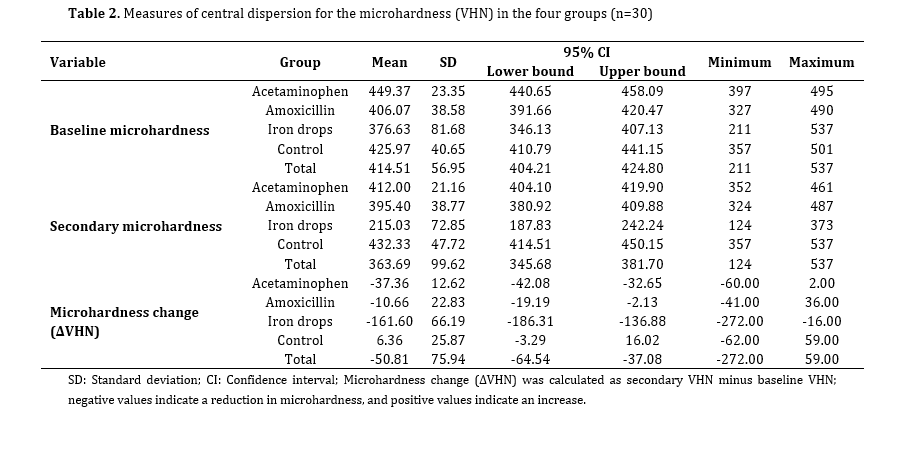
Table 3. Pairwise comparisons of the groups regarding microhardness change
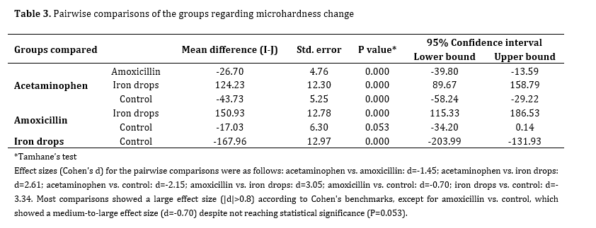
Table 4. Within-group comparison of microhardness
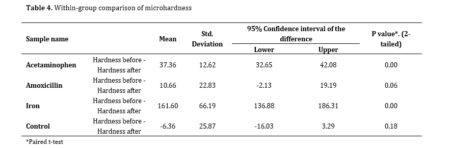
Figure 2. SEM micrographs of the specimens at x1500 magnification: (A) acetaminophen; (B) amoxicillin; (C) iron drop; (D) control
Discussion
This study assessed the effect of commonly used pediatric medications on primary enamel microhardness. The results showed that the microhardness change was significantly greater in the iron drops than in the acetaminophen, amoxicillin, and control groups. The microhardness change was also significantly greater in the acetaminophen group than in the amoxicillin group. The reduction in microhardness was only significant in the acetaminophen and iron drops groups. Thus, the null hypothesis of the study was rejected for acetaminophen and iron drops, but not for amoxicillin.
Rocha et al. [11] evaluated the effect of four analgesics (ibuprofen, Magnopyrol, paracetamol, Tylenol) on primary enamel erosion. They immersed primary molars in the respective medications 4 times a day for 3 days. Titratable acidity was also measured, and microhardness was quantified by the Knoop hardness test under a force of 35 g for 5 seconds. The results showed that only Magnopyrol resulted in a significant reduction in microhardness compared to the control group, and the change in microhardness of the other study groups was not significant; however, in the present study, acetaminophen syrup (Velotyl) resulted in a significant reduction in microhardness compared to the control group. The reason for the difference in the results is the difference in methodology and study groups. In the present study, the titratable acidity of acetaminophen syrup was not measured and it had a higher pH (pH=5.3) than paracetamol (pH=4.4) and Tylenol (pH=4.3). Also, the Vickers test was used to calculate microhardness, which is different from the Knoop hardness test. According to Jung and Jun [19], the higher the buffering capacity of a drug, the greater the rate of erosion, and the slower the drug is neutralized by salivary remineralizing agents, causing more damage. The higher erosion caused by acetaminophen syrup despite its higher pH compared to paracetamol and Tylenol may plausibly be related to its higher buffering capacity, although buffering capacity was not directly measured in the present study, and this explanation remains speculative. Alqarni [20] assessed primary enamel erosion caused by multivitamin syrup, amoxicillin-clavulanic acid, ibuprofen, and acetaminophen. They used a profilometer to assess surface roughness. They showed that the studied drugs significantly increased the enamel surface roughness of primary teeth, with different intensities compared to the control group. Despite the differences in the study groups and the method of assessing dental erosion, their results are somewhat consistent with the present findings, indicating that liquid pediatric medications with acidic pH may lead to a significant increase in surface roughness, and increase the caries incidence. Similarly, in the present study, both analgesics and antibiotics with low pH led to a decrease in microhardness. Singana and Suma [24] evaluated the cariogenic and erosive potential of liquid pediatric medications (including paracetamol, amoxicillin clavulanate, and various antitussives, antihistamines, anticonvulsants, and nutritional supplements) on primary teeth. They immersed the teeth in the respective medications for 1 minute, 10 minutes, and 8 hours. They showed that, except for paracetamol, the studied drugs increased calcium dissolution to different extents, and there was no significant relationship between the pH of the drug and the rate of calcium dissolution. However, in the present study, acetaminophen syrup resulted in a significant reduction in microhardness. In their study, the pH of paracetamol and amoxicillin syrups was reported to be 6.07 and 4.54, respectively. In the present study, the pH of acetaminophen and amoxicillin syrups was reported to be 5.3 and 5.89, respectively. Differences in immersion time and study methodology, as well as pH below the critical threshold (approximately 5.5) [25] for the initiation of enamel demineralization, are the reasons for the difference in the results. This level of acidity (pH < 5.5) can cause enamel erosion in the absence of bacterial activity, but the composition of the acid, buffering capacity, presence of calcium ions, and duration of contact with tooth are more important than pH [19]. This finding is consistent with a SEM-based study by Mahmoud and Omar [26], which similarly found that all tested pediatric liquid medications produced observable erosive damage to primary enamel regardless of pH; notably, antibiotic syrups with pH values above the commonly cited critical threshold of 5.5 (5.68 and 6.22) still showed an irregular, pitted, and rough enamel surface with numerous pores under SEM, comparable in severity to more acidic formulations. Tupalli and colleagues [23] assessed the erosion potential of various liquid pediatric medications including analgesics, antibiotics, antiepileptics, multivitamins, and cough suppressants. Primary teeth were immersed once for 1 minute, 10 minutes, or 8 hours. Changes in PES were assessed by SEM. The results showed that within 1 minute, an etched prism pattern was observed in the paracetamol and amoxicillin syrup groups, which was consistent with the findings of the present study; however, after 10 minutes and 8 hours of immersion, a different pattern in the form of crater formation was observed. In the present study, acetaminophen syrup had a lower pH (5.3) than paracetamol (6.4) reported in the aforementioned study. The difference in the results can be attributed to the differences in experimental design, especially in terms of immersion time, as well as the different pH of the drugs. Chelating agents present in some liquid pediatric medications, such as sodium salts of various amino acids (alanine, aspartame, glutamate) and lactate, at neutral or near-neutral pH, can remove calcium from enamel, resulting in erosive patterns observed under SEM, and this process is independent of pH [23].
According to the findings reported by Sahebnazar et al. [27], iron drops in general can reduce enamel microhardness. Among them, the Irofant product with lower pH and higher titratable acidity showed the most detrimental effect. In contrast, Sideral iron drops had the least negative effect on enamel microhardness of primary teeth. Lipiferr Forte iron drops used in the present study also have the same type of liposomal formulation and are structurally similar to the aforementioned product. In the present study, Lipiferr Forte caused a significant reduction in primary enamel microhardness. However, the extent of this reduction was smaller compared to the iron drops reported in the study by Sahebnazar et al. [27]. In line with the present study, Elkhodary et al. [4] found that Feromin iron syrup resulted in a significant decrease in enamel microhardness of primary teeth.
Taken together, these findings indicate that the type of drug product, excipients, pH, buffering capacity, titratable acidity, and chemical properties may also plausibly contribute to the severity of changes in enamel microhardness; however, as some of these factors were not directly assessed in the present study, this interpretation remains speculative and warrants confirmation in future studies that measure them directly. Various studies have also pointed to a possible association between dental caries and frequent use of oral liquid medications [14,28,29]. The presence of sucrose in medications leads to a decrease in dental plaque pH and also acts as a substrate for the proliferation of oral bacteria [14] which may contribute to an increased risk of dental caries; however, caries development was not directly assessed in the present study.
Knowledge about the erosive and cariogenic potential of commonly used liquid medications in children is of great importance, especially in Iran where prescription of these medications is highly common. The results of such studies can play an important role in increasing the knowledge level of dentists and pediatricians and help them in a more informed choice of medication and provide preventive recommendations. Also, the provision of appropriate information to parents about the possible effects of these medications on tooth enamel, including reduction of microhardness, increased risk of caries, and discoloration, can be effective in promoting children's oral health. Given the effect of iron drops on tooth discoloration, direct contact with tooth surfaces can be reduced by dripping the medicine on the tip of the tongue, rinsing the child's mouth with water after taking the medicine, diluting it with natural apple juice, which has been shown to significantly reduce the adverse effect of iron drops on primary enamel microhardness compared with the undiluted form [27], and brushing the teeth after taking the medicine. Educating and providing preventive strategies to parents, along with prescribing medication, can prevent unwanted dental complications and reduce parents’ concerns.
The discrepancies between the present findings and those of previous studies are likely attributable to differences in formulation chemistry rather than pH alone. Variations in viscosity may alter the contact time and diffusion of acidic components at the enamel surface, while chelating agents, such as EDTA in the acetaminophen formulation used here, can directly bind surface calcium and promote demineralization independently of pH. Differences in citrate or sucrose concentration across formulations, which act as buffering and cariogenic agents respectively, could further account for the divergent erosive potentials reported across studies, even among medications with similar pH values. These formulation-level differences, rather than a single physicochemical parameter, likely explain why studies using ostensibly similar drug classes have reported inconsistent findings regarding enamel erosion.
One limitation of this study was the failure to assess titratable acidity, since instantaneous pH only indicates the level of acidity at a specific moment. Buffering capacity indicates the ability of a solution to maintain pH against changes and its resistance to neutralization by the saliva, and is a more accurate indicator of the total acids present in the product and the amount of base required to bring the pH to a neutral level. Also, this study was conducted in vitro on extracted teeth. The results of in vitro studies are not always generalizable to clinical conditions. Other commonly used drugs with high prevalence of use, as well as confounding factors such as concentration, viscosity, acidity, and more precise drug compositions, can be investigated in future studies. In vivo studies are also recommended to obtain more generalizable results.
Conclusion
Within the limitations of this in vitro study, the results showed that iron drops followed by acetaminophen caused a significant reduction in primary enamel microhardness. The microhardness reduction by amoxicillin did not reach statistical significance. These findings should be interpreted cautiously and should not be directly extrapolated to clinical conditions, as important oral factors such as acquired pellicle, mastication, fluoride exposure and oral biofilm were not incorporated into this experimental study.
Type of Study: Original article |
Subject:
pediatric
References
1. Memè L, Nardelli P, Chieppa S, Del Vecchio M, Cardarelli F, Sguera N, et al. Recent advances in the prevention and treatment of dental erosion: a narrative review. Oral Implantol (Rome). 2024 Dec;16(3.1 Suppl):261-72. [DOI:10.11138/oi163.1suppl261-272]
2. Al-Dlaigan YH, Al-Meedania LA, Anil S. The influence of frequently consumed beverages and snacks on dental erosion among preschool children in Saudi Arabia. Nutr J. 2017 Dec;16(1):80. [DOI:10.1186/s12937-017-0307-9] [PMID] [PMCID]
3. Tocolini DG, Dalledone M, Brancher JA, de Souza JF, Gonzaga CC. Evaluation of the erosive capacity of children's beverages on primary teeth enamel: an in vitro study. J Clin Exp Dent. 2018 Apr;10(4):e383-7. [DOI:10.4317/jced.54546] [PMID] [PMCID]
4. Elkhodary H, Alzughaibi O, Farsi D, Bastawy H, Bayoumi R, Felemban O. The impact of long-term use of pediatric liquid medications on primary tooth enamel: an in vitro study. J Clin Pediatr Dent. 2025 Sep;49(5):119-28. [DOI:10.22514/jocpd.2025.106]
5. Kulkarni P, Anand A, Bansal A, Jain A, Tiwari U, Agrawal S. Erosive effects of pediatric liquid medicinal syrups on primary enamel: An in vitro comparative study. Indian J Dent. 2016 Sep;7(3):131-3. [DOI:10.4103/0975-962X.189338] [PMID] [PMCID]
6. Al Humaid J. Sweetener content and cariogenic potential of pediatric oral medications: a literature review. Int J Health Sci (Qassim). 2018 May;12(3):75-82.
7. Gowdar IM, Aldamigh SA, Alnafisah AM, Wabran MS, Althwaini AS, Alothman TA. Acidogenic evaluation of pediatric medications in Saudi Arabia. J Pharm Bioallied Sci. 2020 Aug;12(Suppl 1):S146-51. [DOI:10.4103/jpbs.JPBS_46_20] [PMID] [PMCID]
8. Scatena C, Galafassi D, Gomes-Silva JM, Borsatto MC, Serra MC. In vitro erosive effect of pediatric medicines on deciduous tooth enamel. Braz Dent J. 2014 Jan-Feb;25(1):22-7. [DOI:10.1590/0103-6440201302344] [PMID]
9. Al-Olimy Y, El-Hosary A, El-Dosoky A. Erosive effects of different pediatric medicaments on primary teeth enamel with and without remineralizing agents. Tanta Dent J. 2024 Jan;21(1):87-93. [DOI:10.4103/tdj.tdj_63_23]
10. Coutinho LS, Sande AC, Nunes NN, Oliveira RS, Campos ED. Cariogenic and erosive potential of pediatric medicines and vitamin supplements. Rev Odontol UNESP. 2022 May;51:e20220006. [DOI:10.1590/1807-2577.00622]
11. Rocha CT, Nogueira ASB, Almeida JdA, Rodrigues LKA, Neves BG. Erosive effect of analgesics on primary tooth enamel-an in vitro study. Pesqui Bras Odontopediatria Clín Integr. 2022;22:e210149. [DOI:10.1590/pboci.2022.066]
12. Bobillot M, Delannoy V, Trouillard A, Kinowski JM, Sanchez-Ballester NM, Soulairol I. Potentially Harmful Excipients: State of the Art for Oral Liquid Forms Used in Neonatology and Pediatrics Units. Pharmaceutics. 2024 Jan;16(1):119. [DOI:10.3390/pharmaceutics16010119] [PMID] [PMCID]
13. Fatih MT, Mahmood MK, Kurda HA, Tassery H, Lan R, Tardivo D, et al. Pediatric liquid medications and dental caries: A narrative review. Health Sci Rep. 2025 Jul;8(7):e71115. [DOI:10.1002/hsr2.71115] [PMID] [PMCID]
14. Zhang A, Yang J, Wang X, Xehesbek B, Zhang J, Hu X, et al. Mechanisms of caries induced by sugars: a narratives review from microbial metabolism to oral ecological imbalance and public health strategies for caries prevention. Front Cell Infect Microbiol. 2026 Jun;16:1834886. [DOI:10.3389/fcimb.2026.1834886] [PMID] [PMCID]
15. Xavier AF, Moura EF, Azevedo WF, Vieira FF, Abreu MH, Cavalcanti AL. Erosive and cariogenicity potential of pediatric drugs: study of physicochemical parameters. BMC Oral Health. 2013 Dec;13(1):71. [DOI:10.1186/1472-6831-13-71] [PMID] [PMCID]
16. Vahedi P, Bagheri N, Khoramian Tusi S, Mohammadian M. Comparative evaluation of the effects of four types of iron supplements on primary teeth discoloration: an in vitro study. BMC Oral Health. 2025 Sep;25(1):1428. [DOI:10.1186/s12903-025-06846-x] [PMID] [PMCID]
17. Tabatabaei Rad AS, Tavassoli-Hojjati S, Hoda RS, Aghaei S. Efficacy of remineralizing agents for prevention of microhardness reduction and change in mineral content of enamel in anterior primary teeth after exposure to iron drop. J Dent (Shiraz). 2025 Jun;26(2):112-20.
18. Alharran AM, Almutairi EY, Alazemi WB, Alnejadah FA, Hasan HS, AlOufan L, et al. Safety and efficacy of liposomal iron for iron deficiency anemia in pediatric population: A systematic review and meta-analysis of randomized controlled trials. Eur J Pediatr. 2026 May;185(6):437. [DOI:10.1007/s00431-026-07108-w] [PMID]
19. Jung EH, Jun MK. Evaluation of the erosive and cariogenic potential of over-the-counter pediatric liquid analgesics and antipyretics. Children (Basel). 2021 Jul;8(7):611. [DOI:10.3390/children8070611] [PMID] [PMCID]
20. Alqarni AS. Comparative evaluation of erosive impact of pediatric drugs on primary tooth enamel: an in vitro study. World J Dent. 2025 Aug;16(7):639-42. [DOI:10.5005/jp-journals-10015-2591]
21. Hemmati S, Tari H, Mosavi J, Shakerian M. Evaluation of three types of iron drop on enamel microhardness and resistance of primary tooth in acidic environment: an in vitro study. J Mashhad Dent Sch. 2022 Sep;46(3):279-87.
22. Hadidi F, Haghgoo R, Kameli S, Ahmadvand M. Evaluation of remineralizing effects of CPP-ACP and nanohydroxyapatite on erosive lesions of enamel in deciduous teeth after exposure to acetaminophen syrup: an in vitro study. Open Dent J. 2022 Sep;16(1):e221020221029220. [DOI:10.2174/18742106-v16-e2206272]
23. Tupalli AR, Satish B, Shetty BR, Battu S, Kumar JP, Nagaraju B. Evaluation of the erosive potential of various pediatric liquid medicaments: an in-vitro study. J Int Oral Health. 2014 Feb;6(1):59-65.
24. Singana T, Suma NK. An in vitro assessment of cariogenic and erosive potential of pediatric liquid medicaments on primary teeth: a comparative study. Int J Clin Pediatr Dent. 2020 Nov-Dec;13(6):595-9. [DOI:10.5005/jp-journals-10005-1824] [PMID] [PMCID]
25. Arora R, Mukherjee U, Arora V. Erosive potential of sugar free and sugar containing pediatric medicines given regularly and long term to children. Indian J Pediatr. 2012 Jun;79(6):759-63. [DOI:10.1007/s12098-011-0543-5] [PMID]
26. Mahmoud EF, Omar OM. Erosive and cariogenic potential of various pediatric liquid medicaments on primary tooth enamel: a SEM study. Dent Med Probl. 2018 Jul-Sep;55(3):247-54. [DOI:10.17219/dmp/91539] [PMID]
27. Sahebnazar N, Tavassoli-Hojjati S, Aghaei S. Effect of sucrosomial® iron and iron drop diluted with natural fruit juice on microhardness of primary enamel. Front Dent. 2022 Oct;19:35. [DOI:10.18502/fid.v19i35.11247] [PMID] [PMCID]
28. Mukundan D, Vignesh R, Ravindran V. Comparative evaluation on the effects of three pediatric syrups on microhardness, roughness and staining of the primary teeth enamel: an in-vitro study. Cureus. 2023 Jul;15(7):e42633. [DOI:10.7759/cureus.42764]
29. Yilmaz N, Baygin Ö, Cakiroglu T, Tüzüner T, Değer O. In vitro evaluation of the effects of frequently prescribed pediatric drugs on the microhardness of permanent tooth enamel. Dent Med Probl. 2019 Apr-Jun;56(2):159-65. [DOI:10.17219/dmp/105562] [PMID]
Send email to the article author
| Rights and permissions | |
 |
This work is licensed under a Creative Commons Attribution-NonCommercial 4.0 International License. |