
Journal of Research in Dental
and Maxillofacial Sciences

Volume 11, Issue 3 (9-2026)
J Res Dent Maxillofac Sci 2026, 11(3): 204-212 |
Back to browse issues page


Ethics code: IR.SBMU.DRC.REC.1400.129


Download citation:
BibTeX | RIS | EndNote | Medlars | ProCite | Reference Manager | RefWorks
Send citation to:


BibTeX | RIS | EndNote | Medlars | ProCite | Reference Manager | RefWorks
Send citation to:
Alizad Rahvar M, Safi Y, Tehranchi A, Khodadadi F, Javadian Z. Correlation of Cleft Palate Volume with Nasal Cavity Volume in Patients with Unilateral Cleft Lip and Palate Using Cone-Beam Computed Tomography. J Res Dent Maxillofac Sci 2026; 11 (3) :204-212
URL: http://jrdms.dentaliau.ac.ir/article-1-1518-en.html
URL: http://jrdms.dentaliau.ac.ir/article-1-1518-en.html


1- Department of Oral and Maxillofacial Radiology, School of Dentistry, Razi Herbal Medicines Research Center, Lorestan University of Medical Sciences, Khorramabad, Iran
2- Department of Oral and Maxillofacial Radiology, School of Dentistry, Shahid Beheshti University of Medical Sciences, Tehran, Iran
3- Department of Orthodontics, School of Dentistry, Shahid Beheshti University of Medical Sciences, Tehran, Iran
4- Department of Pediatric Dentistry, School of Dentistry, Tehran University of Medical Sciences, Tehran, Iran
5- Department of Pediatric Dentistry, School of Dentistry, Tehran University of Medical Sciences, Tehran, Iran ,zahra.j13769@gmail.com
2- Department of Oral and Maxillofacial Radiology, School of Dentistry, Shahid Beheshti University of Medical Sciences, Tehran, Iran
3- Department of Orthodontics, School of Dentistry, Shahid Beheshti University of Medical Sciences, Tehran, Iran
4- Department of Pediatric Dentistry, School of Dentistry, Tehran University of Medical Sciences, Tehran, Iran
5- Department of Pediatric Dentistry, School of Dentistry, Tehran University of Medical Sciences, Tehran, Iran ,
Full-Text [PDF 639 kb]
(99 Downloads)
| Abstract (HTML) (258 Views)
Table 1. Measures of central dispersion for the CPV and NCV (in cubic millimeters) and age of patients
Table 2. Correlation of CPV and NCV in males and females in different age groups
Table 3. Results of linear regression (enter method) regarding the effects of age, CPV, gender, and cleft side on NCV (dependent variable)
Table 4. Comparison of NCV of UCLP patients of the present study with healthy controls of the study by Farzal et al. [1] (age range of 8-12 years)
Discussion
Full-Text: (90 Views)
Abstract
Background and Aim: This study aimed to assess the association of cleft palate volume (CPV) and nasal cavity volume (NCV) in patients with unilateral cleft lip and palate (UCLP) using cone-beam computed tomography (CBCT).
Materials and Methods: This cross-sectional study evaluated the CBCT scans of 70 Caucasian patients with UCLP between 5 and 15 years. All patients had undergone CBCT after primary lip and palate repair and orthodontic expansion, and before eruption of their permanent canine teeth and alveolar graft surgery. The NCV was measured using the volume measurement tool of On-Demand 3D application while CPV was measured using Mimics software and the respective anatomical landmarks. The Pearson’s correlation coefficient was used to analyze the correlation between CPV and NCV, and linear regression was applied to analyze the effects of age, gender, CPV, and cleft side on NCV (alpha=0.05).
Results: A significant correlation was found between the mean CPV (528.81±210.91 mm3) and the mean NCV (11925.68±3429.08 mm3), such that the NCV decreased with an increase in CPV (r=-0.308, P=0.009). The correlation of CPV and NCV was inversely significant in boys (r=-0.350, P=0.029) but not in girls (r=-0.360, P=0.060). A significant inverse correlation existed between CPV and NCV in boys between 5 and 9 years (r=-0.573, P=0.008). Linear regression showed that CPV and gender significantly affected the NCV (P<0.05).
Conclusion: The results showed a reduction in NCV by an increase in CPV in patients with UCLP; this finding can play a prominent role in treatment planning and prognosis of such patients.
Keywords: Cone-Beam Computed Tomography; Nasal Cavity; Cleft Palate
Introduction
Materials and Methods: This cross-sectional study evaluated the CBCT scans of 70 Caucasian patients with UCLP between 5 and 15 years. All patients had undergone CBCT after primary lip and palate repair and orthodontic expansion, and before eruption of their permanent canine teeth and alveolar graft surgery. The NCV was measured using the volume measurement tool of On-Demand 3D application while CPV was measured using Mimics software and the respective anatomical landmarks. The Pearson’s correlation coefficient was used to analyze the correlation between CPV and NCV, and linear regression was applied to analyze the effects of age, gender, CPV, and cleft side on NCV (alpha=0.05).
Results: A significant correlation was found between the mean CPV (528.81±210.91 mm3) and the mean NCV (11925.68±3429.08 mm3), such that the NCV decreased with an increase in CPV (r=-0.308, P=0.009). The correlation of CPV and NCV was inversely significant in boys (r=-0.350, P=0.029) but not in girls (r=-0.360, P=0.060). A significant inverse correlation existed between CPV and NCV in boys between 5 and 9 years (r=-0.573, P=0.008). Linear regression showed that CPV and gender significantly affected the NCV (P<0.05).
Conclusion: The results showed a reduction in NCV by an increase in CPV in patients with UCLP; this finding can play a prominent role in treatment planning and prognosis of such patients.
Keywords: Cone-Beam Computed Tomography; Nasal Cavity; Cleft Palate
Introduction
Cleft lip and palate (CLP) is a common inherited malformation [1]. It is one of the most common congenital facial anomalies [2]. The prevalence of CLP varies by geographic, racial, environmental, and socioeconomic factors [3, 4]. CLP has a complex etiology and is largely due to the interaction of genetic and environmental factors [2]. Three-quarters of CLP cases are unilateral, commonly affecting the left side [5]. CLP is associated with numerous functional, esthetic, and psychosocial complications [6].
The management of CLP patients requires a multidisciplinary approach to achieve the best outcome [6, 7]. It is essential to start treatment as early as possible for these patients [8]. Accurate assessment of cleft morphology before surgical and orthodontic treatment is essential for optimal treatment planning [2].
In addition to its mesiodistal dimension, the cleft defect has a three-dimensional depth within the alveolar bone; therefore, assessment of cleft palate volume (CPV) is important before alveolar bone grafting and orthodontic treatment [9, 10]. The CPV at the onset of orthodontic treatment differs from that in infancy because multiple surgeries performed during this period affect CPV [2].
Anatomical deformities associated with unilateral CLP (UCLP), including nasal septal deviation, inferior turbinate hypertrophy, mucosal thickening, nasal floor deformities, and reduced nasal cavity dimensions, may contribute to nasal airway obstruction [1, 11, 12]. Compared with the general population, individuals with CLP exhibit greater nasal airway resistance and a higher prevalence of mouth breathing, which may adversely affect craniofacial growth and function [1]. Therefore, assessment of the nasal airway, particularly nasal cavity volume (NCV), is an important component of the diagnosis and treatment planning of patients with UCLP, as appropriate surgical and orthodontic management may improve airway function [1].
Advances in cone-beam computed tomography (CBCT) have enabled accurate three-dimensional (3D) assessment of the nasal cavity without the magnification and superimposition associated with two-dimensional imaging. CBCT provides high-resolution 3D images with relatively low radiation exposure and allows precise evaluation of nasal airway morphology and volume, making it the preferred imaging modality for assessing craniofacial structures and planning treatment in patients with CLP [13-15].
Because the palate forms the floor of the nasal cavity, cleft morphology directly affects nasal cavity anatomy. Larger alveolar and palatal defects may consequently be associated with alterations in the size and morphology of the nasal cavity. Furthermore, growth disturbances and repeated surgical interventions in UCLP influence both the maxillary complex and adjacent nasal structures. Consequently, CPV and NCV may be anatomically and developmentally related, although this relationship has not yet been adequately investigated.
Cone-beam computed tomography (CBCT) has become the preferred imaging modality for evaluating craniofacial structures in patients with CLP because it enables accurate three-dimensional (3D) assessment of both hard tissues and the nasal airway. Compared with conventional two-dimensional imaging, CBCT provides high-resolution 3D images with relatively low radiation exposure, without magnification or superimposition, allowing more precise evaluation of nasal cavity volume (NCV), cleft morphology, and other anatomical structures. Consequently, CBCT facilitates comprehensive diagnosis and treatment planning before surgical and orthodontic interventions in patients with CLP [8, 13-15].
Although several CBCT studies have evaluated either CPV or NCV in patients with UCLP, the relationship between these two three-dimensional anatomical parameters has not been adequately investigated. Since the palate constitutes the floor of the nasal cavity and both structures are affected by craniofacial growth and surgical repair, understanding this relationship may improve preoperative assessment and treatment planning. Therefore, this study aimed to investigate the correlation between CPV and NCV in patients with non-syndromic UCLP using CBCT images.
Materials and Methods
The management of CLP patients requires a multidisciplinary approach to achieve the best outcome [6, 7]. It is essential to start treatment as early as possible for these patients [8]. Accurate assessment of cleft morphology before surgical and orthodontic treatment is essential for optimal treatment planning [2].
In addition to its mesiodistal dimension, the cleft defect has a three-dimensional depth within the alveolar bone; therefore, assessment of cleft palate volume (CPV) is important before alveolar bone grafting and orthodontic treatment [9, 10]. The CPV at the onset of orthodontic treatment differs from that in infancy because multiple surgeries performed during this period affect CPV [2].
Anatomical deformities associated with unilateral CLP (UCLP), including nasal septal deviation, inferior turbinate hypertrophy, mucosal thickening, nasal floor deformities, and reduced nasal cavity dimensions, may contribute to nasal airway obstruction [1, 11, 12]. Compared with the general population, individuals with CLP exhibit greater nasal airway resistance and a higher prevalence of mouth breathing, which may adversely affect craniofacial growth and function [1]. Therefore, assessment of the nasal airway, particularly nasal cavity volume (NCV), is an important component of the diagnosis and treatment planning of patients with UCLP, as appropriate surgical and orthodontic management may improve airway function [1].
Advances in cone-beam computed tomography (CBCT) have enabled accurate three-dimensional (3D) assessment of the nasal cavity without the magnification and superimposition associated with two-dimensional imaging. CBCT provides high-resolution 3D images with relatively low radiation exposure and allows precise evaluation of nasal airway morphology and volume, making it the preferred imaging modality for assessing craniofacial structures and planning treatment in patients with CLP [13-15].
Because the palate forms the floor of the nasal cavity, cleft morphology directly affects nasal cavity anatomy. Larger alveolar and palatal defects may consequently be associated with alterations in the size and morphology of the nasal cavity. Furthermore, growth disturbances and repeated surgical interventions in UCLP influence both the maxillary complex and adjacent nasal structures. Consequently, CPV and NCV may be anatomically and developmentally related, although this relationship has not yet been adequately investigated.
Cone-beam computed tomography (CBCT) has become the preferred imaging modality for evaluating craniofacial structures in patients with CLP because it enables accurate three-dimensional (3D) assessment of both hard tissues and the nasal airway. Compared with conventional two-dimensional imaging, CBCT provides high-resolution 3D images with relatively low radiation exposure, without magnification or superimposition, allowing more precise evaluation of nasal cavity volume (NCV), cleft morphology, and other anatomical structures. Consequently, CBCT facilitates comprehensive diagnosis and treatment planning before surgical and orthodontic interventions in patients with CLP [8, 13-15].
Although several CBCT studies have evaluated either CPV or NCV in patients with UCLP, the relationship between these two three-dimensional anatomical parameters has not been adequately investigated. Since the palate constitutes the floor of the nasal cavity and both structures are affected by craniofacial growth and surgical repair, understanding this relationship may improve preoperative assessment and treatment planning. Therefore, this study aimed to investigate the correlation between CPV and NCV in patients with non-syndromic UCLP using CBCT images.
Materials and Methods
This cross-sectional study evaluated the CBCT scans of 70 Caucasian patients with non-syndromic UCLP between 5 and 15 years of age presenting to the School of Dentistry, Shahid Beheshti University of Medical Sciences. All patients had undergone CBCT after primary lip and palate repair and orthodontic expansion, and before eruption of their permanent canine teeth and alveolar graft surgery. The patients had been treated using the conventional lip and palate repair technique, including lip repair between 3-6 months of age, and palate repair between 8-18 months of age without any neonatal orthopedics. They underwent orthodontic treatment including maxillary arch expansion in preparation for alveolar graft surgery, and underwent 3D CBCT imaging of the maxilla before surgery. In all patients, the alveolar bone grafting procedure was performed after CBCT and before the permanent canine erupted into the alveolar cleft area. Ethical approval was obtained from the Institutional Review Board of Shahid Beheshti Dental School (IR.SBMU.DRC.REC.1400.129). Also, patients and their parents/legal guardians had consented to the use of their CBCT scans for research purposes at the time of imaging.
Eligibility criteria:
The inclusion criteria were Caucasian non-syndromic UCLP patients in the age range of 5-15 years who had undergone palatal expansion and CBCT before alveolar graft surgery, and it was possible to extract volumetric data in DICOM file from their CBCT scans.
The exclusion criteria were patients who had not undergone primary surgery, CBCT scans with motion or metal artifacts or poor quality, inability to reconstruct images due to file problems (file could not be opened in OnDemand software), and patients with permanent canines erupted into their cleft site.
Accordingly, 75 CBCT scans were collected from 4 radiology centers in Tehran that had CBCT scans of patients with UCLP. From this number of samples, CBCT scans of 70 patients were finally selected according to the eligibility criteria.
Sample size:
The required sample size was 70 CBCT scans considering alpha=0.05, study power of 80%, and correlation coefficient (r) of 0.33 using the following formula:
N = [(Zα+Zβ)/C]2 + 3=70
Where the standard normal deviate for α=Zα=1.96, the standard normal deviate for β=Zβ=0.84, r=0.33, and C=0.5.
Data collection:
Each CBCT scan was converted into DICOM format and imported into On-Demand 3D Application software version 10.0.1 (Cybermed, South Korea) for measurement of NCV. Three-dimensional reconstructions were generated, and anatomical landmarks including the posterior nasal spine, dorsum sellae, and rhinion were identified. The posterior boundary of the nasal cavity was defined according to previously described criteria using the posterior nasal spine and the line connecting the rhinion and dorsum sellae [1]. After exclusion of the paranasal sinuses and other structures, the NCV was calculated using the software’s volume measurement tool. The volume occupied by the turbinates was automatically excluded by the software (Figures 1 and 2).
Figure 1. Scan data of each patient in three main sagittal, coronal, and axial slices and the 3D view
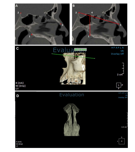
Figure 2. Anatomical landmarks defined to outline the nasal cavity. A. 1: posterior nasal spine, 2: dorsum sellae at midline, 3: rhinion. B. 4: Point of intersection of a line perpendicular to the horizon passing through the posterior nasal spine and the line joining the rhinion and dorsum sellae. C. Posterior part of the posterior nasal spine was removed and the posterior border of the nasal cavity was determined; the line connecting the rhinion and the dorsum sellae is shown in green. D. Airway after manual removal of sinuses and leaving the nasal cavity
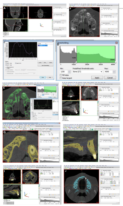
CPV was measured using Mimics Innovation Suite software version 17.0.0.435 (Materialise, Leuven, Belgium). CBCT images were segmented using threshold-based methods to isolate bone structures and generate three-dimensional models. The segmentation thresholds were individually adjusted for each scan to ensure accurate identification of the cortical bone boundaries. The CPV was calculated by subtracting the volume of the cleft defect from the reconstructed alveolar bone volume. To determine the cleft boundaries, the inferior border was defined at the level of the cementoenamel junction of the central incisor adjacent to the cleft, while the superior border corresponded to the upper border of the hard palate. The buccal, palatal, mesial, and distal boundaries were defined based on the adjacent alveolar bone anatomy. Any supernumerary tooth located within the cleft area was removed before volume calculation to avoid underestimation of CPV. The final cleft volume was obtained using Boolean subtraction between the reconstructed bone model with and without the cleft defect (Figure 3).
For comparison of NCV between individuals with UCLP and unaffected individuals, the historical control group from the study by Farzal et al. [1] was used. To achieve age comparability, only subjects aged 8–12 years were included according to the age range of the control group in that study [1].
The use of different software reflected the distinct measurement protocols for the two anatomical volumes.
Measurement reliability:
To assess intra-examiner reliability, 18 CBCT scans were re-examined after 2 weeks by the same examiner who had performed the initial measurements. Considering the intraclass correlation coefficient of 0.811 with a 95% confidence interval of 0.562-0.925, the reliability of the measurements was confirmed. Also, paired t-test was used to evaluate the difference between the two measurements, which showed no significant difference (P=0.734).
Eligibility criteria:
The inclusion criteria were Caucasian non-syndromic UCLP patients in the age range of 5-15 years who had undergone palatal expansion and CBCT before alveolar graft surgery, and it was possible to extract volumetric data in DICOM file from their CBCT scans.
The exclusion criteria were patients who had not undergone primary surgery, CBCT scans with motion or metal artifacts or poor quality, inability to reconstruct images due to file problems (file could not be opened in OnDemand software), and patients with permanent canines erupted into their cleft site.
Accordingly, 75 CBCT scans were collected from 4 radiology centers in Tehran that had CBCT scans of patients with UCLP. From this number of samples, CBCT scans of 70 patients were finally selected according to the eligibility criteria.
Sample size:
The required sample size was 70 CBCT scans considering alpha=0.05, study power of 80%, and correlation coefficient (r) of 0.33 using the following formula:
N = [(Zα+Zβ)/C]2 + 3=70
Where the standard normal deviate for α=Zα=1.96, the standard normal deviate for β=Zβ=0.84, r=0.33, and C=0.5.
Data collection:
Each CBCT scan was converted into DICOM format and imported into On-Demand 3D Application software version 10.0.1 (Cybermed, South Korea) for measurement of NCV. Three-dimensional reconstructions were generated, and anatomical landmarks including the posterior nasal spine, dorsum sellae, and rhinion were identified. The posterior boundary of the nasal cavity was defined according to previously described criteria using the posterior nasal spine and the line connecting the rhinion and dorsum sellae [1]. After exclusion of the paranasal sinuses and other structures, the NCV was calculated using the software’s volume measurement tool. The volume occupied by the turbinates was automatically excluded by the software (Figures 1 and 2).
Figure 1. Scan data of each patient in three main sagittal, coronal, and axial slices and the 3D view
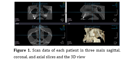
Figure 2. Anatomical landmarks defined to outline the nasal cavity. A. 1: posterior nasal spine, 2: dorsum sellae at midline, 3: rhinion. B. 4: Point of intersection of a line perpendicular to the horizon passing through the posterior nasal spine and the line joining the rhinion and dorsum sellae. C. Posterior part of the posterior nasal spine was removed and the posterior border of the nasal cavity was determined; the line connecting the rhinion and the dorsum sellae is shown in green. D. Airway after manual removal of sinuses and leaving the nasal cavity
CPV was measured using Mimics Innovation Suite software version 17.0.0.435 (Materialise, Leuven, Belgium). CBCT images were segmented using threshold-based methods to isolate bone structures and generate three-dimensional models. The segmentation thresholds were individually adjusted for each scan to ensure accurate identification of the cortical bone boundaries. The CPV was calculated by subtracting the volume of the cleft defect from the reconstructed alveolar bone volume. To determine the cleft boundaries, the inferior border was defined at the level of the cementoenamel junction of the central incisor adjacent to the cleft, while the superior border corresponded to the upper border of the hard palate. The buccal, palatal, mesial, and distal boundaries were defined based on the adjacent alveolar bone anatomy. Any supernumerary tooth located within the cleft area was removed before volume calculation to avoid underestimation of CPV. The final cleft volume was obtained using Boolean subtraction between the reconstructed bone model with and without the cleft defect (Figure 3).
For comparison of NCV between individuals with UCLP and unaffected individuals, the historical control group from the study by Farzal et al. [1] was used. To achieve age comparability, only subjects aged 8–12 years were included according to the age range of the control group in that study [1].
The use of different software reflected the distinct measurement protocols for the two anatomical volumes.
Measurement reliability:
To assess intra-examiner reliability, 18 CBCT scans were re-examined after 2 weeks by the same examiner who had performed the initial measurements. Considering the intraclass correlation coefficient of 0.811 with a 95% confidence interval of 0.562-0.925, the reliability of the measurements was confirmed. Also, paired t-test was used to evaluate the difference between the two measurements, which showed no significant difference (P=0.734).
Statistical analysis:
The obtained data were recorded and analyzed using SPSS 26 (SPSS Inc., IL, USA). The Kolmogorov-Smirnov test confirmed normal distribution of the data; thus, using the Pearson’s correlation test, the correlation between NCV and CPV was examined in the entire study population and then separately by gender and age groups. Linear regression (enter method) was used to analyze the effects of age, CPV, gender, and cleft side on NCV (dependent variable). An independent t-test was used to compare NCV in patients with UCLP and healthy controls of the study by Farzal et al. [1] as a historical control.
Results
The obtained data were recorded and analyzed using SPSS 26 (SPSS Inc., IL, USA). The Kolmogorov-Smirnov test confirmed normal distribution of the data; thus, using the Pearson’s correlation test, the correlation between NCV and CPV was examined in the entire study population and then separately by gender and age groups. Linear regression (enter method) was used to analyze the effects of age, CPV, gender, and cleft side on NCV (dependent variable). An independent t-test was used to compare NCV in patients with UCLP and healthy controls of the study by Farzal et al. [1] as a historical control.
Results
The CBCT scans of 70 patients including 39 males (55.71%) and 28 females (40%), were evaluated. Gender of 3 patients (4.28%) had not been disclosed. The mean age of the participants was 8.80±3.05 years (range 5 to 15 years). The cleft was in the left side in 50 (71.4%) and in the right side in 20 (28.6%) patients.
Table 1 presents the measures of central dispersion for the CPV and NCV (in cubic millimeters) and age of patients. The Pearson’s correlation test showed a significant inverse correlation between the CPV and NCV in the entire study population (r=-0.308, P=0.009). The correlation of CPV and NCV was inversely significant in males (r=-0.350, P=0.029) but not in females (r=-0.360, P=0.060).
Table 2 presents the correlation of CPV and NCV in males and females in different age groups. As shown, a significant inverse correlation existed between CPV and NCV in boys between 5 and 9 years (r=-0.573, P=0.008). This correlation was not significant in girls between 5 and 9 years, and in boys/girls between 10 and 15 years (P>0.05).
Table 3 shows the results of linear regression (enter method) regarding the effects of age, CPV, gender, and cleft side on NCV (dependent variable). As shown, NCV was affected by the CPV (P=0.016), such that per each 1 mm3 increase in CPV, the NCV decreased by 6.37 mm3. Also, gender had a significant effect on NCV (P=0.025), such that females had a smaller NCV than males by 2221.25 units. Age and cleft side had no significant effect on NCV (P>0.05).
Independent t-test was used to compare NCV in patients with UCLP and healthy controls (8-12 years) of the study by Farzal et al. [1] as historical control, and showed no significant difference (Table 4, t=0.43, P=0.66).
Table 1 presents the measures of central dispersion for the CPV and NCV (in cubic millimeters) and age of patients. The Pearson’s correlation test showed a significant inverse correlation between the CPV and NCV in the entire study population (r=-0.308, P=0.009). The correlation of CPV and NCV was inversely significant in males (r=-0.350, P=0.029) but not in females (r=-0.360, P=0.060).
Table 2 presents the correlation of CPV and NCV in males and females in different age groups. As shown, a significant inverse correlation existed between CPV and NCV in boys between 5 and 9 years (r=-0.573, P=0.008). This correlation was not significant in girls between 5 and 9 years, and in boys/girls between 10 and 15 years (P>0.05).
Table 3 shows the results of linear regression (enter method) regarding the effects of age, CPV, gender, and cleft side on NCV (dependent variable). As shown, NCV was affected by the CPV (P=0.016), such that per each 1 mm3 increase in CPV, the NCV decreased by 6.37 mm3. Also, gender had a significant effect on NCV (P=0.025), such that females had a smaller NCV than males by 2221.25 units. Age and cleft side had no significant effect on NCV (P>0.05).
Independent t-test was used to compare NCV in patients with UCLP and healthy controls (8-12 years) of the study by Farzal et al. [1] as historical control, and showed no significant difference (Table 4, t=0.43, P=0.66).
Table 1. Measures of central dispersion for the CPV and NCV (in cubic millimeters) and age of patients
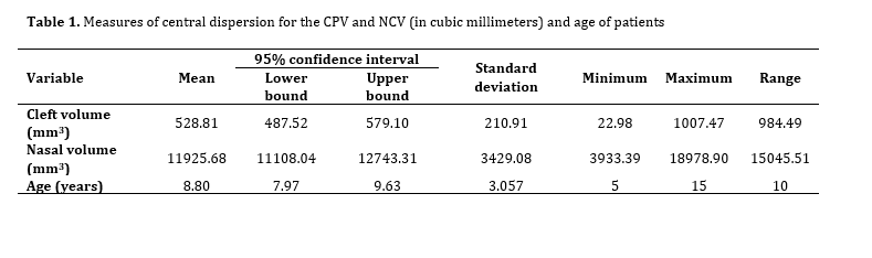
Table 2. Correlation of CPV and NCV in males and females in different age groups
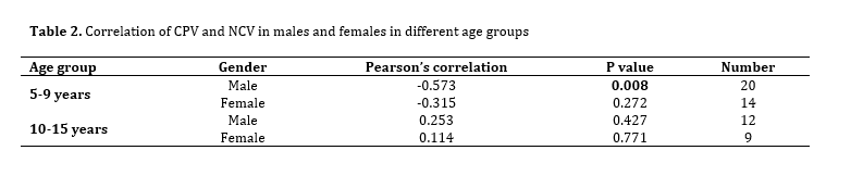
Table 3. Results of linear regression (enter method) regarding the effects of age, CPV, gender, and cleft side on NCV (dependent variable)
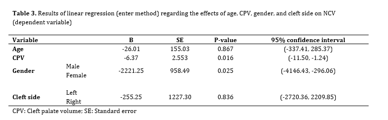
Table 4. Comparison of NCV of UCLP patients of the present study with healthy controls of the study by Farzal et al. [1] (age range of 8-12 years)
![Table 4. Comparison of NCV of UCLP patients of the present study with healthy controls of the study by Farzal et al. [1] (age range of 8-12 years)](./files/site1/images/20263/javadian_t4.png)
Discussion
This study demonstrated a significant inverse correlation between CPV and NCV in patients with non-syndromic UCLP, suggesting that larger cleft defects may be associated with reduced nasal cavity dimensions. Since the palate forms the anatomical floor of the nasal cavity, a larger cleft defect may reflect greater disruption of maxillary and nasal development, potentially explaining the observed inverse relationship between CPV and NCV. The findings remained consistent in male patients, whereas the lack of significance in females and some age subgroups may be related to limited sample sizes and variations in craniofacial growth patterns.
Regarding the relationship between the CP and nasal cavity in individuals with UCLP, only one study was found [16] that used acoustic rhinometry to investigate the relationship between cleft size in infancy and size and function of the nose in adulthood. They concluded that the size and function of the nasal airway on the cleft side were reduced compared to the non-cleft side, but no relationship was found between the initial cleft size in infancy and nasal size and function in adulthood. Comparing cleft width rather than volume, measuring cleft width and nasal volume at two different times, and using acoustic rhinometry as the gold standard compared to CBCT imaging could be factors that differed from our study and might have affected the results. In addition, some of the subjects in their study had undergone minimal rhinoplasty. In our study, unlike previous studies, the On-Demand software volume calculation tool was used to calculate the NCV three-dimensionally, while previous studies that used CBCT scans to assess nasal volume used Dolphin and Mimics software for this purpose.
Starbuck et al. [13] divided the samples into subgroups based on the type of cleft (right unilateral, left unilateral, bilateral), age group, gender, and whether or not alveolar graft surgery was performed. Unlike Starbuck et al. [13], who reported effects of age and cleft type on NCV, the present study did not identify significant associations with these variables. Differences in sample characteristics, age distribution, and measurement protocols may explain these findings.
In order to compare the NCV in individuals with UCLP with individuals without CLP, the control group of the study by Farzal et al. [1] was used due to the similarity in boundaries determined for measuring the NCV, and patients aged 8 to 12 years were compared in the two studies. No significant difference was reported in the NCV of the two groups, while in the study by Farzal et al. [1], the NCV of individuals with unilateral and bilateral CLP was significantly smaller than that of the control group. Such different results could be due to the unequal number of samples and different gender distribution in the two groups, the difference in the software used to assess the NCV in the two studies, and the fact that maxillary expansion was performed for the patients in the present study (it can increase the internal dimensions of the nose in children with UCLP by 26%) [17].
In the present study, the mean NCV in 70 UCLP patients was estimated to be 11925.68±3429.08 mm3. In previous studies, the mean NCV in such patients ranged from 6.6±1.9 cm3 to 14.7±2.2 cm3 [18, 19]. These variations can be attributed to different definitions of anatomical boundaries, the use of different software programs for volumetric measurements, different imaging methods, different sample sizes, and different age ranges of patients in the studies. Other confounding factors include the type and timing of surgeries performed before volume measurement, and orthodontic interventions, including whether or not maxillary expansion had been performed. For example, the mean NCV in UCLP patients reported by Farzal et al. [1] was smaller than that in the present study despite the similar range determined, which could be due to the different age range and the maxillary expansion performed in our study patients.
Previous studies have reported reduced nasal cavity dimensions on the cleft side in individuals with UCLP, although findings regarding total nasal airway volume have been inconsistent [1, 11, 13, 17, 20, 21]. These discrepancies may be related to variations in anatomical definitions, imaging modalities, segmentation methods, age groups, and previous orthodontic or surgical interventions. In the present study, the exclusion of turbinate volume and nasopharyngeal structures, as well as previous maxillary expansion in some patients, may have influenced the measured NCV.
This study had several limitations. The retrospective design and exclusion of scans with inadequate quality may have introduced selection bias. The limited sample size, particularly in subgroup analyses, may have reduced statistical power. The absence of an internal control group limited direct comparison with individuals without CLP. Future studies including larger cohorts, functional airway assessments, and detailed information regarding previous surgical interventions are recommended.
Conclusion
Regarding the relationship between the CP and nasal cavity in individuals with UCLP, only one study was found [16] that used acoustic rhinometry to investigate the relationship between cleft size in infancy and size and function of the nose in adulthood. They concluded that the size and function of the nasal airway on the cleft side were reduced compared to the non-cleft side, but no relationship was found between the initial cleft size in infancy and nasal size and function in adulthood. Comparing cleft width rather than volume, measuring cleft width and nasal volume at two different times, and using acoustic rhinometry as the gold standard compared to CBCT imaging could be factors that differed from our study and might have affected the results. In addition, some of the subjects in their study had undergone minimal rhinoplasty. In our study, unlike previous studies, the On-Demand software volume calculation tool was used to calculate the NCV three-dimensionally, while previous studies that used CBCT scans to assess nasal volume used Dolphin and Mimics software for this purpose.
Starbuck et al. [13] divided the samples into subgroups based on the type of cleft (right unilateral, left unilateral, bilateral), age group, gender, and whether or not alveolar graft surgery was performed. Unlike Starbuck et al. [13], who reported effects of age and cleft type on NCV, the present study did not identify significant associations with these variables. Differences in sample characteristics, age distribution, and measurement protocols may explain these findings.
In order to compare the NCV in individuals with UCLP with individuals without CLP, the control group of the study by Farzal et al. [1] was used due to the similarity in boundaries determined for measuring the NCV, and patients aged 8 to 12 years were compared in the two studies. No significant difference was reported in the NCV of the two groups, while in the study by Farzal et al. [1], the NCV of individuals with unilateral and bilateral CLP was significantly smaller than that of the control group. Such different results could be due to the unequal number of samples and different gender distribution in the two groups, the difference in the software used to assess the NCV in the two studies, and the fact that maxillary expansion was performed for the patients in the present study (it can increase the internal dimensions of the nose in children with UCLP by 26%) [17].
In the present study, the mean NCV in 70 UCLP patients was estimated to be 11925.68±3429.08 mm3. In previous studies, the mean NCV in such patients ranged from 6.6±1.9 cm3 to 14.7±2.2 cm3 [18, 19]. These variations can be attributed to different definitions of anatomical boundaries, the use of different software programs for volumetric measurements, different imaging methods, different sample sizes, and different age ranges of patients in the studies. Other confounding factors include the type and timing of surgeries performed before volume measurement, and orthodontic interventions, including whether or not maxillary expansion had been performed. For example, the mean NCV in UCLP patients reported by Farzal et al. [1] was smaller than that in the present study despite the similar range determined, which could be due to the different age range and the maxillary expansion performed in our study patients.
Previous studies have reported reduced nasal cavity dimensions on the cleft side in individuals with UCLP, although findings regarding total nasal airway volume have been inconsistent [1, 11, 13, 17, 20, 21]. These discrepancies may be related to variations in anatomical definitions, imaging modalities, segmentation methods, age groups, and previous orthodontic or surgical interventions. In the present study, the exclusion of turbinate volume and nasopharyngeal structures, as well as previous maxillary expansion in some patients, may have influenced the measured NCV.
This study had several limitations. The retrospective design and exclusion of scans with inadequate quality may have introduced selection bias. The limited sample size, particularly in subgroup analyses, may have reduced statistical power. The absence of an internal control group limited direct comparison with individuals without CLP. Future studies including larger cohorts, functional airway assessments, and detailed information regarding previous surgical interventions are recommended.
Conclusion
The present study showed a reduction in NCV with an increase in CPV. This issue can play a significant role in treatment planning of patients with UCLP. With preventive management in infants born with a larger cleft volume, further consequences of reduced NCV and subsequent increase in respiratory problems can be prevented. Despite the need to verify the present findings in prospective studies on well-matched samples, we believe that this study paved the way to increase the success rate of treatment in patients with UCLP.
Type of Study: Original article |
Subject:
pediatric
References
1. Farzal Z, Walsh J, Lopes de Rezende Barbosa G, Zdanski CJ, Davis SD, Superfine R, et al. Volumetric nasal cavity analysis in children with unilateral and bilateral cleft lip and palate. Laryngoscope. 2016 Jun;126(6):1475-80. [DOI:10.1002/lary.25543] [PMID] [PMCID]
2. Kasiri N, Bayani M, Mohammad-Rabei E, Ebrahimizadeh H. Correlation between alveolar cleft volume and alveolar bone quality in patients with unilateral cleft lip and palate: A cone-beam computed tomography study. J Stomatol Oral Maxillofac Surg. 2022 Jun;123(3):e69-75. [DOI:10.1016/j.jormas.2021.06.013] [PMID]
3. Brydges HT, Laspro M, Verzella AN, Alcon A, Schechter J, Cassidy MF, et al. Contemporary Prevalence of Oral Clefts in the US: Geographic and Socioeconomic Considerations. J Clin Med. 2024 Apr;13(9):2570. [DOI:10.3390/jcm13092570] [PMID] [PMCID]
4. Adeyemi K, Chang V, Ifeanacho E, Sunner R. Socioemotional development of children with cleft lip with or without cleft palate (CL/P) across socioeconomic backgrounds and potential impacts on families: Narrative review. Child Health Inter Litr Disc J. 2025 Sep;4(1):9-15. [DOI:10.15173/child.v4i1.3956]
5. de Ladeira PR, Alonso N. Protocols in cleft lip and palate treatment: systematic review. Plast Surg Int. 2012;2012:562892. [DOI:10.1155/2012/562892] [PMID] [PMCID]
6. Borah MP, Sharma N, Borah T. Cleft lip and palate (CLP): An invisible disability or impairment? TISS J Disabil Stud Res. 2024 Jun;4(1):76-102. [DOI:10.2139/ssrn.4887838]
7. Kaiser JK, Kamble RH, Nambiar K, Nerurkar S, Suchak D. Comprehensive multidisciplinary management of bilateral cleft lip and palate. Cureus. 2024 Jan;16(1):e52643. [DOI:10.7759/cureus.52643]
8. Castilho JF, Ribeiro IDC, Andrade ALCDÁ, Oliveira DD, Lima ILDA. Multidisciplinary treatment in a patient with Bilateral cleft lip and palate: A case report. Iran J Orthod. 2024 Dec;19(2):1-12.
9. Sehrawat K, Kumar S, Harikrishnan P. Three-dimensional evaluation of alveolar cleft volume and nasomaxillary form in patients with unilateral cleft lip and palate. Cleft Palate Craniofac J. 2025 Jan;62(1):5-12. [DOI:10.1177/10556656231196057] [PMID]
10. Vicente A, Wiedel AP, Becker M, Brogårdh-Roth S, Shi XQ, Hellén-Halme K. Quantitative assessment of cleft volume and evaluation of cleft's impact on adjacent anatomical structures using CBCT imaging. Oral Radiol. 2024 Apr;40(2):295-303. [DOI:10.1007/s11282-023-00736-0] [PMID] [PMCID]
11. Pimenta LA, de Rezende Barbosa GL, Pretti H, Emodi O, van Aalst J, Rossouw PE, et al. Three-dimensional evaluation of nasopharyngeal airways of unilateral cleft lip and palate patients. Laryngoscope. 2015 Mar;125(3):736-9. [DOI:10.1002/lary.24895] [PMID]
12. Frank-Ito DO, Carpenter DJ, Cheng T, Avashia YJ, Brown DA, Glener A, et al. Computational analysis of the mature unilateral cleft lip nasal deformity on nasal patency. Plast Reconstr Surg Glob Open. 2019 May;7(5):e2244. [DOI:10.1097/GOX.0000000000002244] [PMID] [PMCID]
13. Starbuck JM, Friel MT, Ghoneima A, Flores RL, Tholpady S, Kula K. Nasal airway and septal variation in unilateral and bilateral cleft lip and palate. Clin Anat. 2014 Oct;27(7):999-1008. [DOI:10.1002/ca.22428] [PMID]
14. Tso HH, Lee JS, Huang JC, Maki K, Hatcher D, Miller AJ. Evaluation of the human airway using cone-beam computerized tomography. Oral Surg Oral Med Oral Pathol Oral Radiol Endod. 2009 Nov;108(5):768-76. [DOI:10.1016/j.tripleo.2009.05.026] [PMID]
15. Al-Fahdawi MA, Farid MM, El-Fotouh MA, El-Kassaby MA. Cone-beam computed tomography analysis of the nasopharyngeal airway in nonsyndromic cleft lip and palate subjects. Cleft Palate Craniofac J. 2017 Mar;54(2):202-9. [DOI:10.1597/15-134] [PMID]
16. Reiser E, Andlin-Sobocki A, Mani M, Holmström M. Initial size of cleft does not correlate with size and function of nasal airway in adults with unilateral cleft lip and palate. J Plast Surg Hand Surg. 2011 Jun;45(3):129-35. [DOI:10.3109/2000656X.2011.576839] [PMID]
17. Trindade IE, Castilho RL, Sampaio-Teixeira AC, Trindade-Suedam IK, Silva-Filho OG. Effects of orthopedic rapid maxillary expansion on internal nasal dimensions in children with cleft lip and palate assessed by acoustic rhinometry. J Craniofac Surg. 2010 Mar;21(2):306-11. [DOI:10.1097/SCS.0b013e3181cf5f5f] [PMID]
18. Dos Inocentes RJM, Marzano-Rodrigues MN, de Espíndola GG, García-Usó M, Yatabe-Ioshida MS, Trindade IEK, et al. Adults with unilateral cleft lip and palate present reduced internal nasal volumes: Findings of a three-dimensional morphometric assessment in cone-beam computed tomography scans. J Craniofac Surg. 2021 Jan-Feb 01;32(1):e15-9. [DOI:10.1097/SCS.0000000000006820] [PMID]
19. Hassegawa CA, Garcia-Usó MA, Yatabe-Ioshida MS, Trindade IEK, Fukushiro AP, Carreira DGG, et al. Internal nasal dimensions of children with unilateral cleft lip and palate and maxillary atresia: comparison between acoustic rhinometry technique and cone-beam computed tomography. Codas. 2021 May;33(3):e20200099.
20. Friel MT, Starbuck JM, Ghoneima AM, Murage K, Kula KS, Tholpady S, et al. Airway obstruction and the unilateral cleft lip and palate deformity: Contributions by the bony septum. Ann Plast Surg. 2015 Jul;75(1):37-43. [DOI:10.1097/SAP.0000000000000027] [PMID]
21. Ertaş Ü, Ataol M. Evaluation of nasal airway volume of operated unilateral cleft lip and palate patients compared with skeletal Class III individuals. Cleft Palate Craniofac J. 2019 Jan;56(1):15-20. [DOI:10.1177/1055665618774024] [PMID]
Send email to the article author
| Rights and permissions | |
 |
This work is licensed under a Creative Commons Attribution-NonCommercial 4.0 International License. |