
Journal of Research in Dental
and Maxillofacial Sciences

Volume 11, Issue 3 (9-2026)
J Res Dent Maxillofac Sci 2026, 11(3): 242-250 |
Back to browse issues page

Ethics code: IR.ZUMS.REC.1403.187


Download citation:
BibTeX | RIS | EndNote | Medlars | ProCite | Reference Manager | RefWorks
Send citation to:


BibTeX | RIS | EndNote | Medlars | ProCite | Reference Manager | RefWorks
Send citation to:
Aghmasheh F, Hekmat B, Mozafar A. Association between Anatomical Variations of the Osteomeatal Complex and Pathological Conditions of the Maxillary Sinus on Cone-Beam Computed Tomography Images. J Res Dent Maxillofac Sci 2026; 11 (3) :242-250
URL: http://jrdms.dentaliau.ac.ir/article-1-1464-en.html
URL: http://jrdms.dentaliau.ac.ir/article-1-1464-en.html


1- Department of Oral and Maxillofacial Radiology, School of Dentistry, Zanjan University of Medical Sciences, Zanjan, Iran.
2- Department of Oral and Maxillofacial Radiology, School of Dentistry, Zanjan University of Medical Sciences, Zanjan, Iran. ,bahar_hekmat70@yahoo.com
2- Department of Oral and Maxillofacial Radiology, School of Dentistry, Zanjan University of Medical Sciences, Zanjan, Iran. ,
Full-Text [PDF 403 kb]
(4 Downloads)
| Abstract (HTML) (14 Views)
Table 3. Distribution of maxillary sinus pathologies by gender
Table 4. Distribution of anatomical variations in patients with and without mucosal thickening
Table 5. Distribution of anatomical variations in patients with and without mucous retention cyst
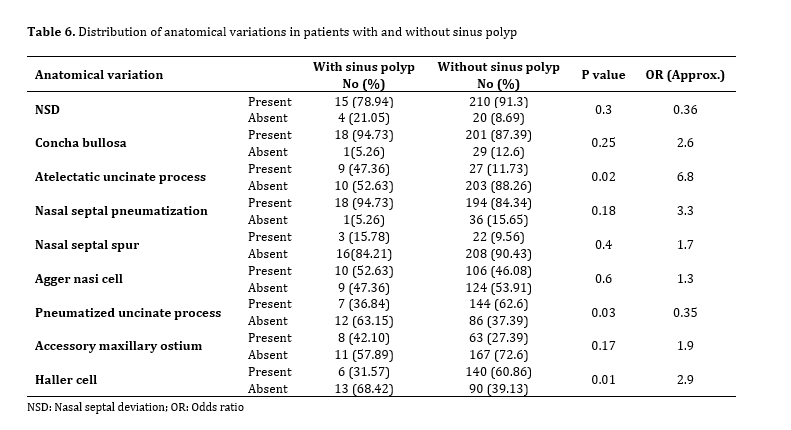
Table 6. Distribution of anatomical variations in patients with and without sinus polyp
Discussion
Full-Text: (1 Views)
Abstract
Background and Aim: The osteomeatal complex (OMC) plays a critical role in the ventilation and drainage of the paranasal sinuses, particularly the maxillary sinus. Anatomical variations in this region may predispose individuals to impaired drainage and development of various pathologies. This study aimed to investigate the relationship between anatomical variations of the OMC and maxillary sinus pathology using cone-beam computed tomography (CBCT).
Materials and Methods: This analytical cross-sectional study evaluated 249 CBCT scans of the sinonasal region of patients referred to the Oral and Maxillofacial Radiology Department of Zanjan Dental School during 2023-2024. All scans were assessed for the presence of anatomical variations in the OMC and pathological findings in the maxillary sinuses. Statistical analysis was performed using the Chi-square test, odds ratios (OR), and post-hoc power analysis, with a significance level set at 0.05.
Results: The findings revealed a significant association between the presence of nasal septal deviation (NSD) and the presence of mucosal thickening (P=0.03, OR=1.83). Additionally, the presence of pneumatized uncinate process (P=0.03, OR=6), atelectatic uncinate process (P=0.02, OR=6.8), and Haller cells (P=0.01, OR=2.9) had a significant association with the occurrence of sinus polyps. Other anatomical variations investigated did not demonstrate a statistically significant relationship with maxillary sinus pathologies (P>0.05). Furthermore, the prevalence of mucosal thickening was significantly higher in males compared to females (P=0.03).
Conclusion: Certain anatomical variations of the OMC may contribute to the dysfunction of the maxillary sinus and development of pathological conditions such as polyps.
Keywords: Cone-Beam Computed Tomography; Maxillary Sinus; Paranasal Sinuses
Introduction
Materials and Methods: This analytical cross-sectional study evaluated 249 CBCT scans of the sinonasal region of patients referred to the Oral and Maxillofacial Radiology Department of Zanjan Dental School during 2023-2024. All scans were assessed for the presence of anatomical variations in the OMC and pathological findings in the maxillary sinuses. Statistical analysis was performed using the Chi-square test, odds ratios (OR), and post-hoc power analysis, with a significance level set at 0.05.
Results: The findings revealed a significant association between the presence of nasal septal deviation (NSD) and the presence of mucosal thickening (P=0.03, OR=1.83). Additionally, the presence of pneumatized uncinate process (P=0.03, OR=6), atelectatic uncinate process (P=0.02, OR=6.8), and Haller cells (P=0.01, OR=2.9) had a significant association with the occurrence of sinus polyps. Other anatomical variations investigated did not demonstrate a statistically significant relationship with maxillary sinus pathologies (P>0.05). Furthermore, the prevalence of mucosal thickening was significantly higher in males compared to females (P=0.03).
Conclusion: Certain anatomical variations of the OMC may contribute to the dysfunction of the maxillary sinus and development of pathological conditions such as polyps.
Keywords: Cone-Beam Computed Tomography; Maxillary Sinus; Paranasal Sinuses
Introduction
The maxillary sinus originates from the inward growth of the nasal mucosal lining and represents the largest paranasal sinus. During development, it typically enlarges to occupy a substantial portion of the maxillary bone. This sinus is connected to the nasal cavity through the maxillary ostium and plays an important role in ventilation and regulation of intranasal air pressure [1,2]. The osteomeatal complex (OMC) should be regarded not as a single anatomical entity but as an integrated functional system consisting of multiple interrelated structures. These components include the maxillary sinus ostium, ethmoidal infundibulum, uncinate process, hiatus semilunaris, anterior ethmoidal foramen, ethmoidal bulla, and the middle meatus [2]. Paranasal sinuses develop as a result of nasal mucosal invagination into the adjacent facial bones, a process that explains the wide range of anatomical variations observed in this area. Common variations include concha bullosa, Agger nasi cells, Haller cells, deviations or structural alterations of the uncinate process such as pneumatization or atelectasis, nasal septal pneumatization, and nasal septal spurs [3,4].
Although traditional radiographic methods such as plain radiographs can be used to assess the paranasal sinuses, computed tomography (CT) remains the preferred modality for precise anatomical evaluation. Progress in CT imaging and nasal endoscopic techniques has significantly improved the understanding of the complex anatomy of the OMC and enhanced the diagnostic differentiation of sinus disorders. Nevertheless, the relatively high radiation exposure associated with conventional CT has encouraged the exploration of alternative imaging techniques that maintain diagnostic reliability while reducing radiation dose [3-5]. Cone-beam computed tomography (CBCT) has emerged as an advanced imaging technique capable of producing high-resolution images with accurate anatomical detail. Compared with conventional CT, CBCT is associated with a markedly lower radiation dose, reduced cost, and a more compact imaging system. These advantages make CBCT particularly suitable for diagnostic purposes and treatment planning in the maxillofacial region [6-8]. Anatomical variations of the paranasal sinuses are of clinical importance, as they may contribute to the development of inflammatory conditions and are frequently encountered during diagnostic and therapeutic procedures involving the sinonasal area [3]. Accordingly, the present study was designed to evaluate the association between anatomical variations of the OMC and pathological changes of the maxillary sinuses on CBCT images.
Materials and Methods
Although traditional radiographic methods such as plain radiographs can be used to assess the paranasal sinuses, computed tomography (CT) remains the preferred modality for precise anatomical evaluation. Progress in CT imaging and nasal endoscopic techniques has significantly improved the understanding of the complex anatomy of the OMC and enhanced the diagnostic differentiation of sinus disorders. Nevertheless, the relatively high radiation exposure associated with conventional CT has encouraged the exploration of alternative imaging techniques that maintain diagnostic reliability while reducing radiation dose [3-5]. Cone-beam computed tomography (CBCT) has emerged as an advanced imaging technique capable of producing high-resolution images with accurate anatomical detail. Compared with conventional CT, CBCT is associated with a markedly lower radiation dose, reduced cost, and a more compact imaging system. These advantages make CBCT particularly suitable for diagnostic purposes and treatment planning in the maxillofacial region [6-8]. Anatomical variations of the paranasal sinuses are of clinical importance, as they may contribute to the development of inflammatory conditions and are frequently encountered during diagnostic and therapeutic procedures involving the sinonasal area [3]. Accordingly, the present study was designed to evaluate the association between anatomical variations of the OMC and pathological changes of the maxillary sinuses on CBCT images.
Materials and Methods
This cross-sectional study was conducted following approval by the Ethics Committee of Zanjan University of Medical Sciences (IR.ZUMS.REC.1403.187). CBCT scans of patients referred to the Oral and Maxillofacial Radiology Department of Zanjan Dental School during 2023-2024 for evaluation of the maxillary sinuses were retrospectively reviewed.
Considering a 95% confidence level (CI) and a sampling error of 6.15%, and based on a study by Shokri et al. [8], the total sample size was calculated to be 249 participants using the following formula:
n = Z² × P (1 − P) / d²
n= (1.96) ²×0.586× (1 − 0.586)/ (0.0615) ² = 249
Where:
n= required sample size
Z = 1.96 (95% CI)
P = estimated proportion
d = sampling error (6.15%)
Images of inadequate quality, scans belonging to individuals younger than 18 years, and cases with a history of surgery, trauma, cleft anomalies, or benign or malignant tumors affecting the sinonasal region were excluded. Ultimately, CBCT images of 249 patients, including 143 females and 106 males, met the inclusion criteria and were analyzed.
All scans were acquired using a NewTom 3G CBCT scanner (Quantitative Radiology, Verona, Italy). Reconstructed images were examined on a 20-inch monitor (LG, Seoul, Korea) under dim ambient lighting conditions and evaluated in axial, coronal, and sagittal planes.
All image analyses were independently performed by two experienced, board-certified oral and maxillofacial radiologists, each with more than 10 years of clinical experience, using NNT Viewer software (NewTom, Verona, Italy). In case of disagreement between the two observers, another oral and maxillofacial radiologist was consulted as a third observer.
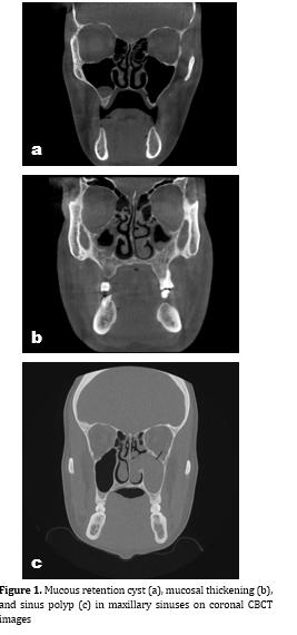
The observers assessed the presence of OMC anatomical variations as well as pathological findings within the maxillary sinuses. The assessed anatomical variations included concha bullosa, atelectatic uncinate process, pneumatized uncinate process, nasal septal deviation (NSD), septal pneumatization, septal spur, anterior agger nasi cells, accessory maxillary ostium, and Haller cells. In addition, maxillary sinuses were evaluated for pathological conditions such as mucosal thickening, mucous retention cysts, mucoceles, and sinus polyps (Figure 1).
Pathological findings of the maxillary sinuses were assessed based on CBCT radiographic characteristics. Schneiderian membrane thickening greater than 3 mm was considered pathological mucosal thickening. Partial or complete opacification of the sinus cavity associated with mucosal thickening was recorded as a radiographic sign of sinus mucosal inflammatory changes. Mucous retention cysts were identified as dome-shaped, well-defined, non-corticated radiopaque lesions arising from the sinus wall or floor. Sinus polyps were diagnosed as soft tissue radiopacities associated with adjacent mucosal thickening. Mucoceles were identified by complete sinus opacification accompanied by expansion and alteration of the normal sinus contours. All findings were evaluated exclusively according to CBCT imaging criteria described in previous radiologic studies [2,9].
Figure 1. Mucous retention cyst (a), mucosal thickening (b), and sinus polyp (c) in maxillary sinuses on coronal CBCT images
All collected data were documented in a standardized checklist and analyzed using SPSS version 26 (IBM Corp., Armonk, NY, USA). Descriptive statistics were calculated. Categorical variables were compared using the chi-square test. Associations were quantified using odds ratios (ORs) with 95% confidence intervals (CIs). A post hoc power analysis was performed to assess the statistical power of the study, with a target power of >80%. P<0.05 was considered statistically significant.
Results
Considering a 95% confidence level (CI) and a sampling error of 6.15%, and based on a study by Shokri et al. [8], the total sample size was calculated to be 249 participants using the following formula:
n = Z² × P (1 − P) / d²
n= (1.96) ²×0.586× (1 − 0.586)/ (0.0615) ² = 249
Where:
n= required sample size
Z = 1.96 (95% CI)
P = estimated proportion
d = sampling error (6.15%)
Images of inadequate quality, scans belonging to individuals younger than 18 years, and cases with a history of surgery, trauma, cleft anomalies, or benign or malignant tumors affecting the sinonasal region were excluded. Ultimately, CBCT images of 249 patients, including 143 females and 106 males, met the inclusion criteria and were analyzed.
All scans were acquired using a NewTom 3G CBCT scanner (Quantitative Radiology, Verona, Italy). Reconstructed images were examined on a 20-inch monitor (LG, Seoul, Korea) under dim ambient lighting conditions and evaluated in axial, coronal, and sagittal planes.
All image analyses were independently performed by two experienced, board-certified oral and maxillofacial radiologists, each with more than 10 years of clinical experience, using NNT Viewer software (NewTom, Verona, Italy). In case of disagreement between the two observers, another oral and maxillofacial radiologist was consulted as a third observer.
The observers assessed the presence of OMC anatomical variations as well as pathological findings within the maxillary sinuses. The assessed anatomical variations included concha bullosa, atelectatic uncinate process, pneumatized uncinate process, nasal septal deviation (NSD), septal pneumatization, septal spur, anterior agger nasi cells, accessory maxillary ostium, and Haller cells. In addition, maxillary sinuses were evaluated for pathological conditions such as mucosal thickening, mucous retention cysts, mucoceles, and sinus polyps (Figure 1).
Pathological findings of the maxillary sinuses were assessed based on CBCT radiographic characteristics. Schneiderian membrane thickening greater than 3 mm was considered pathological mucosal thickening. Partial or complete opacification of the sinus cavity associated with mucosal thickening was recorded as a radiographic sign of sinus mucosal inflammatory changes. Mucous retention cysts were identified as dome-shaped, well-defined, non-corticated radiopaque lesions arising from the sinus wall or floor. Sinus polyps were diagnosed as soft tissue radiopacities associated with adjacent mucosal thickening. Mucoceles were identified by complete sinus opacification accompanied by expansion and alteration of the normal sinus contours. All findings were evaluated exclusively according to CBCT imaging criteria described in previous radiologic studies [2,9].
Figure 1. Mucous retention cyst (a), mucosal thickening (b), and sinus polyp (c) in maxillary sinuses on coronal CBCT images
All collected data were documented in a standardized checklist and analyzed using SPSS version 26 (IBM Corp., Armonk, NY, USA). Descriptive statistics were calculated. Categorical variables were compared using the chi-square test. Associations were quantified using odds ratios (ORs) with 95% confidence intervals (CIs). A post hoc power analysis was performed to assess the statistical power of the study, with a target power of >80%. P<0.05 was considered statistically significant.
Results
The inter-class correlation coefficient was calculated to assess interobserver reliability, which was found to be 96%.
Based on the data obtained from CBCT images, various anatomical indices of the OMC showed a diverse distribution among the studied cases. The results indicated that NSD and concha bullosa were the most prevalent anatomical variations, observed in 90% and 88% of the cases, respectively. In contrast, atelectatic uncinate process was identified in only 14.5% of the patients.
Nasal septal spur was present in 56.2% of the cases, while nasal septal pneumatization was observed in 85.1% of the scans. Agger nasi cells were detected in 46.6% and pneumatized uncinate process in 60.6% of the patients. The accessory maxillary ostium was reported in 28.5% of the cases, and Haller cells were present in 58.6% of the examined images. Table 1 summarizes these findings.
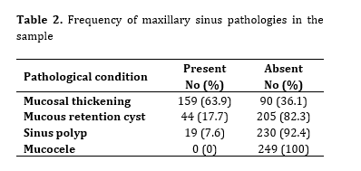
In the assessment of pathological conditions of the maxillary sinuses, mucosal thickening was the most frequently observed pathology, with a prevalence of 63.9%. Mucous retention cysts were identified in 17.7% of the cases, while sinus polyps were reported in 7.6% of the sample. No cases of mucoceles were observed in the studied population. Table 2 summarizes these findings.
Table 1. Frequency of anatomical variations of the OMC
Table 2. Frequency of maxillary sinus pathologies in the sample
Based on the data obtained from CBCT images, various anatomical indices of the OMC showed a diverse distribution among the studied cases. The results indicated that NSD and concha bullosa were the most prevalent anatomical variations, observed in 90% and 88% of the cases, respectively. In contrast, atelectatic uncinate process was identified in only 14.5% of the patients.
Nasal septal spur was present in 56.2% of the cases, while nasal septal pneumatization was observed in 85.1% of the scans. Agger nasi cells were detected in 46.6% and pneumatized uncinate process in 60.6% of the patients. The accessory maxillary ostium was reported in 28.5% of the cases, and Haller cells were present in 58.6% of the examined images. Table 1 summarizes these findings.
In the assessment of pathological conditions of the maxillary sinuses, mucosal thickening was the most frequently observed pathology, with a prevalence of 63.9%. Mucous retention cysts were identified in 17.7% of the cases, while sinus polyps were reported in 7.6% of the sample. No cases of mucoceles were observed in the studied population. Table 2 summarizes these findings.
Table 1. Frequency of anatomical variations of the OMC
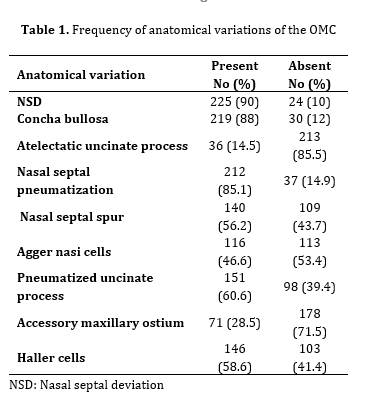
Table 2. Frequency of maxillary sinus pathologies in the sample
The Chi-square test showed a statistically significant difference between males and females in the prevalence of mucosal thickening (P=0.03). No statistically significant differences were observed for mucous retention cysts (P=0.22) or sinus polyps (P=0.12). The post-hoc power analysis indicated that the study had sufficient power (>80%) to detect the observed differences in pathological conditions of the maxillary sinuses between genders. Table 3 summarizes these findings.
A comparison between patients with and without mucosal thickening showed a statistically significant difference only for NSD (P=0.03). No statistically significant differences were found for concha bullosa (P=0.07), atelectatic uncinate process (P=0.64), nasal septal pneumatization (P=0.95), nasal septal spur (P=0.08), agger nasi cells (P=0.60), pneumatized uncinate process (P=0.60), accessory maxillary ostium (P=0.90), and Haller cells (P=0.87). OR analysis indicated that NSD was associated with increased odds of mucosal thickening (OR = 1.83), while other anatomical variations showed no meaningful associations. Table 4 summarizes these findings.
Among patients with and without mucous retention cysts, no statistically significant differences were observed for NSD (P=0.5), concha bullosa (P=0.5), atelectatic uncinate process (P=0.8), nasal septal pneumatization (P=0.15), nasal septal spur (P=0.21), Agger nasi cells (P=0.9), pneumatized uncinate process (P=0.1), accessory maxillary ostium (P=0.7) and Haller cells (P=0.3). Table 5 summarizes these findings. OR analysis showed weak associations, with the highest values observed for nasal septal pneumatization (OR=1.92) and pneumatized uncinate process (OR=1.69), although these effects were weak (Table 5).
Among patients with and without sinus polyps, statistically significant differences were observed for atelectatic uncinate process (P=0.02), pneumatized uncinate process (P=0.03), and Haller cells (P=0.01). No statistically significant differences were found for NSD (P=0.30), concha bullosa (P=0.25), nasal septal pneumatization (P=0.18), nasal septal spur (P=0.40), Agger nasi cells (P=0.60), and accessory maxillary ostium (P=0.18). Table 6 summarizes these findings. OR analysis indicated that atelectatic uncinate process (OR=6.8), pneumatized uncinate process (OR=6), and Haller cells (OR=2.9) were associated with increased odds of sinus polyps. The remaining anatomical variations showed weak or negligible associations (Table 6).
A comparison between patients with and without mucosal thickening showed a statistically significant difference only for NSD (P=0.03). No statistically significant differences were found for concha bullosa (P=0.07), atelectatic uncinate process (P=0.64), nasal septal pneumatization (P=0.95), nasal septal spur (P=0.08), agger nasi cells (P=0.60), pneumatized uncinate process (P=0.60), accessory maxillary ostium (P=0.90), and Haller cells (P=0.87). OR analysis indicated that NSD was associated with increased odds of mucosal thickening (OR = 1.83), while other anatomical variations showed no meaningful associations. Table 4 summarizes these findings.
Among patients with and without mucous retention cysts, no statistically significant differences were observed for NSD (P=0.5), concha bullosa (P=0.5), atelectatic uncinate process (P=0.8), nasal septal pneumatization (P=0.15), nasal septal spur (P=0.21), Agger nasi cells (P=0.9), pneumatized uncinate process (P=0.1), accessory maxillary ostium (P=0.7) and Haller cells (P=0.3). Table 5 summarizes these findings. OR analysis showed weak associations, with the highest values observed for nasal septal pneumatization (OR=1.92) and pneumatized uncinate process (OR=1.69), although these effects were weak (Table 5).
Among patients with and without sinus polyps, statistically significant differences were observed for atelectatic uncinate process (P=0.02), pneumatized uncinate process (P=0.03), and Haller cells (P=0.01). No statistically significant differences were found for NSD (P=0.30), concha bullosa (P=0.25), nasal septal pneumatization (P=0.18), nasal septal spur (P=0.40), Agger nasi cells (P=0.60), and accessory maxillary ostium (P=0.18). Table 6 summarizes these findings. OR analysis indicated that atelectatic uncinate process (OR=6.8), pneumatized uncinate process (OR=6), and Haller cells (OR=2.9) were associated with increased odds of sinus polyps. The remaining anatomical variations showed weak or negligible associations (Table 6).
Table 3. Distribution of maxillary sinus pathologies by gender
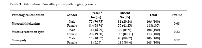
Table 4. Distribution of anatomical variations in patients with and without mucosal thickening
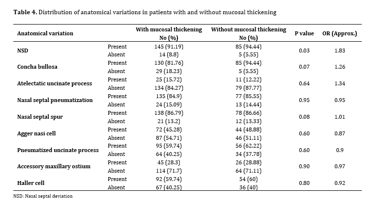
Table 5. Distribution of anatomical variations in patients with and without mucous retention cyst
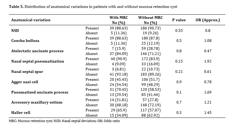
Table 6. Distribution of anatomical variations in patients with and without sinus polyp
Discussion
The OMC is a critical anatomical region responsible for adequate ventilation and drainage of the paranasal sinuses, especially the maxillary sinus. Structural variations within this complex may compromise normal drainage pathways and predispose individuals to the development of sinonasal disorders such as sinusitis, mucous retention cysts, and sinus polyps. Accordingly, the present study sought to assess the relationship between OMC anatomical variations and maxillary sinus pathologies using CBCT imaging, with the aim of determining whether these variations could function as predictive indicators of sinus disease. The findings of this study demonstrated a statistically significant association between both atelectatic and pneumatized uncinate processes and the occurrence of sinus polyps. This relationship may be explained by mechanical obstruction or narrowing of the sinus drainage pathways. In contrast, other anatomical variations, including concha bullosa, agger nasi cells, Haller cells, accessory maxillary ostium, septal spurs, and nasal septal pneumatization, did not show significant correlations with sinusitis, polyps, or mucous retention cysts.
A significantly higher prevalence of mucosal thickening was observed among male patients, which is consistent with the results reported by Shi et al. [10]. The increased susceptibility in males may be influenced by sex-related anatomical or functional factors, such as greater exposure to environmental pollutants, differences in breathing patterns, hormonal influences—including the potential protective effect of estrogen in females—and variations in health-related behaviors. However, this observation contrasts with the findings of Ference et al. [11], who reported nearly twice the prevalence of inflammatory disease in females compared to males. Such inconsistencies may be attributed to differences in study populations, environmental conditions, lifestyle factors, occupational exposure, and other confounding variables.
A significant association was also identified between NSD and mucosal thickening, suggesting that septal deviation may adversely affect maxillary sinus ventilation and drainage, thereby contributing to acute or chronic inflammatory processes. Potential mechanisms include partial obstruction of the sinus ostium, asymmetrical airflow dynamics, and altered pressure gradients within the OMC, all of which may promote microbial growth and mucosal inflammation. This finding aligns with the results reported by Karataş et al. [12], who demonstrated a higher prevalence of maxillary sinusitis in individuals with moderate to severe NSD. Conversely, Smith et al. [13] found no significant association between NSD and maxillary sinusitis, proposing that factors such as immune response, viral infections, or allergic conditions may play a more dominant role, and that NSD alone may not be sufficient to induce sinusitis. Differences in sample size, diagnostic criteria, or the severity of NSD may explain these conflicting results.
In the present study, concha bullosa was identified in 88% of the study population, indicating a high prevalence. Despite this, no statistically significant association was found between concha bullosa and any of the evaluated sinus pathologies. This suggests that concha bullosa alone may not represent an independent risk factor; rather, its clinical impact may become relevant when combined with other anatomical or environmental contributors such as NSD, narrowing of the infundibulum, or obstructive cells including Haller or Agger nasi cells. This observation is consistent with several previous studies [14,15], although it contrasts with the findings of Ahmmed et al. [16] and Tiwari and Goyal [17], who reported that an association between concha bullosa and impaired sinus drainage can lead to inflammatory disease. Variations in population characteristics, size or type of concha bullosa, and differences in controlling for confounding anatomical factors may account for these discrepancies.
An atelectatic uncinate process was observed in 14.5% of the evaluated cases and demonstrated a statistically significant association with sinus polyps, representing one of the most prominent pathomorphological indicators identified in this study. Such structural alterations may lead to narrowing or obstruction of the osteomeatal drainage pathway, resulting in mucus accumulation, localized hypoxia, and persistent inflammation, which may ultimately contribute to polyp formation [14]. This finding is in agreement with several previous reports [8,15], although it contradicts the results of Parks [6], who reported no significant relationship between uncinate process morphology and sinus pathology. This discrepancy may be explained by methodological differences, as Parks [6] grouped all uncinate variations, whereas the present study specifically focused on the atelectatic form, which has a greater potential to mechanically disrupt drainage. Additionally, Parks [6] evaluated mucosal thickening as the primary pathological outcome, while the present study specifically assessed polyp formation. Haller cells were detected in 58.6% of the cases, a prevalence comparable to that reported by Shokri et al. [8]. While no significant association was observed between Haller cells and mucosal thickening or mucous retention cysts, a statistically significant association was found between Haller cells and sinus polyps. This suggests that Haller cells may contribute to localized ventilation impairment and chronic inflammatory processes, ultimately leading to polypoid changes. This interpretation is supported by Yan et al. [18], who identified Haller cells as one of the most frequent obstructive components of the OMC associated with sinus polyps.
No cases of maxillary sinus mucoceles were identified on the CBCT images analyzed in this study, resulting in a zero prevalence rate. This finding is consistent with the results reported by Huzaifa and Soni [19]. In contrast, de Martin Francischetto et al. [20] reported a higher prevalence of mucoceles in pediatric and adolescent populations, suggesting that age-related factors and differences in study populations may account for this variation. Several other anatomical variations, including nasal septal pneumatization, nasal septal spurs, Agger nasi cells, accessory maxillary ostia, and pneumatized uncinate processes, were frequently observed but did not demonstrate statistically significant associations with sinus pathology. It should be emphasized that the presence of an anatomical variation does not necessarily equate to functional impairment. Many of these variants may still permit adequate ventilation and drainage unless accompanied by additional risk factors such as allergic conditions, recurrent infections, or previous surgical interventions. These findings are consistent with the conclusions reported by Salari et al. [21] and Taghilo and Halimi [22].
In this study, sinus abnormalities were assessed solely based on CBCT radiographic findings, without correlating them with patients’ clinical symptoms, nasal endoscopy findings, or other clinical diagnostic assessments. Therefore, the identified radiographic findings should not be considered definitive clinical diagnoses of sinusitis. In addition, due to the retrospective nature of the study, information on some potential confounding factors, including allergic diseases, recurrent upper respiratory tract infections, smoking, and other systemic or environmental factors, was unavailable or could not be adequately controlled. Future studies are recommended to simultaneously evaluate and correlate CBCT radiographic findings with patients’ clinical symptoms and, where possible, nasal endoscopic findings, to more accurately clarify the relationship between anatomical variations and the clinical status of the maxillary sinus.
Conclusion
A significantly higher prevalence of mucosal thickening was observed among male patients, which is consistent with the results reported by Shi et al. [10]. The increased susceptibility in males may be influenced by sex-related anatomical or functional factors, such as greater exposure to environmental pollutants, differences in breathing patterns, hormonal influences—including the potential protective effect of estrogen in females—and variations in health-related behaviors. However, this observation contrasts with the findings of Ference et al. [11], who reported nearly twice the prevalence of inflammatory disease in females compared to males. Such inconsistencies may be attributed to differences in study populations, environmental conditions, lifestyle factors, occupational exposure, and other confounding variables.
A significant association was also identified between NSD and mucosal thickening, suggesting that septal deviation may adversely affect maxillary sinus ventilation and drainage, thereby contributing to acute or chronic inflammatory processes. Potential mechanisms include partial obstruction of the sinus ostium, asymmetrical airflow dynamics, and altered pressure gradients within the OMC, all of which may promote microbial growth and mucosal inflammation. This finding aligns with the results reported by Karataş et al. [12], who demonstrated a higher prevalence of maxillary sinusitis in individuals with moderate to severe NSD. Conversely, Smith et al. [13] found no significant association between NSD and maxillary sinusitis, proposing that factors such as immune response, viral infections, or allergic conditions may play a more dominant role, and that NSD alone may not be sufficient to induce sinusitis. Differences in sample size, diagnostic criteria, or the severity of NSD may explain these conflicting results.
In the present study, concha bullosa was identified in 88% of the study population, indicating a high prevalence. Despite this, no statistically significant association was found between concha bullosa and any of the evaluated sinus pathologies. This suggests that concha bullosa alone may not represent an independent risk factor; rather, its clinical impact may become relevant when combined with other anatomical or environmental contributors such as NSD, narrowing of the infundibulum, or obstructive cells including Haller or Agger nasi cells. This observation is consistent with several previous studies [14,15], although it contrasts with the findings of Ahmmed et al. [16] and Tiwari and Goyal [17], who reported that an association between concha bullosa and impaired sinus drainage can lead to inflammatory disease. Variations in population characteristics, size or type of concha bullosa, and differences in controlling for confounding anatomical factors may account for these discrepancies.
An atelectatic uncinate process was observed in 14.5% of the evaluated cases and demonstrated a statistically significant association with sinus polyps, representing one of the most prominent pathomorphological indicators identified in this study. Such structural alterations may lead to narrowing or obstruction of the osteomeatal drainage pathway, resulting in mucus accumulation, localized hypoxia, and persistent inflammation, which may ultimately contribute to polyp formation [14]. This finding is in agreement with several previous reports [8,15], although it contradicts the results of Parks [6], who reported no significant relationship between uncinate process morphology and sinus pathology. This discrepancy may be explained by methodological differences, as Parks [6] grouped all uncinate variations, whereas the present study specifically focused on the atelectatic form, which has a greater potential to mechanically disrupt drainage. Additionally, Parks [6] evaluated mucosal thickening as the primary pathological outcome, while the present study specifically assessed polyp formation. Haller cells were detected in 58.6% of the cases, a prevalence comparable to that reported by Shokri et al. [8]. While no significant association was observed between Haller cells and mucosal thickening or mucous retention cysts, a statistically significant association was found between Haller cells and sinus polyps. This suggests that Haller cells may contribute to localized ventilation impairment and chronic inflammatory processes, ultimately leading to polypoid changes. This interpretation is supported by Yan et al. [18], who identified Haller cells as one of the most frequent obstructive components of the OMC associated with sinus polyps.
No cases of maxillary sinus mucoceles were identified on the CBCT images analyzed in this study, resulting in a zero prevalence rate. This finding is consistent with the results reported by Huzaifa and Soni [19]. In contrast, de Martin Francischetto et al. [20] reported a higher prevalence of mucoceles in pediatric and adolescent populations, suggesting that age-related factors and differences in study populations may account for this variation. Several other anatomical variations, including nasal septal pneumatization, nasal septal spurs, Agger nasi cells, accessory maxillary ostia, and pneumatized uncinate processes, were frequently observed but did not demonstrate statistically significant associations with sinus pathology. It should be emphasized that the presence of an anatomical variation does not necessarily equate to functional impairment. Many of these variants may still permit adequate ventilation and drainage unless accompanied by additional risk factors such as allergic conditions, recurrent infections, or previous surgical interventions. These findings are consistent with the conclusions reported by Salari et al. [21] and Taghilo and Halimi [22].
In this study, sinus abnormalities were assessed solely based on CBCT radiographic findings, without correlating them with patients’ clinical symptoms, nasal endoscopy findings, or other clinical diagnostic assessments. Therefore, the identified radiographic findings should not be considered definitive clinical diagnoses of sinusitis. In addition, due to the retrospective nature of the study, information on some potential confounding factors, including allergic diseases, recurrent upper respiratory tract infections, smoking, and other systemic or environmental factors, was unavailable or could not be adequately controlled. Future studies are recommended to simultaneously evaluate and correlate CBCT radiographic findings with patients’ clinical symptoms and, where possible, nasal endoscopic findings, to more accurately clarify the relationship between anatomical variations and the clinical status of the maxillary sinus.
Conclusion
The present study indicated that specific anatomical variations of the OMC, most notably NSD, atelectatic uncinate process, and Haller cells were significantly associated with maxillary sinus pathologies, including mucosal thickening and sinus polyps. These structural variations may disrupt normal sinus ventilation and drainage, thereby increasing susceptibility to inflammatory conditions. In contrast, commonly encountered variations such as concha bullosa and agger nasi cells did not show independent associations with sinus disease and are more likely to contribute to pathology only when combined with other anatomical or environmental factors.
Type of Study: Original article |
Subject:
Radiology
References
1. Whyte A, Boeddinghaus R. The maxillary sinus: physiology, development and imaging anatomy. Dentomaxillofac Radiol. 2019 Dec;48(8):201. [DOI:10.1259/dmfr.20190205] [PMID]
2. Lam E, Mallya S, editors. White and Pharoah's oral radiology: principles and interpretation. 9th ed. St. Louis (MO): Elsevier; 2025. Chapter 26, Paranasal sinus abnormalities. p. 400.
3. Roman RA, Hedeşiu M, Gersak M, Fidan F, Băciuţ G, Băciuţ M. Assessing the prevalence of paranasal sinuses anatomical variants in patients with sinusitis using Cone Beam Computer Tomography. Clujul Med. 2016 Jul;89(3):423. [DOI:10.15386/cjmed-598] [PMID] [PMCID]
4. Wippold FJ. Head and neck imaging: the role of CT and MRI. J Magn Reson Imaging. 2007 Mar;25(3):453-65. [DOI:10.1002/jmri.20838] [PMID]
5. Mendiratta V, Baisakhiya N, Singh D, Datta G, Mittal A, Mendiratta P. Sinonasal anatomical variants: CT and endoscopy study and its correlation with extent of disease. Indian J Otolaryngol Head Neck Surg. 2016 Sep;68(3):352-8. [DOI:10.1007/s12070-015-0920-x] [PMID] [PMCID]
6. Parks ET. Cone beam computed tomography for the nasal cavity and paranasal sinuses. Dent Clin North Am. 2014 Jul;58(3):627-51. [DOI:10.1016/j.cden.2014.04.003] [PMID]
7. Khojastepour L, Mirhadi S, Mesbahi SA. Anatomical variations of ostiomeatal complex in CBCT of patients seeking rhinoplasty. J Dent. 2015 Mar;16(1):42.
8. Shokri A, Faradmal MJ, Hekmat B. Correlations between anatomical variations of the nasal cavity and ethmoidal sinuses on cone-beam computed tomography scans. Imaging Sci Dent. 2019 Jun;49(2):103. [DOI:10.5624/isd.2019.49.2.103] [PMID] [PMCID]
9. Küçükkurt S. Maxillary sinus pathologies in dental implant candidates: CBCT-based prevalence and odontogenic risk factors. BMC Oral Health. 2025 May;25(1):776. [DOI:10.1186/s12903-025-06079-y] [PMID] [PMCID]
10. Shi JB, Fu QL, Zhang H, Cheng L, Wang YJ, Zhu DD, et al. Epidemiology of chronic rhinosinusitis: results from a cross‐sectional survey in seven C hinese cities. Allergy. 2015 May;70(5):533-9. [DOI:10.1111/all.12577] [PMID] [PMCID]
11. Ference EH, Tan BK, Hulse KE, Chandra RK, Smith SB, Kern RC, et al. Commentary on gender differences in prevalence, treatment, and quality of life of patients with chronic rhinosinusitis. Allergy Rhinol. 2015 Jun;6(2):ar-2015. [DOI:10.2500/ar.2015.6.0120] [PMID] [PMCID]
12. Karataş D, Koç A, Yüksel F, Doğan M, Bayram A, Cihan MC. The effect of nasal septal deviation on frontal and maxillary sinus volumes and development of sinusitis. J Craniofac Surg. 2015 Jul;26(5):1508-12. [DOI:10.1097/SCS.0000000000001809] [PMID]
13. Smith KD, Edwards PC, Saini TS, Norton NS. The prevalence of concha bullosa and nasal septal deviation and their relationship to maxillary sinusitis by volumetric tomography. Int J Dent. 2010 May;20 (1):404-7. [DOI:10.1155/2010/404982] [PMID] [PMCID]
14. Akay G, Yaman D, Karadağ Ö, Güngör K. Evaluation of the relationship of dimensions of maxillary sinus drainage system with anatomical variations and sinusopathy: cone-beam computed tomography findings. Med Princ Pract. 2020 Nov;29(4):354-63. [DOI:10.1159/000504963] [PMID] [PMCID]
15. Alizadeh F, Jamalpour M, Shokri A, Ebrahimi A, Irani AD. Investigation of the relationship between nasal cavity variations and pathological conditions of maxillary sinuses in cone beam computed tomography. Sci Rep. 2024 May;24(2):12-23. [DOI:10.21203/rs.3.rs-4732532/v1]
16. Ahmmed SU, Khan MN, Hossain MZ, Mridha MK, Bhuiyan AP, Ahmed KS. Study of prevalence of concha bullosa, nasal septal deviation and sinusitis based on CT findings. Bangladesh J Otorhinolaryngol. 2020 Jul;26(1):18-23. [DOI:10.3329/bjo.v26i1.47948]
17. Tiwari R, Goyal R. Role of concha bullosa in chronic rhinosinusitis. Indian J Otolaryngol Head Neck Surg. 2019 Mar;71(1):128-31. [DOI:10.1007/s12070-018-1497-y] [PMID] [PMCID]
18. Yan H, Bao X, Jiang T, Li T, Fu W, Guo M, et al. The anatomical variations of paranasal sinuses may be related to the formation of antrochoanal polyp by computed tomography imaging study. Quant Imaging Med Surg. 2024 Jan;14(1):592. [DOI:10.21037/qims-23-1034] [PMID] [PMCID]
19. Huzaifa M, Soni A. Mucocele and ranula [Internet]. In: StatPearls. Treasure Island (FL): StatPearls Publishing; 2023 Jul 24.Available from: https://www.ncbi. nlm.nih.gov/ books/NBK560855/
20. de Martin Francischetto MD, Gomes FC, Calvo AF, Floriano I, Tedesco TK, Imparato JC, et al. Prevalence of oral mucocele in children and adolescents: systematic review and meta-analysis. Res Soc Dev. 2022 Aug;11(10):700. [DOI:10.33448/rsd-v11i10.32933]
21. Salari A, Monir SE, Ostovarrad F, Samadnia AH, Alavi FN. The frequency of maxillary sinus pathologic findings in cone-beam computed tomography images of patients candidate for dental implant treatment. J Adv Periodontol Implant Dent. 2021 Feb;13(1):2. 22. Taghiloo H, Halimi Z. The frequencies of different types of nasal septum deviation and their effect on increasing the thickness of maxillary sinus mucosa. J Dent Res Dent Clin Dent Prospects. 2019 Oct;13(3):208. [DOI:10.34172/japid.2021.001] [PMID] [PMCID]
Send email to the article author
| Rights and permissions | |
 |
This work is licensed under a Creative Commons Attribution-NonCommercial 4.0 International License. |