Journal of Research in Dental
and Maxillofacial Sciences
Volume 11, Issue 2 (6-2026)
J Res Dent Maxillofac Sci 2026, 11(2): 156-163 |
Back to browse issues page

Ethics code: IR.IAU.DENTAL.REC.1398.022
Clinical trials code: invitro study
Download citation:
BibTeX | RIS | EndNote | Medlars | ProCite | Reference Manager | RefWorks
Send citation to:


BibTeX | RIS | EndNote | Medlars | ProCite | Reference Manager | RefWorks
Send citation to:
Farzandi N, Farnia F, Aghaei S, Saati K, Tavassoli-Hojjati S. Effect of Several Iron Drops with Different Dilutions on Color and Structure of Primary Teeth. J Res Dent Maxillofac Sci 2026; 11 (2) :156-163
URL: http://jrdms.dentaliau.ac.ir/article-1-1107-en.html
URL: http://jrdms.dentaliau.ac.ir/article-1-1107-en.html


1- Department of Pediatric Dentistry, Faculty of Dentistry, Tehran Medical Sciences, Islamic Azad University, Tehran, Iran
2- Department of Restorative Dentistry, Faculty of Dentistry, Tehran Medical Sciences, Islamic Azad University, Tehran, Iran.
3- Department of Pediatric Dentistry, Faculty of Dentistry, Tehran Medical Sciences, Islamic Azad University, Tehran, Iran ,tavasolisara@yahoo.com
2- Department of Restorative Dentistry, Faculty of Dentistry, Tehran Medical Sciences, Islamic Azad University, Tehran, Iran.
3- Department of Pediatric Dentistry, Faculty of Dentistry, Tehran Medical Sciences, Islamic Azad University, Tehran, Iran ,
Full-Text [PDF 740 kb]
(12 Downloads)
| Abstract (HTML) (13 Views)
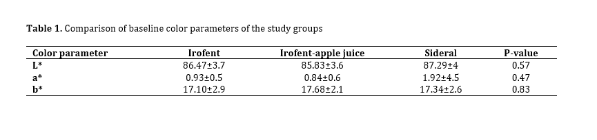
Table 1. Comparison of baseline color parameters of the study groups
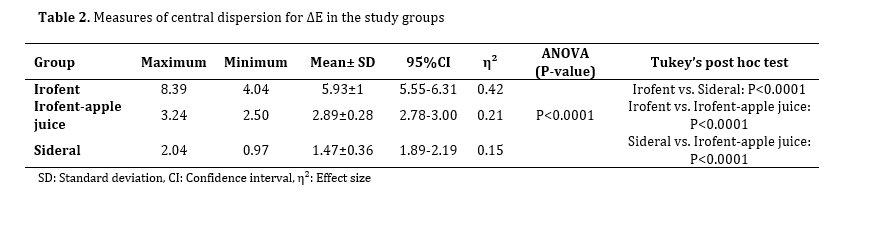
Table 2. Measures of central dispersion for ∆E in the study groups
Full-Text: (4 Views)
Abstract
Background and Aim: Discoloration of primary teeth caused by iron supplementation often discourages parents from using it for their children. This study aimed to evaluate the effects of Sideral iron drops with a sucrosomial structure, Irofent iron drops, and Irofent diluted with natural apple juice on the color and structural integrity of primary tooth enamel.
Materials and Methods: In this in vitro study, 45 sound primary anterior teeth were randomly divided into three groups of 15 for treatment with Sideral, Irofent, and a mixture of Irofent and natural apple juice. A digital spectrophotometer measured the initial tooth color. Teeth were immersed in their respective iron solutions for 5 minutes at 37°C, rinsed with distilled water, and their final color was recorded. One sample from each group underwent scanning electron microscopy (SEM) to assess surface structural changes. Quantitative data were analyzed using one-way ANOVA. P<0.05 was considered significant.
Results: Color change (∆E) was significantly greater in the Irofent group compared to Irofent–apple juice and Sideral groups (P<0.001). The Irofent–apple juice group showed significantly more discoloration than Sideral (P<0.001). SEM revealed enamel surface cracks in Irofent and Irofent–apple juice groups; whereas the Sideral group showed no defects.
Conclusion: Sideral iron drops did not cause significant discoloration or structural damage to primary enamel in this in vitro study. Combining Irofent with natural apple juice may reduce its adverse effects.
Keywords: Tooth Discoloration; Dental Enamel; Iron; Tooth, Deciduous; Microscopy, Electron
Introduction
The discoloration caused by oral iron supplements often leads to parental complaints. Also, iron-induced discoloration may be mistaken for dental caries [1-3]. Because of this concern, some parents give up using this essential mineral supplement for their children [3-5]. The destructive effects of anemia caused by iron deficiency in children remain for years, adversely affecting the children’s mental, physical, and behavioral performance [6-8]. Irofent iron drops are commonly used in Iran due to easy access and reasonable price [1, 9-11]. However, it can change the color, structure, and hardness of primary teeth, especially anterior teeth in the esthetic zone [1, 11, 12]. To mitigate these dental side effects, clinicians often recommend diluting iron drops with fruit juice [12-14]. Clinicians recommend daily consumption of apple juice from 6 months of age [13, 14].
Recently, Sideral iron drops were introduced to the market. It has been claimed that the sucrosomial structure of Sideral prevents iron from coming into contact with teeth and causing discoloration [15]. In sucrosomial formulation, iron is covered by a phospholipid membrane. The material of the phospholipid membrane is similar to the material of the cell wall, and its iron is easily absorbed through the intestinal wall cells. Therefore, in addition to helping absorb more iron, it has a pleasant taste with minimal digestive complications [16-18]. Previous studies have documented the adverse effects of Irofent iron drops on color of primary enamel [1, 19-21]. But to the best of the authors’ knowledge, no previous study has investigated the effect of diluting iron drops with juice and Sideral iron drops on color of primary tooth enamel. Thus, the purpose of this study was to compare the effect of Irofent iron drops and its mixture with natural apple juice, as well as Sideral iron drops, on the color and structure of primary tooth enamel.
Materials and Methods
This in vitro study was approved by the ethics committee of the university (IR.IAU.DENTAL.REC.1398.022). The sample size was calculated based on a study by Sahebnazar et al. [15] using one-way ANOVA. The effect size (Cohen’s f) was estimated to be approximately 0.4, and based on one-way ANOVA power analysis (α=0.05, power=90%, number of groups=3), the minimum required sample size was determined to be 15 samples per group.
A total of 45 sound primary anterior teeth, including maxillary and mandibular incisors and canines, which had been extracted due to orthodontic reasons or natural exfoliation (tooth mobility) and did not have any caries, cracks, fracture, restoration, or coronal hypoplasia, were selected after examination under a stereomicroscope (Olympus, Tokyo, Japan) at x10 magnification [1, 4, 11, 20]. Also, less than 3 months had passed since their extraction [10, 11]. The teeth were stored in 0.9% saline at room temperature. To assess color change (∆E) after exposure to iron drops compared with baseline (before exposure), the baseline color of primary teeth was measured using VITA Easy-Shade compact spectrophotometer (Vita Zahnfabrik, Bad Säckingen, Germany) [22]. To prepare the samples, the teeth were dissected at the cementoenamel junction with a diamond disc (Jota, Switzerland), the pulp tissue was completely removed, and the pulp chamber was filled with wax [1, 20, 23, 24]. Then, a paper label was placed on the labial surface of the teeth with dimensions of 4 x 4 mm, and the surrounding area was covered with colorless nail varnish [1, 11, 20]. Then, the paper label was removed and the baseline color was evaluated by VITA Easy-Shade spectrophotometer in daylight [2, 25, 26]. Before measuring the color of the samples, the spectrophotometer (Vita Zahnfabrik, Bad Säckingen, Germany) was calibrated using the reference block. To evaluate the tooth color, the handpiece probe was placed at a 90-degree angle in contact with the surface of the center of the tooth in ambient light, and then L*1, b*1 and a*1 values of the CIE L*a*b* color space were recorded. Spectrophotometric color measurements were repeated three times for each sample, and the mean value was recorded. The intra-examiner reliability was assessed by reassessing 10% of the samples, yielding an intraclass correlation coefficient of >0.90, indicating excellent repeatability [27].
After measuring the baseline (primary) color of the teeth, 45 samples were randomly divided into 3 groups of 15. Random numbers were used in PASS software to randomly distribute teeth in groups. The examiner who performed color measurements was blinded to the group assignment. Blinding was maintained until statistical analysis was completed [28].
Using the one-way ANOVA statistical test, lack of difference in baseline color of the study groups was ensured. Before the exposure of teeth to the iron drops in the groups, the pH of the solutions was measured twice by a pH-meter (Metrohm 827 pH lab, Germany). The pH of Sideral (SiderAL, Junia Pharma, Italy) was 4.74±0.02, the pH of Irofent (Kharazmi Pharmaceutical Co., Iran) was 1.98±0.02, the pH of apple juice alone was 4.47±0.03, and the pH of the combination of Irofent (Kharazmi Pharmaceutical Co., Iran) and apple juice was 2.52±0.03. The study groups were as follows:
Group 1: The samples were exposed to 15 drops of Irofent (Kharazmi Pharmaceutical Co., Iran).
Group 2: The samples were exposed to a combination of 15 drops of Irofent with 15 drops of filtered natural apple juice. To prepare apple juice, red apples were peeled and passed through a strainer 5-6 times until it became completely smooth and clear.
Group 3: The samples were exposed to 15 drops of Sideral iron drops (Junia Pharma, Italy).
The samples of each group were placed in the respective volume of iron drops for 5 minutes at a temperature of 37°C in a shaker incubator (IKA, Roentgen, Germany) [1, 7, 15, 21, 29, 30]. After 5 minutes, the samples in all groups were removed from the solution and washed with distilled water [7, 11]. The samples were blindly re-evaluated by VITA Easy-Shade spectrophotometer in the same lighting conditions as before, and the L*2, a*2 , and b*2 parameters were recorded [2, 25, 26].
L* represents the degree of lightness, which is between 0 (black) and 100 (white); a* and b* are signs of chroma and show the degree of greenness (negative a*) and redness (positive a*), and the degree of blueness (negative b*) and yellowness (positive b*) [6, 31]. The magnitude of ∆E in each sample was calculated using the following formula [2, 25, 31]:
∆E = [ (L1-L2)2 + (a1-a2)2 + (b1-b2)2]1/2
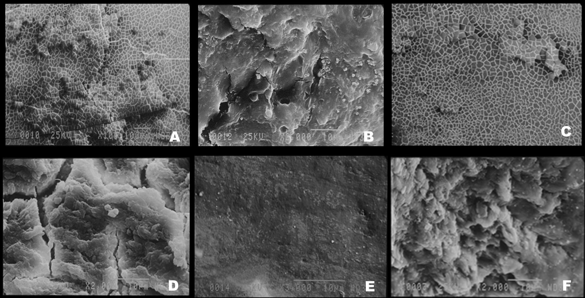
From each group, one sample was randomly selected for examination under a scanning electron microscope (SEM). For this purpose, the sample was connected to a cylindrical gold ingot and subjected to gold sputter coating in a coating machine (Fine Coat Ion Sputter JFC-1100; JEOL Ltd., Tokyo, Japan) by the gas plasma discharge method in an argon gas environment [32]. The samples were coated with gold and placed inside the special chamber of SEM (JEOL company, model JXA-840, Japan) with a magnification range of 10 to 100,000 times to examine the enamel surface structure [33]. Finally, the SEM images were recorded by a photographic camera (Mamiya, Japan) [33]. The normality of data distribution was verified using the Shapiro–Wilk test (W range: 0.95–0.98, P>0.10 for all variables), and homogeneity of variances was confirmed with the Levene’s test (P>0.20). Thus, comparisons were made with one-way ANOVA, followed by Tukey’s post-hoc test for multiple comparisons using SPSS version 22. P<0.05 was considered statistically significant [11].
Results
A total of 45 sound primary anterior teeth, including maxillary and mandibular incisors and canines, which had been extracted due to orthodontic reasons or natural exfoliation (tooth mobility) and did not have any caries, cracks, fracture, restoration, or coronal hypoplasia, were selected after examination under a stereomicroscope (Olympus, Tokyo, Japan) at x10 magnification [1, 4, 11, 20]. Also, less than 3 months had passed since their extraction [10, 11]. The teeth were stored in 0.9% saline at room temperature. To assess color change (∆E) after exposure to iron drops compared with baseline (before exposure), the baseline color of primary teeth was measured using VITA Easy-Shade compact spectrophotometer (Vita Zahnfabrik, Bad Säckingen, Germany) [22]. To prepare the samples, the teeth were dissected at the cementoenamel junction with a diamond disc (Jota, Switzerland), the pulp tissue was completely removed, and the pulp chamber was filled with wax [1, 20, 23, 24]. Then, a paper label was placed on the labial surface of the teeth with dimensions of 4 x 4 mm, and the surrounding area was covered with colorless nail varnish [1, 11, 20]. Then, the paper label was removed and the baseline color was evaluated by VITA Easy-Shade spectrophotometer in daylight [2, 25, 26]. Before measuring the color of the samples, the spectrophotometer (Vita Zahnfabrik, Bad Säckingen, Germany) was calibrated using the reference block. To evaluate the tooth color, the handpiece probe was placed at a 90-degree angle in contact with the surface of the center of the tooth in ambient light, and then L*1, b*1 and a*1 values of the CIE L*a*b* color space were recorded. Spectrophotometric color measurements were repeated three times for each sample, and the mean value was recorded. The intra-examiner reliability was assessed by reassessing 10% of the samples, yielding an intraclass correlation coefficient of >0.90, indicating excellent repeatability [27].
After measuring the baseline (primary) color of the teeth, 45 samples were randomly divided into 3 groups of 15. Random numbers were used in PASS software to randomly distribute teeth in groups. The examiner who performed color measurements was blinded to the group assignment. Blinding was maintained until statistical analysis was completed [28].
Using the one-way ANOVA statistical test, lack of difference in baseline color of the study groups was ensured. Before the exposure of teeth to the iron drops in the groups, the pH of the solutions was measured twice by a pH-meter (Metrohm 827 pH lab, Germany). The pH of Sideral (SiderAL, Junia Pharma, Italy) was 4.74±0.02, the pH of Irofent (Kharazmi Pharmaceutical Co., Iran) was 1.98±0.02, the pH of apple juice alone was 4.47±0.03, and the pH of the combination of Irofent (Kharazmi Pharmaceutical Co., Iran) and apple juice was 2.52±0.03. The study groups were as follows:
Group 1: The samples were exposed to 15 drops of Irofent (Kharazmi Pharmaceutical Co., Iran).
Group 2: The samples were exposed to a combination of 15 drops of Irofent with 15 drops of filtered natural apple juice. To prepare apple juice, red apples were peeled and passed through a strainer 5-6 times until it became completely smooth and clear.
Group 3: The samples were exposed to 15 drops of Sideral iron drops (Junia Pharma, Italy).
The samples of each group were placed in the respective volume of iron drops for 5 minutes at a temperature of 37°C in a shaker incubator (IKA, Roentgen, Germany) [1, 7, 15, 21, 29, 30]. After 5 minutes, the samples in all groups were removed from the solution and washed with distilled water [7, 11]. The samples were blindly re-evaluated by VITA Easy-Shade spectrophotometer in the same lighting conditions as before, and the L*2, a*2 , and b*2 parameters were recorded [2, 25, 26].
L* represents the degree of lightness, which is between 0 (black) and 100 (white); a* and b* are signs of chroma and show the degree of greenness (negative a*) and redness (positive a*), and the degree of blueness (negative b*) and yellowness (positive b*) [6, 31]. The magnitude of ∆E in each sample was calculated using the following formula [2, 25, 31]:
∆E = [ (L1-L2)2 + (a1-a2)2 + (b1-b2)2]1/2
From each group, one sample was randomly selected for examination under a scanning electron microscope (SEM). For this purpose, the sample was connected to a cylindrical gold ingot and subjected to gold sputter coating in a coating machine (Fine Coat Ion Sputter JFC-1100; JEOL Ltd., Tokyo, Japan) by the gas plasma discharge method in an argon gas environment [32]. The samples were coated with gold and placed inside the special chamber of SEM (JEOL company, model JXA-840, Japan) with a magnification range of 10 to 100,000 times to examine the enamel surface structure [33]. Finally, the SEM images were recorded by a photographic camera (Mamiya, Japan) [33]. The normality of data distribution was verified using the Shapiro–Wilk test (W range: 0.95–0.98, P>0.10 for all variables), and homogeneity of variances was confirmed with the Levene’s test (P>0.20). Thus, comparisons were made with one-way ANOVA, followed by Tukey’s post-hoc test for multiple comparisons using SPSS version 22. P<0.05 was considered statistically significant [11].
Results
The study was conducted on 45 samples in 3 groups (n=15) of Sideral, Irofent, and Irofent-apple juice. There was no statistically significant difference in L*1, a*1, and b*1 among the three groups at the beginning of the study as shown by one-way ANOVA (Table 1). Table 2 shows measures of central dispersion for ∆E of the study groups. One-way ANOVA showed a significant difference in ∆E among the study groups (P<0.0001). The results of pairwise comparisons showed that the ∆E of the Irofent group was significantly higher than the Irofent-natural apple juice group (P<0.0001), and the Irofent-natural apple juice group showed a significant difference with the Sideral group in this regard (P<0.0001). In other words, ∆E was significantly higher in the Irofent group than the Irofent-natural apple juice group, and ∆E of the Irofent-natural apple juice group was significantly higher than that of the Sideral group.
A ∆E greater than 3.3 was considered clinically perceptible [28]. In our study, the ∆E of the samples exposed to Irofent exceeded this threshold, whereas the ∆E of those exposed to Irofent-apple juice or Sideral remained below this threshold.
SEM findings are shown in Figure 1 (a, b, c, d, e, f). As can be seen, in teeth that were exposed to Irofent iron drops and Irofent iron drops plus natural apple juice, numerous microcracks, fractures and increased porosity were visible (Figure 1a and 1c); whereas such cracks were not seen in teeth exposed to Sideral iron drops (Figure 1e).
Table 1. Comparison of baseline color parameters of the study groups
Table 2. Measures of central dispersion for ∆E in the study groups
Given the prevalence of anemia among Iranian children and the necessity of iron intake during infancy, which overlaps with the eruption of primary teeth, it is imperative to identify a supplement that does not adversely affect dental health. Recent innovations, like Sideral iron drops with sucrosomial formulation, promise to avoid the negative dental impacts seen with other iron drops. Diluting iron drops to mitigate side effects has also been suggested. This study examined the effects of Irofent iron drops, Sideral iron drops, and a combination of Irofent with natural apple juice on color and enamel structural changes of primary teeth. The findings revealed that while all groups exhibited discoloration, the Irofent group showed significantly more discoloration than the Irofent-natural apple juice group, and more than the Sideral group. Mehran et al. [1] found similar results, indicating that Irofent caused more discoloration and structural alterations in primary teeth than other iron drops in their study. Pani et al. [19] and Shojaipour et al.[23] also reported increased discoloration with iron drops. The inclusion of citric acid in iron supplements, intended to enhance iron absorption, contributes to increased enamel solubility due to its acidic nature [21]. Irofent's low pH of 1.98 exacerbates this effect [10]. Studies by Hekmatfar et al. [30] and Babaei et al. [21] support these findings, showing that Irofent's lower pH leads to a more acidic environment and increased enamel erosion. Shabzendedar et al. [20] Mehran et al. [1] and Nazemisalman et al. [28] further corroborate the link between iron supplement acidity and enamel discoloration.
The discoloration observed with Irofent is likely due to its acidic pH (1.98) and the presence of ferrous iron (Fe²⁺), which, upon contact with enamel and environmental oxygen, can oxidize and precipitate as ferric hydroxide or oxide compounds. These compounds, particularly Fe(OH)₃ and Fe₂O₃, deposit on the enamel surface and cause yellowish-brown to dark discoloration. Furthermore, in the presence of sulfur-containing compounds (from saliva or proteins), ferrous iron can react to form black-colored ferrous sulfide (FeS), further intensifying the discoloration. The low-pH environment can also enhance enamel demineralization, increasing surface roughness and facilitating stain retention [28].
The sucrosomial formulation of Sideral iron drops encloses ferric pyrophosphate within a phospholipid bilayer coated with a sucrester matrix, forming a “sucrosome.” This structure mimics the intestinal epithelium, allowing paracellular absorption via M-cells without direct contact with the oral cavity. Unlike liposomal iron, which uses only a phospholipid bilayer, sucrosomial iron adds a sugar-based outer layer, enhancing stability, bioavailability, and mucosal tolerance [34].
Moreover, Sideral's formulation excludes citric acid and instead includes milk proteins and tricalcium phosphate, which may help buffer its effects on enamel and reduce mineral loss [16]. Its higher pH (4.74) also contributes to its milder effect on teeth [34].
Diluting Irofent with natural apple juice (pH 4.47) significantly reduced enamel discoloration compared to undiluted Irofent. Although iron absorption is generally improved with acidic beverages like fruit juices, apple juice and orange juice are traditionally recommended [21]. Scientific literature suggests that apple juice offers similar absorption efficacy with potentially less erosive damage to enamel. As daily apple juice intake is often recommended from 6 months of age, its use as a diluent may reduce Irofent's acidity and minimize enamel contact, thereby mitigating discoloration [21]. However, industrial acidic juices, with added preservatives and low pH, are not suitable for this purpose [21].
In the present study, the ∆E value was 5.93 for Irofent, 2.89 for Irofent–apple juice, and 1.47 for Sideral. Clinically, a ∆E above 3.3 is perceptible to the human eye, suggesting that both Sideral and the diluted form of Irofent are more acceptable alternatives in terms of esthetics [35].
SEM analysis revealed increased porosity, microcracks, and enamel surface degradation in the Irofent and Irofent–apple juice groups, whereas the Sideral group exhibited minimal surface alterations. These structural changes can be attributed to the acidic pH of Irofent (1.98), which promotes enamel demineralization and softening, particularly in primary teeth with thinner and less mineralized enamel. The low pH facilitates the dissolution of hydroxyapatite crystals, leading to surface roughening and microstructural damage. The diluted Irofent still exhibited damage, although to a lesser extent, due to its moderately acidic environment. In contrast, Sideral’s higher pH (4.74) and the protective nature of its sucrosomial coating reduce direct contact of iron with the enamel surface, minimizing both chemical erosion and physical degradation. These observations support the hypothesis that acidity and formulation type are key contributors to enamel damage observed under SEM. These findings are consistent with observations by Mehran et al. [1] and Pasdar et al. [10], who also noted more severe enamel changes following Irofent exposure.
One potential limitation of this study that raised concerns was the short 5-minute exposure time used for iron application. We acknowledge that this timeframe does not replicate the cumulative and intermittent exposure occurring clinically. However, this 5-minute protocol has been used in multiple in vitro studies [1,10,21,29,30], and provides a standardized method for simulating acute exposure and allowing meaningful comparison between formulations under controlled conditions. Since no clear standard exists for exposure duration under in vitro conditions of studies on drug–tooth interactions, many authors have adopted 5 minutes as a practical, reproducible model for evaluating discoloration and surface effects [1, 9, 10, 15, 21, 29, 30].
Sideral’s lower iron concentration (7 mg/mL) compared to Irofent (25 mg/mL) raises the question of whether its reduced discoloration is solely due to the sucrosomial structure or also due to lower iron content. Future studies should address this confounding variable through controlled-dose comparisons.
Extrapolating in vitro results to clinical outcomes remains challenging due to the absence of standard guidelines. Factors such as salivary flow, buffering capacity, dietary habits, and oral hygiene may modulate the extent of discoloration observed in real-world use. While some studies report widespread esthetic complaints among children using iron supplements, the clinical relevance of in vitro-based discoloration remains under investigation [21].
Conclusion
Within the limitations of this short-term in vitro study and the small SEM subsample, the following results were obtained: Irofent iron drops caused a distinct enamel discoloration, whereas Sideral iron drops resulted in only slight discoloration that remained below the clinically perceptible threshold (ΔE < 3.3), suggesting that Sideral may represent a safer alternative. Dilution of Irofent iron drops with natural apple juice showed a potential to further reduce discoloration; however, this effect requires confirmation in future in vivo studies before clinical recommendations can be made.
Type of Study: Original article |
Subject:
pediatric
References
1. Mehran M, Bassir MM, Jafari S. Effect of two kinds of iron drops on the discoloration, atomic absorption and structural changes of primary teeth enamel. J Dent Med. 2009 Mar;21(4):200-7.
2. AlSaleh S, Labban M, AlHariri M, Tashkandi E. Evaluation of self shade matching ability of dental students using visual and instrumental means. J Dent. 2012 Jul;40:e82-7. [DOI:10.1016/j.jdent.2012.01.009] [PMID]
3. Öner Özdaş D, Akkuş Güldü G, Akdağ B, Zorlu S, Tosun T. Use of laser for discoloration of primary teeth due to Iron supplement syrup. Lasers Dent Sci. 2025 Apr;9(1):15. [DOI:10.1007/s41547-025-00290-4]
4. Khosravi H, Hosseinzadeh Shamsi Anar M, Shariati Bafghi S, Mozaffari Khosravi V. Prevalence of eating disorders and obesity in high school girl students in Yazd, 2010-2011. Toloo-e-Behdasht. 2011;10(1):38-49.
5. Christofides A, Asante KP, Schauer C, Sharieff W, Owusu‐Agyei S, Zlotkin S. Multi‐micronutrient Sprinkles including a low dose of iron provided as microencapsulated ferrous fumarate improves haematologic indices in anaemic children: a randomized clinical trial. Matern Child Nutr. 2006 Jul;2(3):169-80. [DOI:10.1111/j.1740-8709.2006.00060.x] [PMID] [PMCID]
6. FesharakiNia A, SharifZadeh G. Effective factors on mothers' performance regarding supplementary iron-drop taking by their children in Birjand. J Birjand Univ Med Sci. 2006 Oct;13(3):9-15.
7. Sulieman M. An overview of tooth discoloration: extrinsic, intrinsic and internalized stains. Dent Update. 2005 Oct;32(8):463-71. [DOI:10.12968/denu.2005.32.8.463] [PMID]
8. Gingoyon A, Borkhoff CM, Koroshegyi C, Mamak E, Birken CS, Maguire JL, et al. Chronic iron deficiency and cognitive function in early childhood. Pediatrics. 2022 Dec;150(6):e2021055926. [DOI:10.1542/peds.2021-055926] [PMID]
9. Tabari M, Alaghemand H, Rabiee M, Khefri S, Ahadi MS, Nikpour MR. The effect of silicone oil and nano-hydroxyapatite/chitosan powder on microhardness and surface structure of primary teeth enamel after iron drop consumption. J Dent Sch. 2013 Jul;31(2):138-47.
10. Pasdar N, Alaghehmand H, Mottaghi F, Tavassoli M. Experimental study of iron and multivitamin drops on enamel microhardness of primary tooth. J Int Soc Prev Community Dent. 2015 Nov;5(6):518-24. [DOI:10.4103/2231-0762.170524] [PMID] [PMCID]
11. Rad AST, Tavassoli-Hojjati S, Hoda RS, Aghaei S. Efficacy of remineralizing agents for prevention of microhardness reduction and change in mineral content of enamel in anterior primary teeth after exposure to iron drop. J Dent. 2025 Jun;26(2):112.
12. Nunn JH, Ng SK, Sharkey I, Coulthard M. The dental implications of chronic use of acidic medicines in medically compromised children. Pharm World Sci. 2001 Jun;23(3):118-9. [DOI:10.1023/A:1011202409386] [PMID]
13. Hotz C, Gibson R. Complementary feeding practices and dietary intakes from complementary foods amongst weanlings in rural Malawi. Eur J Clin Nutr. 2001 Oct;55(10):841-9. [DOI:10.1038/sj.ejcn.1601239] [PMID]
14. Parratt JA, Tighe MP. Medicines update: oral iron for the management of iron deficiency anaemia in infants and children. Arch Dis Child Educ Pract Ed. 2025 Dec;110(6):284-8. [DOI:10.1136/archdischild-2024-327253] [PMID]
15. Sahebnazar N, Tavassoli-Hojjati S, Aghaei S. Effect of Sucrosomial® Iron and Iron Drop Diluted with Natural Fruit Juice on Microhardness of Primary Enamel. Front Dent. 2022 Oct;19:35. [DOI:10.18502/fid.v19i35.11247] [PMID] [PMCID]
16. Gomez-Ramirez S, Brilli E, Tarantino G, Munoz M. Sucrosomial® iron: a new generation iron for improving oral supplementation. Pharmaceuticals (Basel). 2018 Oct;11(4):97. [DOI:10.3390/ph11040097] [PMID] [PMCID]
17. Micheletto M, Gaio E, Tedesco E, Di Maira G, Mantovan E, Zanella M, et al. Intestinal absorption study of a granular form of ferric pyrophosphate. Metabolites. 2022 May;12(5):463. [DOI:10.3390/metabo12050463] [PMID] [PMCID]
18. Szostak‐Paluch K, Drabik D, Jędruchniewicz N, Dwornikowska‐Dąbrowska M. In vitro studies of a novel liposomal formulation for safe and efficient iron delivery. Eur J Lipid Sci Technol. 2024 Feb;126(2):2300217. [DOI:10.1002/ejlt.202300217]
19. Pani SC, Alenazi FM, Alotain AM, Alanazi HD, Alasmari AS. Extrinsic tooth staining potential of high dose and sustained release iron syrups on primary teeth. BMC Oral Health. 2015 Aug;15(1):90. [DOI:10.1186/s12903-015-0072-0] [PMID] [PMCID]
20. Shabzendehdar M, Makarem A, Orafai H, Khashayarmanesh Z, Ebrahimzadeh S. Comparsion of primary enamel discoloration caused by the use of three different iron drops (An in vitro study). Journal of Mashhad Dental School. 2006;30(3, 4):247-54.
21. Babaei N, Molaei T, Belyad S, Hekmatfar S. Relationship of pH and the viscosity of five different iron supplements with the absorption of iron ions and enamel discoloration in the anterior primary teeth (an in vitro study). Dental Research Journal. 2021;18(1):7-8. [DOI:10.4103/1735-3327.310036] [PMID] [PMCID]
22. Tayebi S, Esmaeilzade M, Rezai LS, Fotovat F, Vosogh RN, Faregh N. Color change of primary teeth following using 4 types of iron supplements available in the Iranian Pharmacopeia. Avicenna Journal of Dental Research. 2019;11(2):66-71. [DOI:10.34172/ajdr.2019.12]
23. Shojaipour R, Khazaeli P, Mahmodi T. Adsorption rate of iron onto primary incisor teeth following the application of three iron drops. Journal of Kerman University of Medical sciences. 2010;16(1):42-84.
24. Alkayakh T, Aldarewesh A. Effect of Dental Posts used in Restoring Badly Broken Primary teeth. Libyan Journal of Medical Research. 2024;18(1):65-71. [DOI:10.54361/LJM18-1.07]
25. Lee B-S, Huang S-H, Chiang Y-C, Chien Y-S, Mou C-Y, Lin C-P. Development of in vitro tooth staining model and usage of catalysts to elevate the effectiveness of tooth bleaching. Dental materials. 2008;24(1):57-66. [DOI:10.1016/j.dental.2007.01.012] [PMID]
26. Kutkut N, Jordi M, Almalki A, Conejo J, Anadioti E, Blatz M. Comparison of the accuracy and reliability of instrumental shade selection devices and visual shade selection: An in vitro study. Journal of Esthetic and Restorative Dentistry. 2025;37(2):477-84. [DOI:10.1111/jerd.13311] [PMID]
27. Lokhande RS, Nagarathna P, Deoghare A, Chhatani N, Busi S, Malladi S. Effect of Different Iron Supplements on Color Stability of Nanocomposite Restorative Materials. International Journal of Clinical Pediatric Dentistry. 2024;17(3):274. [DOI:10.5005/jp-journals-10005-2790] [PMID] [PMCID]
28. Nazemisalman B, Mohseni M, Darvish S, Farsadeghi M, Luchian I. Effects of Iron Salts on Demineralization and Discoloration of Primary Incisor Enamel Subjected to Artificial Cariogenic Challenge versus Saline Immersion. Healthcare. 2023;11(4):14. [DOI:10.3390/healthcare11040569] [PMID] [PMCID]
29. Salman NR, El-Tekeya MM, Bakry NS, Soliman S. Remineralization effect of fluoride varnish containing casein phosphopeptide amorphous calcium phosphate on caries-like lesions in primary teeth (in vitro study). Alexandria Dental Journal. 2019;44(1):13-6. [DOI:10.21608/adjalexu.2019.57568]
30. Hekmatfar S, Piraneh H, Jafari K. Evaluation of the relationship between pH and titrable acidity of five different of iron supplements with the absorption of iron ions in the anterior primary teeth (an in vitro study). Dental research journal. 2018;15(5):367. [DOI:10.4103/1735-3327.240473] [PMID] [PMCID]
31. Naffah N, Ounsi H, Ozcan M, Salameh Z. Evaluation of the color stability of three resin-ceramic materials using a spectrophotometer and a digital photography software. Contemporary Clinical Dentistry. 2024;15(1):44-50. [DOI:10.4103/ccd.ccd_656_18] [PMID] [PMCID]
32. Awad MM, Albedaiwi L, Almahdy A, Khan R, Silikas N, Hatamleh MM, et al. Effect of universal adhesives on microtensile bond strength to hybrid ceramic. BMC oral health. 2019;19:1-7. [DOI:10.1186/s12903-019-0865-7] [PMID] [PMCID]
33. Atalay C, Koc Vural U, Miletic I, Gurgan S. Shear bond strengths of two newly marketed self‐adhesive resin cements to different substrates: a light and scanning electron microscopy evaluation. Microscopy research and technique. 2022;85(5):1694-702. [DOI:10.1002/jemt.24031] [PMID]
34. Giordano G, Napolitano M, Di Battista V, Lucchesi A. Oral high-dose sucrosomial iron vs intravenous iron in sideropenic anemia patients intolerant/refractory to iron sulfate: a multicentric randomized study. Annals of Hematology. 2021;100(9):2173-9. [DOI:10.1007/s00277-020-04361-3] [PMID] [PMCID]
35. Ozgur B, Unverdi GE, Ertan A, Cehreli Z. Effectiveness and color stability of resin infiltration on demineralized and hypomineralized (MIH) enamel in children: six-month results of a prospective trial. Operative dentistry. 2023;48(3):258-67. [DOI:10.2341/22-041-C] [PMID]
Send email to the article author
| Rights and permissions | |
 |
This work is licensed under a Creative Commons Attribution-NonCommercial 4.0 International License. |