

BibTeX | RIS | EndNote | Medlars | ProCite | Reference Manager | RefWorks
Send citation to:
URL: http://jrdms.dentaliau.ac.ir/article-1-103-en.html


2- Oral & Maxillofacial Pathologist, Toronto, Canada.
3- Assistant Professor, Oral & Maxillofacial Pathology Dept, Dental Branch of Tehran, Islamic Azad University, Tehran, Iran. ,
4- Dentist
5- Student of Dentistry, Dental Branch of Tehran, Islamic Azad University, Tehran, Iran.
Abstract
Background & aim: Increase of micronucleus in buccal mucosa and it’s correlation with cancerogenesis is now considerable. Recently, occupation in dental laboratories is propounded as an affective factor. So this study was aimed to evaluate the effect of occupational exposure on buccal mucosa cells in dental laboratories technicians.
Material & methods: This historical-cohort study was conducted on 16 male dental laboratories technicians and 16 male persons as control group. All samples were matched through age and based on exclusions criteria following as recent viral patient, use of special medicine, smoke cigarette and drink alcohol, dangerous occupation and history of radiotherapy. Buccal mucosal cells are sampled by plastic spatula and they were stained through Papanicolaou method and the micronucleus frequencies were evaluated by light microscope under 400 magnification. T-test statistical analysis were used and significant level of was considered lower than 0.05
Result: The micronucleus frequencies were 69±70 and 27±8.6 in case and control samples, respectively; which was 2.6 times more. T-test statistical analysis showed that the difference of micronucleus frequencies was significant. (P<0.001)
Conclusion: This study showed that the frequency of cells with micronucleus in buccal mucosa of occupied people in dental laboratories are 2.6 times more than the unoccupied individuals. So being in this profession by itself may be a risk factor to induction of oral malignant transformation.
Key words: micronucleus, oral cancer, dental laboratories technician
Introduction
One of notable apprehensions of dental laboratories technicians is scattering dust of their job and settle expose with organic solvent that it’s an important trouble for technician’s health(1). Dental technicians use various materials in laboratory procedures of dental prosthesis. Metal alloys (Cobalt, Chromium, and Nickel) and monomers and polymers based on MMA (methyl methacrylate) are used in most of dental laboratories. The combination of Chromium and Nickel is known to be correlated with lung cancer. Also Cobalt and its combinations may be carcinogenic, too (2). Exposure to MMA is companion with asthma, contact dermatitis and headache (3). On the other hand, Silzbach et al. reported pneumoconiosis in dental technicians (4). Therefore dental technicians are recommended to use mask, eye keeper and public and local ventilation systems (5).
Micronucleus (MN) is very small nucleus body which is divided from main nucleus in interphase session during cell cycle. MNs are separated from main nucleus and still added to cell main nucleus and they also have complete chromosomes or pieces of them. MNs are one of the biological signs of genotoxicity in human erythrocytes, lymphocytes, reticulocytes and buccal mucosal cells. The increase in MNs rate has been a criterion for measuring and diagnosing of aneugenicity and clastogenicity (3-5) and also for studying genotoxicity of various chemicals (6-8). Therefore evaluating of MN has used for studying clastogenes and anuploidgenes effects after injuries caused by occupational and peripheral factors in human epidemiology and animal experiment (9).
Many researches have been studied the effects and disadvantages of laboratory materials that show various results (9, 10). There is no comprehensive study based on our knowledge of existing references, in assessment of the diagnosing value of MN assessing test of buccal mucosa in dental technicians.
Attending the lack of information and regarding to the importance of early diagnosing of DNA injury leading to cancerogenesis, this study was aimed to evaluate frequency of MNs in buccal mucosa of dental technician, in dental branch of Tehran, Islamic Azad University at 2013.
Material & methods
This historical-cohort study was designed following as a pilot study with the confidence rate of 95% and power of analysis of 90%. 16 dental laboratories technicians and 16 no occupied persons were chosen as case and control groups, respectively. After taking the ethical committee agreement, the case group was selected from dental laboratory staff with at least 1 year history of working in laboratories in Dental branch of Tehran, Islamic Azad University. The laboratory was equipped with ordinary ventilation systems. The case and control groups were men in order to exclude gender parameter and selected from the people whom have never been in laboratories living in Tehran and these groups were matched through age, and socio-economic factors. Information was collected and registered after getting consents for participating in this study. Individuals with recent viral disease, taking specific medications, smoking cigarette, using alcohol, addiction to narcotics, risky occupations and radiotherapy exposures were excluded.
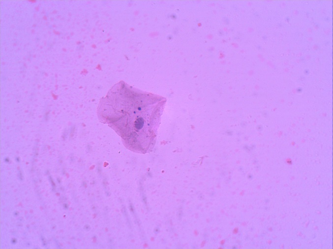
Then, the case samples were requested to wash their mouth with water before sampling their buccal cells. These cells were scarped by wet plastic spatula and scattered on microscopic clean glass slides and the smears were fixed by Pathofix spray ( Padtan Teb co., Tehran, Iran), dried in room temperature then papanicolaou staining was used for cytologic assessment. MN assessment was done using light microscope with 400 magnification. Criteria used by Talbert et al. were also used for MN’s evaluating (11). 500 cells were counted for each samples and frequency of MN was considered in each samples.(12) (Fig 1)
Statistical analysis using T-test was approached on the average frequency of MN. Significant level lower than 0.05 was set in this study.
Fig1: Papanicolaou staining of buccal mucosa showed a cell with MNs using light microscope with 400 magnification.
Result
The present study was conducted on 16 male dental laboratory technicians with age average of 36±19 and 16 male control samples with age average of 39±88 years old. Frequency of MN in buccal mucosa cells was shown in table 1.
Table1. Frequency of MN in buccal mucosa cells
The micronucleus frequencies were 69±70 and 27±8.6 in case and control samples, respectively; which was 2.6 times more than the control group and T-test showed that the difference of MN frequency was significant (P<0.001).
Variation coefficient or homogenous rate of MN in both groups was approximately the same and it was about 25%.
Discussion
.jpg)
The present study showed the frequency of MN was significantly higher than control samples. Scattering of dust made by technical and procedures and exposure with organic solutions can be affect dental technician’s health (1). Different materials are used for prosthetic procedures that contain metal alloys (cobalt, chromium, nickel) and monomers and MMA-base polymers. Exposure with combinations of chromium and nickel was reported to correlation with cancer. It is also studied that cobalt and its combinations may be carcinogenic to human (2).
Attending to expensive and complicated treatment procedures, also prolonged and challenging rehabilitation of malignant lesions, nowadays, research methods are based on early diagnosis and more important, screening of high risk population relating to oral cancerogenesis. On the other hand, the non-invasive methods as micronucleus assessment of buccal mucosa may be helpful in detection of any nuclear changes can be indicator of dysplastic alteration of oral mucosa before clinical manifestations. Therefore this study was based on assessment of MN frequency in buccal mucosa of occupied individuals in dental laboratories and it showed that the average percent of cells with MN in case group was more than control one significantly. This finding is in line with a previous study showing that individuals, who work in dental laboratories, presented more MN frequencies in their cells (2, 3, 4). Azhar et al. studied cell toxicity of methyl methacrylate monomers in dental technicians using MN test in buccal mucous. They studied people who work dental prostheses producing laboratories and they had been exposed with Methylmethacrylate and it was concluded that MN frequency in occupied individuals doesn’t differ from unoccupied ones (10). It was also shown in other studies that the occupation period is an important and dangerous factor which increases the MN rate (13).
Use of convenient ventilation system can be the eventual reason for the difference between our study and Azhar’s. Another important issue is the consideration of safety principles like washing the hands and using masks and gloves in laboratory. Washing the hands in an effective way to decrease the skin absorption of organic solutions and monomers (10). Ishikawa et al. study showed that washing hands in work environment decreases the rate of MN significantly however use of gloves doesn’t have significant effect. Although, in this study individuals use surgical mask which are convenient for protecting against liquid while using filtered and gas masks is recommended for gas and dusts. In spite of that, he suggested that using mask is better than not using them (9). Also, that Ishikawa’s study had been done on peripheral blood cells not mucobuccal cells and its showed that occupation as a dental technician was risk factor that significantly increased MN frequency (14).
Staining methods used in diagnosing MNs in exofoliative cells of oral mucous varies in different researches and contain Fleulgen-fast green fluorescent colors such as diamindo-2-phenylnodole DAPI, acridine orange, ropidium iodide, aygrunwald Giemsa, papanicolaou(14). Fleulgen-fast green is more used by researches due to its specificity for DNA and its clear coloring of cytoplasm that makes it easier to count MNs (15). While papanicolaou coloring is also used in many recent studies (10, 16, 17) and results were acceptable. This method doesn’t need complex processes such as cell culturing and metaphase preparation and not even DNA specific coloring. In Peradeep et al. research MMA prevented cell growth even in 1 micro liter concentration. Decrease the number of vital cells and increases cell death (18) which support the toxic effect of this material. On the other hand, controversies are found in some studies according to the role of gender, that some of them show more MN rate in men and some of them in women (19-21) that was not comparable with the present study which all samples were men in order to exclude gender variable. Final, it seems majority of the previous studies in line with our results concluded in increasing of MN frequency of buccal mucosa cells in this population. However, another research with greater sample volume is recommended.
Conclusion
This study showed that the average percent of cells with MN in buccal mucosa of occupied people in dental laboratories are significantly higher than control samples. So, being in this profession by itself may be a risk factor to induction of nuclear changes of oral mucosa which increased susceptibility of oral malignant transformation.
| Rights and permissions | |
 |
This work is licensed under a Creative Commons Attribution-NonCommercial 4.0 International License. |