Journal of Research in Dental
and Maxillofacial Sciences
Volume 11, Issue 1 (3-2026)
J Res Dent Maxillofac Sci 2026, 11(1): 22-30 |
Back to browse issues page

Ethics code: 1401.122
Download citation:
BibTeX | RIS | EndNote | Medlars | ProCite | Reference Manager | RefWorks
Send citation to:


BibTeX | RIS | EndNote | Medlars | ProCite | Reference Manager | RefWorks
Send citation to:
Roshani F, Jabarifard K, Babaei N, Hekmatfar S. Effect of Different Irrigating Solutions on Pushout Bond Strength of Composite Posts in Primary Teeth: An In Vitro Study. J Res Dent Maxillofac Sci 2026; 11 (1) :22-30
URL: http://jrdms.dentaliau.ac.ir/article-1-986-en.html
URL: http://jrdms.dentaliau.ac.ir/article-1-986-en.html


1- Dental Faculty, Ardabil University of Medical Sciences, Ardabil, Iran
2- Dentist in Private Practice, Ardabil, Iran
3- Department of Pediatric Dentistry, School of Dentistry, Ardabil University of Medical Sciences, Ardabil, Iran ,hekmatfar24@gmail.com
2- Dentist in Private Practice, Ardabil, Iran
3- Department of Pediatric Dentistry, School of Dentistry, Ardabil University of Medical Sciences, Ardabil, Iran ,
Full-Text [PDF 452 kb]
(22 Downloads)
| Abstract (HTML) (42 Views)
Table 1. Measures of central dispersion for the PBS (MPa) of primary anterior teeth in the study groups
Table 2. Pairwise comparison of the irrigant groups regarding their PBS (MPa)
Table 3. Frequency distribution of the failure modes of the composite bond to intracanal dentin in primary anterior teeth
Discussion
A modulus of elasticity range (16-25 GPa) similar to that of dentin, which results in distribution of forces along the root axis, optimal sealing of dentin surface against intraoral fluids, and optimal esthetics make composite resins a favorable restorative material for both patients and dental clinicians [17,18]. Many factors influence the bond strength to dentin, such as generation of the bonding agent, percentage of mineralization, and dentin microstructure. Inter-tubular dentin plays an important role in bond strength as micromechanical retention is due to the penetration of the bonding agent into the collagen of inter-tubular dentin. Dentin structure of the primary root canal contains more tubules and less inter-tubular dentin; therefore, the bond strength of composite to dentin is much more challenging following endodontic treatment [19]. This study evaluated the effect of dentin pretreatment with CHX, saline, NaOCl, and EDTA on PBS of composite post bonded to primary dentin using a self-etch (7th generation) adhesive. Universal adhesives were introduced to overcome the shortcomings of the former generations of one-step self-etch adhesives. They can be used in total-etch and selective-etch modes. In vitro [20] and in vivo [21] studies show acceptable results of universal adhesives. In the present study, a self-etch adhesive system was used for all specimens. In non-cooperative children, it simplifies and shortens the bonding process and is suitable for use. Bahrololoomi and Mehravar [10] recommended a universal adhesive system and a 6th generation self-etch adhesive for bonding of composite resin posts in primary anterior teeth. A common mistake when using a 5th generation adhesive is that collagen fibers collapse when drying the dentin in the etch and rinse protocol, which can prevent adequate penetration of resin monomers, and subsequently reduce the bond strength. Afshar et al. [9] evaluated the PBS of a conventional composite along with 5th, 6th, and 7th generation bonding agents to intracanal dentin of primary anterior teeth, and reported that no statistically significant difference was noted among the groups in this respect. However, the mean bond strength was higher than that in the present study. This difference can be attributed to the different method of preparation of the specimens, and that they did not perform thermocycling. According to Meshki et al. [22], the PBS of 8th generation bonding agents was higher than that of 5th, 6th, and 7th generation bonding agents, especially in self-etch mode compared to total-etch mode, for bonding of composite posts to dentin in primary anterior teeth. Endodontic treatment of the root canal includes mechanical instrumentation and chemical cleaning by using irrigants. The characteristics of irrigants have an influence on bond strength to root canal dentin [23, 24]. NaOCl is the most frequently used chemical irrigant due to its lubrication, disinfection, and tissue dissolution properties, and removal of organic components [25]. Bassir et al. [11] reported that 3.3% NaOCl and 2% CHX irrigants significantly increased the PBS of dentin in primary anterior teeth. In the present study, the NaOCl pretreatment group showed a significantly higher mean PBS. The reason for this result may be that NaOCl changes the organic-inorganic ratio by removing the organic components mainly from the collagen matrix, and enhances the bond strength due to improved resin penetration [26, 27]. Some studies found that 5.25% NaOCl oxidizes some parts of root canal dentin, leading to incomplete polymerization of adhesive resin [28,29]. The wide variation in the results of studies may be explained by the differences in methodologies. The tissue dissolution capacity and debridement properties of NaOCl can be changed by temperature, concentration, and exposure time [30, 31]. CHX, as a cationic biguanide, has an optimal antimicrobial effect at a pH of approximately 5.5 to 7.0; it is adsorbed by the microorganisms’ cell wall, leading to the breakdown of intracellular components [32]. Ng et al. [33] reported a significant increase in the bond strength of fiber posts cemented by resin cements following root canal irrigation with CHX. Therefore, CHX adsorption by dentin causes the penetration of resin into the dentinal tubules, possibly causing a high bond strength. Besides, it has been reported that 0.1-2% CHX can effectively inhibit matrix metalloproteinases, which reduces the degradation of the adhesive interface between composite resin and dentin [33,34].
In the present study, the PBS of CHX and EDTA groups was significantly higher than that of the saline group. According to a study by Retana-Lobo et al. [34], dentin collagen matrix degradation happens because of the slow active release of matrix metalloproteinase-2 (or other proteolytic enzymes) from dentin matrices subjected to mineralization during storage. Milani et al. [16] and Qamar et al. [35] showed that application of CHX increased the bond strength of composite restorations to dentinal walls. In the present study, 10% EDTA was used for 60 seconds before bonding, and as a result, the PBS significantly increased compared to the saline group. EDTA, as a carboxylic acid, has organic materials, which can chelate calcium ions and remove hydroxyapatite with no change in the fibrillar structure of the collagen network or collagen denaturation [36]. The findings of previous reports are also contradictory in this regard. Wang et al. [37] reported a decrease in bond strength using 15% EDTA; whereas, Kasraei et al [38] observed an elevation in the mean bond strength. Variations in the results can be due to the EDTA pH, concentration, and application duration. A concentration of 1.5%-5% is not effective for removal of the smear layer; while, a concentration of 15% used for a longer duration causes complete dissolution of the smear layer, significant widening of the dentinal tubules, and increases the depth of demineralization [39]. Luque-Martinez et al. [40] reported that EDTA increased the immediate microtensile bond strength of sclerotic dentin substrate and prevented degradation when associated with a self-etch adhesive. A strong decrease in the microtensile bond strength was reported after conditioning with 0.5 M EDTA for 5 days. Probably, the chelating effect of EDTA will cause the resin to penetrate the unchanged collagen and increase the bond strength. Since resin penetration can be done only to a certain depth of dentin, continuation of the process of removing minerals and exposing collagen for a longer time by conditioning with EDTA can create a thick hybrid layer with low mineral support, which can be responsible for the reduction in bond strength. This effect seems to be more noticeable in universal adhesives whose mechanism of action is through micromechanical retention of collagen and exposed dentinal tubules [41,15].
The failure mode analysis indicated that most failures were adhesive type and in the saline group, which supported the PBS results. The specimens with low PBS showed adhesive failure. Some studies have confirmed that the failure mode in primary enamel and dentin is usually adhesive and mixed [42,43]. Afshar et al. [9] found that the failure modes of composite to intracanal dentin of primary teeth were significantly different between the total-etch and two-step self-etch systems.
This study had some limitations. In vitro setting can poorly simulate the oral environment. On the other hand, the force applied to the teeth in the oral cavity is a combination of shear, tensile, and compressive forces. The present study only evaluated the PBS through the application of shear force. Thus, generalization of the results to the clinical setting must be done with caution.
Conclusion
The results of this study showed that pretreatment with NaOCl enhanced the PBS of composite posts to root dentin in primary anterior teeth.
Full-Text: (10 Views)
Abstract
Background and Aim: Restoration of anterior teeth is challenging in pediatric dentistry due to the poor retention of restorative material and the small size of the teeth. This study was conducted to investigate the pushout strength (PBS) of intra-canal composite posts in primary anterior teeth after using different root canal irrigants.
Materials and Methods: In this in vitro experimental study, 44 extracted primary anterior teeth were randomly assigned to four groups of (I) sodium hypochlorite (NaOCl), (II) ethylenediaminetetraacetic acid (EDTA), (III) saline, and (IV) chlorhexidine (CHX). Tooth crowns were cut by a high-speed handpiece 1 mm above the cementoenamel junction. After cleaning and shaping, the root canals were obturated, and post spaces were prepared. Composite resin was packed in the 3 mm coronal third of the canal. The teeth were thermocycled, and each tooth was sectioned into 1 mm slices at the mid-root. Next, the specimens were subjected to PBS testing. The mode of failure was determined under a stereomicroscope at ×40 magnification. Data were analyzed using one-way ANOVA and Games-Howell test (alpha=0.05).
Results: Saline showed the lowest (1.09±0.36 MPa), and NaOCl exhibited the highest (8.57±0.15 MPa) PBS (P<0.001). The difference in PBS between the EDTA and CHX groups was not statistically significant (P=0.115). Adhesive failure was more frequent than other failure modes.
Conclusion: The results showed that root canal pretreatment with NaOCl enhanced the PBS of composite posts in primary anterior teeth.
Keywords: Composite Resins; Tooth, Deciduous; Sodium Hypochlorite
Introduction
Early childhood caries often affects the maxillary central and lateral incisors, as well as the maxillary and mandibular first molars, and can destroy the coronal structure of the teeth [1]. Restoration of damaged primary anterior teeth is challenging [2]. The success of primary anterior tooth restoration is influenced by many factors, such as thin enamel, small size of teeth and bonding surface area, age, cooperation of the child, and cost of treatment. Direct restoration with bonding materials is the most common treatment for primary anterior teeth. Composite resins are always preferred for this purpose, due to their optimal esthetics, strengthening of the remaining tooth structure, dentin bonding capacity, and affordability [3,4]. When the remaining tooth structure is insufficient, it would be challenging to provide a restoration with optimal longevity and acceptable esthetics [2]. The use of intracanal posts increases the durability of restorations. Intracanal posts should be fabricated in some cases to achieve sufficient durability of restoration, stabilize the composite crown, and provide sufficient resistance to masticatory forces [5,6]. Composite posts are usually fabricated following pulpectomy by pushing the composite into the canal to obtain a tapered intracanal post [7]. Dentinal tubules have a greater diameter and density, and less mineral content in primary teeth than in permanent teeth [8]. Thus, dental materials used in primary teeth should be able to overcome these limitations and provide maximum retention to ensure optimal durability of composite restorations. A recent development in dentin bonding agents reduced the number of procedural steps from three in the fourth-generation systems to one step in the seventh- and eighth-generation systems [9]. One-step systems simplify the bonding process and appear to be effective for young children with poor cooperation [10].
Ethylenediaminetetraacetic acid (EDTA), chlorhexidine (CHX), and sodium hypochlorite (NaOCl) are the most commonly used endodontic irrigants that provide antimicrobial activity, lubrication, smear layer removal, and tissue dissolution action. The ability of irrigants to create a chemical interaction with dentinal walls may lead to changes in the bond strength of composite posts to dentin [11,12]. The negative effects of NaOCl on the polymerization of resin monomers have been previously investigated [13]. There are also controversial reports on the effects of CHX on the bond strength of adhesives [14]. EDTA causes chelation of calcium and potassium ions, and reduces the modulus of elasticity and flexural strength of dentin, which affects the properties of dentin and ultimately increases the risk of root fracture [15]. Considering all the above, the present study aimed to evaluate the pushout strength (PBS) of intracanal composite posts in primary anterior teeth after using different endodontic irrigants.
Materials and Methods
This in vitro study was performed on 44 primary anterior teeth, extracted due to orthodontic reasons. Ethical approval was received from the Ethics Committee of Ardabil University of Medical Sciences. (IR.ARUMS.REC.1401.122).
The sample size was calculated using the following formula:

Where α=0.05, Z(1-β)=1.28 Z(1-α/2)=1.96, µ1=5.28, µ2=8.40, S1=2.84, and S2=1.26.
Teeth with erosion and/or caries, and those that had undergone endodontic treatment, were excluded. All included teeth had at least two-thirds of the root length. The teeth were stored in saline solution, which was refreshed once a week. Before the experiment, they underwent disinfection by immersion in 0.5% chloramine T solution (Merck KGaA, Darmstadt, Germany) for one week. The tooth crowns were cut at high speed, and under water coolant 1 mm above their cementoenamel junction using a fissure diamond bur (No. 138; Jota AG, Switzerland), and their apex was sealed using a light-cure liner to provide an apical seal, keep the root filling material in the root canal, and standardize the specimens during their preparation. Complete pulpectomy was performed along with irrigation of the canal with saline. The roots were dried with paper points (Meta Biomed, Korea) and then filled with zinc oxide eugenol (Kemdent, UK). The zinc oxide eugenol was covered by one layer (about 1 mm) of self-cure glass ionomer cement (GC, Japan). A post space was prepared 5 mm apical to the cutting surface (4 mm apical to the cementoenamel junction) for placement of composite posts [16]. The specimens were divided into four groups (n=11) of (I) 1% NaOCl, (II) 10% EDTA (Cobalt, Iran), (III) 0.9% saline, and (IV) 2% CHX (Behsa, Iran).
The irrigation procedures were performed for 1 min, and then self-etch adhesive (G-Premio BOND; GC Corp., Japan) was applied on the root canal walls, followed by gentle air-drying for 5 seconds, and light-curing for 10 seconds (as instructed). The root canals were obturated with a conventional composite resin (Tetric N-Ceram; Ivoclar Vivadent, Schaan, Liechtenstein) in all groups using the incremental technique with 2-mm increments, and 20 seconds of light-curing was performed for all layers. The root canals were filled with composite resin until reaching the orifice. Next, the teeth underwent thermocycling for 500 cycles (TC300; Vafaei Industrial, Iran) at 5-55°C (dwell time of 20 seconds, transfer time of 10 seconds). The specimens were mounted in cold-cure acrylic resin blocks. Each tooth was sectioned into 1 mm thick slices at the mid-root (Figure 1). Both sides of all slices were subjected to photography using a Sony camera (Cyber-Shot DSC-HX100v, Japan), and the composite cross-sectional area was calculated. The camera position was the same for all sections. The PBS was measured in a universal testing machine (2050; Zwick/Roell, Ulm, Germany). The specimens were subjected to a force at a crosshead speed of 0.5 mm/min in an apico-coronal direction [10]. Maximum load to the target area causing composite debonding was recorded in Newtons (N). The obtained load in Newtons was divided by the cross-sectional area in square millimeters (mm²) to determine the PBS in megapascals (MPa). The following formula was used to calculate the cross-sectional area:
𝑆 = (A1+A2) × (h/2)
Background and Aim: Restoration of anterior teeth is challenging in pediatric dentistry due to the poor retention of restorative material and the small size of the teeth. This study was conducted to investigate the pushout strength (PBS) of intra-canal composite posts in primary anterior teeth after using different root canal irrigants.
Materials and Methods: In this in vitro experimental study, 44 extracted primary anterior teeth were randomly assigned to four groups of (I) sodium hypochlorite (NaOCl), (II) ethylenediaminetetraacetic acid (EDTA), (III) saline, and (IV) chlorhexidine (CHX). Tooth crowns were cut by a high-speed handpiece 1 mm above the cementoenamel junction. After cleaning and shaping, the root canals were obturated, and post spaces were prepared. Composite resin was packed in the 3 mm coronal third of the canal. The teeth were thermocycled, and each tooth was sectioned into 1 mm slices at the mid-root. Next, the specimens were subjected to PBS testing. The mode of failure was determined under a stereomicroscope at ×40 magnification. Data were analyzed using one-way ANOVA and Games-Howell test (alpha=0.05).
Results: Saline showed the lowest (1.09±0.36 MPa), and NaOCl exhibited the highest (8.57±0.15 MPa) PBS (P<0.001). The difference in PBS between the EDTA and CHX groups was not statistically significant (P=0.115). Adhesive failure was more frequent than other failure modes.
Conclusion: The results showed that root canal pretreatment with NaOCl enhanced the PBS of composite posts in primary anterior teeth.
Keywords: Composite Resins; Tooth, Deciduous; Sodium Hypochlorite
Introduction
Early childhood caries often affects the maxillary central and lateral incisors, as well as the maxillary and mandibular first molars, and can destroy the coronal structure of the teeth [1]. Restoration of damaged primary anterior teeth is challenging [2]. The success of primary anterior tooth restoration is influenced by many factors, such as thin enamel, small size of teeth and bonding surface area, age, cooperation of the child, and cost of treatment. Direct restoration with bonding materials is the most common treatment for primary anterior teeth. Composite resins are always preferred for this purpose, due to their optimal esthetics, strengthening of the remaining tooth structure, dentin bonding capacity, and affordability [3,4]. When the remaining tooth structure is insufficient, it would be challenging to provide a restoration with optimal longevity and acceptable esthetics [2]. The use of intracanal posts increases the durability of restorations. Intracanal posts should be fabricated in some cases to achieve sufficient durability of restoration, stabilize the composite crown, and provide sufficient resistance to masticatory forces [5,6]. Composite posts are usually fabricated following pulpectomy by pushing the composite into the canal to obtain a tapered intracanal post [7]. Dentinal tubules have a greater diameter and density, and less mineral content in primary teeth than in permanent teeth [8]. Thus, dental materials used in primary teeth should be able to overcome these limitations and provide maximum retention to ensure optimal durability of composite restorations. A recent development in dentin bonding agents reduced the number of procedural steps from three in the fourth-generation systems to one step in the seventh- and eighth-generation systems [9]. One-step systems simplify the bonding process and appear to be effective for young children with poor cooperation [10].
Ethylenediaminetetraacetic acid (EDTA), chlorhexidine (CHX), and sodium hypochlorite (NaOCl) are the most commonly used endodontic irrigants that provide antimicrobial activity, lubrication, smear layer removal, and tissue dissolution action. The ability of irrigants to create a chemical interaction with dentinal walls may lead to changes in the bond strength of composite posts to dentin [11,12]. The negative effects of NaOCl on the polymerization of resin monomers have been previously investigated [13]. There are also controversial reports on the effects of CHX on the bond strength of adhesives [14]. EDTA causes chelation of calcium and potassium ions, and reduces the modulus of elasticity and flexural strength of dentin, which affects the properties of dentin and ultimately increases the risk of root fracture [15]. Considering all the above, the present study aimed to evaluate the pushout strength (PBS) of intracanal composite posts in primary anterior teeth after using different endodontic irrigants.
Materials and Methods
This in vitro study was performed on 44 primary anterior teeth, extracted due to orthodontic reasons. Ethical approval was received from the Ethics Committee of Ardabil University of Medical Sciences. (IR.ARUMS.REC.1401.122).
The sample size was calculated using the following formula:
Where α=0.05, Z(1-β)=1.28 Z(1-α/2)=1.96, µ1=5.28, µ2=8.40, S1=2.84, and S2=1.26.
Teeth with erosion and/or caries, and those that had undergone endodontic treatment, were excluded. All included teeth had at least two-thirds of the root length. The teeth were stored in saline solution, which was refreshed once a week. Before the experiment, they underwent disinfection by immersion in 0.5% chloramine T solution (Merck KGaA, Darmstadt, Germany) for one week. The tooth crowns were cut at high speed, and under water coolant 1 mm above their cementoenamel junction using a fissure diamond bur (No. 138; Jota AG, Switzerland), and their apex was sealed using a light-cure liner to provide an apical seal, keep the root filling material in the root canal, and standardize the specimens during their preparation. Complete pulpectomy was performed along with irrigation of the canal with saline. The roots were dried with paper points (Meta Biomed, Korea) and then filled with zinc oxide eugenol (Kemdent, UK). The zinc oxide eugenol was covered by one layer (about 1 mm) of self-cure glass ionomer cement (GC, Japan). A post space was prepared 5 mm apical to the cutting surface (4 mm apical to the cementoenamel junction) for placement of composite posts [16]. The specimens were divided into four groups (n=11) of (I) 1% NaOCl, (II) 10% EDTA (Cobalt, Iran), (III) 0.9% saline, and (IV) 2% CHX (Behsa, Iran).
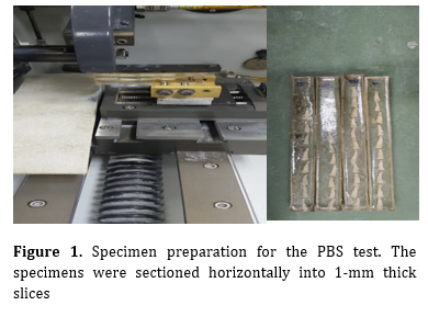
The irrigation procedures were performed for 1 min, and then self-etch adhesive (G-Premio BOND; GC Corp., Japan) was applied on the root canal walls, followed by gentle air-drying for 5 seconds, and light-curing for 10 seconds (as instructed). The root canals were obturated with a conventional composite resin (Tetric N-Ceram; Ivoclar Vivadent, Schaan, Liechtenstein) in all groups using the incremental technique with 2-mm increments, and 20 seconds of light-curing was performed for all layers. The root canals were filled with composite resin until reaching the orifice. Next, the teeth underwent thermocycling for 500 cycles (TC300; Vafaei Industrial, Iran) at 5-55°C (dwell time of 20 seconds, transfer time of 10 seconds). The specimens were mounted in cold-cure acrylic resin blocks. Each tooth was sectioned into 1 mm thick slices at the mid-root (Figure 1). Both sides of all slices were subjected to photography using a Sony camera (Cyber-Shot DSC-HX100v, Japan), and the composite cross-sectional area was calculated. The camera position was the same for all sections. The PBS was measured in a universal testing machine (2050; Zwick/Roell, Ulm, Germany). The specimens were subjected to a force at a crosshead speed of 0.5 mm/min in an apico-coronal direction [10]. Maximum load to the target area causing composite debonding was recorded in Newtons (N). The obtained load in Newtons was divided by the cross-sectional area in square millimeters (mm²) to determine the PBS in megapascals (MPa). The following formula was used to calculate the cross-sectional area:
𝑆 = (A1+A2) × (h/2)
Figure 1. Specimen preparation for the PBS test. The specimens were sectioned horizontally into 1-mm thick slices
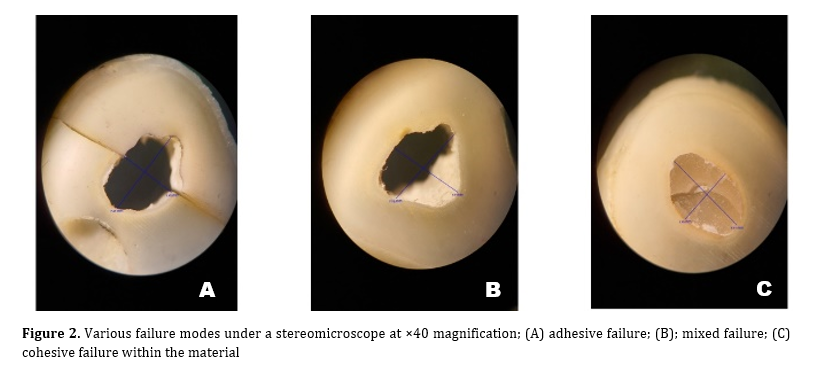
Where A1 is the apical section perimeter of the root canal, H is the height of the root cross-section, S is the cross-sectional area in square millimeters (mm²), and A2 is the root canal coronal section perimeter. The failure mode was determined under a stereomicroscope (Olympus, Japan) at ×40 magnification and classified as: mixed, adhesive, or cohesive (within dentin or composite).
Finally, the PBS values were collected and statistically analyzed. The Kolmogorov-Smirnov test was used to examine the normality of the distribution of the PBS data. One-way ANOVA and Games-Howell test were applied to compare the PBS among and between the groups, respectively (alpha=0.05).
Results
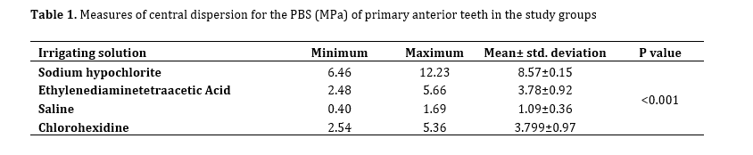
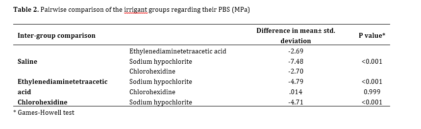
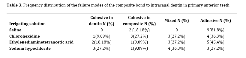
One-way ANOVA showed a significant difference in PBS among the groups (P<0.001; Table 1). Thus, pairwise comparisons were conducted using the Games-Howell test (Table 2). The NaOCl pretreatment group showed a significantly higher PBS than all other groups (P<0.001). The saline group showed a significantly lower PBS than all other groups (P<0.001). The CHX group showed no significant difference compared to the EDTA group (P=0.115). The frequency percentage of failure modes in the four groups is shown in Table 3. The adhesive failure mode had the highest frequency. Cohesive failure in dentin was the least common failure mode (Figure 2).
Where A1 is the apical section perimeter of the root canal, H is the height of the root cross-section, S is the cross-sectional area in square millimeters (mm²), and A2 is the root canal coronal section perimeter. The failure mode was determined under a stereomicroscope (Olympus, Japan) at ×40 magnification and classified as: mixed, adhesive, or cohesive (within dentin or composite).
Finally, the PBS values were collected and statistically analyzed. The Kolmogorov-Smirnov test was used to examine the normality of the distribution of the PBS data. One-way ANOVA and Games-Howell test were applied to compare the PBS among and between the groups, respectively (alpha=0.05).
Results
One-way ANOVA showed a significant difference in PBS among the groups (P<0.001; Table 1). Thus, pairwise comparisons were conducted using the Games-Howell test (Table 2). The NaOCl pretreatment group showed a significantly higher PBS than all other groups (P<0.001). The saline group showed a significantly lower PBS than all other groups (P<0.001). The CHX group showed no significant difference compared to the EDTA group (P=0.115). The frequency percentage of failure modes in the four groups is shown in Table 3. The adhesive failure mode had the highest frequency. Cohesive failure in dentin was the least common failure mode (Figure 2).
Table 1. Measures of central dispersion for the PBS (MPa) of primary anterior teeth in the study groups
Table 2. Pairwise comparison of the irrigant groups regarding their PBS (MPa)
Table 3. Frequency distribution of the failure modes of the composite bond to intracanal dentin in primary anterior teeth
Discussion
A modulus of elasticity range (16-25 GPa) similar to that of dentin, which results in distribution of forces along the root axis, optimal sealing of dentin surface against intraoral fluids, and optimal esthetics make composite resins a favorable restorative material for both patients and dental clinicians [17,18]. Many factors influence the bond strength to dentin, such as generation of the bonding agent, percentage of mineralization, and dentin microstructure. Inter-tubular dentin plays an important role in bond strength as micromechanical retention is due to the penetration of the bonding agent into the collagen of inter-tubular dentin. Dentin structure of the primary root canal contains more tubules and less inter-tubular dentin; therefore, the bond strength of composite to dentin is much more challenging following endodontic treatment [19]. This study evaluated the effect of dentin pretreatment with CHX, saline, NaOCl, and EDTA on PBS of composite post bonded to primary dentin using a self-etch (7th generation) adhesive. Universal adhesives were introduced to overcome the shortcomings of the former generations of one-step self-etch adhesives. They can be used in total-etch and selective-etch modes. In vitro [20] and in vivo [21] studies show acceptable results of universal adhesives. In the present study, a self-etch adhesive system was used for all specimens. In non-cooperative children, it simplifies and shortens the bonding process and is suitable for use. Bahrololoomi and Mehravar [10] recommended a universal adhesive system and a 6th generation self-etch adhesive for bonding of composite resin posts in primary anterior teeth. A common mistake when using a 5th generation adhesive is that collagen fibers collapse when drying the dentin in the etch and rinse protocol, which can prevent adequate penetration of resin monomers, and subsequently reduce the bond strength. Afshar et al. [9] evaluated the PBS of a conventional composite along with 5th, 6th, and 7th generation bonding agents to intracanal dentin of primary anterior teeth, and reported that no statistically significant difference was noted among the groups in this respect. However, the mean bond strength was higher than that in the present study. This difference can be attributed to the different method of preparation of the specimens, and that they did not perform thermocycling. According to Meshki et al. [22], the PBS of 8th generation bonding agents was higher than that of 5th, 6th, and 7th generation bonding agents, especially in self-etch mode compared to total-etch mode, for bonding of composite posts to dentin in primary anterior teeth. Endodontic treatment of the root canal includes mechanical instrumentation and chemical cleaning by using irrigants. The characteristics of irrigants have an influence on bond strength to root canal dentin [23, 24]. NaOCl is the most frequently used chemical irrigant due to its lubrication, disinfection, and tissue dissolution properties, and removal of organic components [25]. Bassir et al. [11] reported that 3.3% NaOCl and 2% CHX irrigants significantly increased the PBS of dentin in primary anterior teeth. In the present study, the NaOCl pretreatment group showed a significantly higher mean PBS. The reason for this result may be that NaOCl changes the organic-inorganic ratio by removing the organic components mainly from the collagen matrix, and enhances the bond strength due to improved resin penetration [26, 27]. Some studies found that 5.25% NaOCl oxidizes some parts of root canal dentin, leading to incomplete polymerization of adhesive resin [28,29]. The wide variation in the results of studies may be explained by the differences in methodologies. The tissue dissolution capacity and debridement properties of NaOCl can be changed by temperature, concentration, and exposure time [30, 31]. CHX, as a cationic biguanide, has an optimal antimicrobial effect at a pH of approximately 5.5 to 7.0; it is adsorbed by the microorganisms’ cell wall, leading to the breakdown of intracellular components [32]. Ng et al. [33] reported a significant increase in the bond strength of fiber posts cemented by resin cements following root canal irrigation with CHX. Therefore, CHX adsorption by dentin causes the penetration of resin into the dentinal tubules, possibly causing a high bond strength. Besides, it has been reported that 0.1-2% CHX can effectively inhibit matrix metalloproteinases, which reduces the degradation of the adhesive interface between composite resin and dentin [33,34].
In the present study, the PBS of CHX and EDTA groups was significantly higher than that of the saline group. According to a study by Retana-Lobo et al. [34], dentin collagen matrix degradation happens because of the slow active release of matrix metalloproteinase-2 (or other proteolytic enzymes) from dentin matrices subjected to mineralization during storage. Milani et al. [16] and Qamar et al. [35] showed that application of CHX increased the bond strength of composite restorations to dentinal walls. In the present study, 10% EDTA was used for 60 seconds before bonding, and as a result, the PBS significantly increased compared to the saline group. EDTA, as a carboxylic acid, has organic materials, which can chelate calcium ions and remove hydroxyapatite with no change in the fibrillar structure of the collagen network or collagen denaturation [36]. The findings of previous reports are also contradictory in this regard. Wang et al. [37] reported a decrease in bond strength using 15% EDTA; whereas, Kasraei et al [38] observed an elevation in the mean bond strength. Variations in the results can be due to the EDTA pH, concentration, and application duration. A concentration of 1.5%-5% is not effective for removal of the smear layer; while, a concentration of 15% used for a longer duration causes complete dissolution of the smear layer, significant widening of the dentinal tubules, and increases the depth of demineralization [39]. Luque-Martinez et al. [40] reported that EDTA increased the immediate microtensile bond strength of sclerotic dentin substrate and prevented degradation when associated with a self-etch adhesive. A strong decrease in the microtensile bond strength was reported after conditioning with 0.5 M EDTA for 5 days. Probably, the chelating effect of EDTA will cause the resin to penetrate the unchanged collagen and increase the bond strength. Since resin penetration can be done only to a certain depth of dentin, continuation of the process of removing minerals and exposing collagen for a longer time by conditioning with EDTA can create a thick hybrid layer with low mineral support, which can be responsible for the reduction in bond strength. This effect seems to be more noticeable in universal adhesives whose mechanism of action is through micromechanical retention of collagen and exposed dentinal tubules [41,15].
The failure mode analysis indicated that most failures were adhesive type and in the saline group, which supported the PBS results. The specimens with low PBS showed adhesive failure. Some studies have confirmed that the failure mode in primary enamel and dentin is usually adhesive and mixed [42,43]. Afshar et al. [9] found that the failure modes of composite to intracanal dentin of primary teeth were significantly different between the total-etch and two-step self-etch systems.
This study had some limitations. In vitro setting can poorly simulate the oral environment. On the other hand, the force applied to the teeth in the oral cavity is a combination of shear, tensile, and compressive forces. The present study only evaluated the PBS through the application of shear force. Thus, generalization of the results to the clinical setting must be done with caution.
Conclusion
The results of this study showed that pretreatment with NaOCl enhanced the PBS of composite posts to root dentin in primary anterior teeth.
Type of Study: Original article |
Subject:
pediatric
Send email to the article author
| Rights and permissions | |
 |
This work is licensed under a Creative Commons Attribution-NonCommercial 4.0 International License. |