Journal of Research in Dental
and Maxillofacial Sciences
Volume 10, Issue 2 (6-2025)
J Res Dent Maxillofac Sci 2025, 10(2): 88-95 |
Back to browse issues page


Ethics code: IR.MUBABOL.REC.1399.486
Download citation:
BibTeX | RIS | EndNote | Medlars | ProCite | Reference Manager | RefWorks
Send citation to:


BibTeX | RIS | EndNote | Medlars | ProCite | Reference Manager | RefWorks
Send citation to:
Maleki S, Khodadadi E, Mahmoudi E, Seyedmajidi S. In Vitro Cleaning Efficacy of Neolix and M3 Immatural Rotary Files in Comparison with Hand Files in Primary Molar Root Canals. J Res Dent Maxillofac Sci 2025; 10 (2) :88-95
URL: http://jrdms.dentaliau.ac.ir/article-1-903-en.html
URL: http://jrdms.dentaliau.ac.ir/article-1-903-en.html


1- Student Research Committee, Babol University of Medical Sciences, Babol, Iran.
2- Dental Materials Research Center, Health Research Institute, Babol University of Medical Sciences, Babol, Iran. ,dr_ekhodadadi@yahoo.com
3- Oral Health Research Center, Health Research Institute, Babol University of Medical Sciences, Babol, Iran.
4- Dental Materials Research Center, Health Research Institute, Babol University of Medical Sciences, Babol, Iran.
2- Dental Materials Research Center, Health Research Institute, Babol University of Medical Sciences, Babol, Iran. ,
3- Oral Health Research Center, Health Research Institute, Babol University of Medical Sciences, Babol, Iran.
4- Dental Materials Research Center, Health Research Institute, Babol University of Medical Sciences, Babol, Iran.
Full-Text [PDF 701 kb]
(532 Downloads)
| Abstract (HTML) (1725 Views)
Background and Aim: This study assessed the cleaning efficacy of Neolix and M3 Immatural rotary files compared with hand K-files in primary molar root canals.
Materials and Methods: This in vitro study evaluated 40 primary maxillary and mandibular molars. After access cavity preparation, Indian ink was injected into the root canals of primary molars. The teeth were then randomly divided into three experimental and one control group (n=30 canals per group). In group 1, the root canals were instrumented with #20 M3 Immatural file and then with #25/4% to the working length. In group 2, the root canals were instrumented with #25/6% Neoniti A1 file. In group 3, hand K-files were used for instrumentation of the mesial canals of mandibular molars and buccal canals of maxillary molars to #25, and palatal canals of maxillary molars and distal canals of mandibular molars to #30. Group 4 served as the control group and was only rinsed with saline with no instrumentation. After clearing, the teeth were inspected under a stereomicroscope. Data were analyzed by the Kruskal-Wallis and Mann-Whitney U tests.
Results: No significant difference was noted among the groups in cleaning of the coronal third of the root canals (P=0.174). In the middle and apical thirds, two rotary systems showed significantly superior cleaning compared with hand files (P=0.031 and P=0.007, respectively), but there was no significant difference between the two rotary files (P>0.05).
Conclusion: Neolix and M3 Immatural files can serve as efficient alternatives to hand files for pulpectomy of primary molars.
Keywords: Instrumentation; Pulpectomy; Root Canal Therapy; Tooth, Deciduous
Introduction
Full-Text: (376 Views)
Abstract
Background and Aim: This study assessed the cleaning efficacy of Neolix and M3 Immatural rotary files compared with hand K-files in primary molar root canals.
Materials and Methods: This in vitro study evaluated 40 primary maxillary and mandibular molars. After access cavity preparation, Indian ink was injected into the root canals of primary molars. The teeth were then randomly divided into three experimental and one control group (n=30 canals per group). In group 1, the root canals were instrumented with #20 M3 Immatural file and then with #25/4% to the working length. In group 2, the root canals were instrumented with #25/6% Neoniti A1 file. In group 3, hand K-files were used for instrumentation of the mesial canals of mandibular molars and buccal canals of maxillary molars to #25, and palatal canals of maxillary molars and distal canals of mandibular molars to #30. Group 4 served as the control group and was only rinsed with saline with no instrumentation. After clearing, the teeth were inspected under a stereomicroscope. Data were analyzed by the Kruskal-Wallis and Mann-Whitney U tests.
Results: No significant difference was noted among the groups in cleaning of the coronal third of the root canals (P=0.174). In the middle and apical thirds, two rotary systems showed significantly superior cleaning compared with hand files (P=0.031 and P=0.007, respectively), but there was no significant difference between the two rotary files (P>0.05).
Conclusion: Neolix and M3 Immatural files can serve as efficient alternatives to hand files for pulpectomy of primary molars.
Keywords: Instrumentation; Pulpectomy; Root Canal Therapy; Tooth, Deciduous
Introduction
Due to the existing concerns about space loss, potential orthodontic problems, and the adverse effect of premature loss of primary teeth on
children’s quality of life, pulpectomy is considered as a more conservative treatment option for primary teeth with irreversible pulpitis or necrotic pulp compared to tooth extraction [1, 2]. Pulpectomy is commonly preferred for such teeth when the root canals are accessible, and there is evidence of normal supporting bone [2, 3]. However, pulp therapy treatments often require the children’s cooperation and may necessitate multiple sessions or the use of general anesthesia [4]. An ideal pulpectomy procedure should be fast and optimally debride, clean, and shape the primary root canals without compromising the root structure or the permanent successor tooth bud [5, 6].
Preparation of primary root canals by using rotary instruments was first suggested by Barr et al. [7] in 1999, and they reported that it is an efficient technique for debridement of irregular primary root canal walls. Rotary instrumentation of the root canal system saves time, improves patient cooperation, and decreases the operator’s fatigue [2, 8]. Moreover, nickel-titanium rotary instruments preserve the original path of the curved canals in primary teeth. However, it should be noted that rotary instruments have high technical sensitivity, and the operator should acquire the necessary skills to use them [9].
Difficult access to root canals due to limited mouth opening in children often complicates the pulpectomy treatment of primary molars. Short rotary files with optimal taper are more suitable for primary teeth. M3 Immatural files (United Dental, Shanghai, China) are short-length (16 mm) rotary files available in three sizes of #20, #25, and #30 with 4% taper, which are suitable for primary teeth. They have shape memory and a convex triangular cross-section [10, 11].
Different single-file rotary systems are available in the market, which can greatly save time. Neoniti A1 (Neolix Creative Dental Instruments, Chatres, La Foret, France) is a single-file rotary system with full-rotation movement. It is available in different tip sizes of #20, #25, and #40. Heat treatment of files in this system results in their flexibility. Also, these files have a non-homogenous rectangular cross-section, which enables better instrumentation of curved canals while preserving their original anatomy [12].
Various techniques are available for assessing the cleaning efficacy of the root canal system, including scanning electron microscopy, micro-computed tomography imaging, stereomicroscopy, and longitudinal tooth sectioning for microscopic analysis [13]. Clearing of tissues is ideal for in vitro evaluation of the root canal system without damaging the tooth structure [14]. Tomar et al. [15] stated that stereomicroscopy offers a highly sensitive and reliable approach for three-dimensional evaluation of the root canal system. Additionally, this technique is more cost-effective compared to alternative methods.
Considering the gap of information regarding the cleaning efficacy of Neolix (#25-0.06) and M3 Immatural rotary files in primary root canals, this study aimed to compare the cleaning efficacy of Neolix and M3 Immatural rotary files with hand K-files for instrumentation of primary molar root canals by the clearing technique. The null hypothesis of the study was that there would be no significant difference in the cleaning efficacy of the files used in different sections of the root canal system.
Materials and Methods
This in vitro experimental study was conducted on primary molars with at least two-thirds of their sound root remaining, and a root length of 7-12 mm. The teeth had been extracted due to over-retention or severe bone resorption as noted on periapical radiographs. The selected teeth had sound crowns and roots without severe curvature, fracture, or resorption. The study protocol was approved by the ethics committee of Babol University of Medical Sciences (IR.MUBABOL.REC.1399.486), and informed consent was obtained from the parents for the use of their children’s extracted teeth for research purposes.
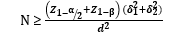
The minimum sample size was calculated to be 15 canals in each group based on the results of a previous study comparing rotary and hand files [16], with a 95% confidence level (α = 0.05), 80% study power (β = 0.2), and an effect size (d) of 0.5. The sample size was determined using the following formula:
Forty primary molars (including 20 maxillary and 20 mandibular primary molars) were evaluated in this study. The teeth were randomly divided into four groups (each group included 10 teeth and 30 canals).
The teeth were immersed in 0.5% sodium hypochlorite (Golrang, Tehran, Iran) for one week for disinfection, and stored in saline until the experiment [16].
Root canal preparation:
Access cavity was prepared in teeth using a round diamond bur (Mani Inc., Tokyo, Japan). After rinsing the root canals with saline (Samen, Mashahd, Iran), a #10 K-file (Mani Inc., Tokyo, Japan) was introduced into the canal until its tip was visible at the apex. The working length was determined 1 mm short of the apex. After introducing the file into the root canal, 1-2 mL of Indian ink was injected into the canal using an insulin syringe until ink leaked out from the apex. Ink injection was then repeated [16].
Study groups:
The teeth were randomly divided into four groups:
Group 1 (n=30 canals): Root canals were instrumented with #20 M3 Immatural file (M3; United Dental, Shanghai, China) and then #25 with 4% taper to the working length at 350 rpm with 1.5 N/cm torque by the single-length technique.
Group 2 (n=30 canals): Root canals were instrumented with Neoniti A1 (Neolix Creative Dental Instruments, Chȃtres-La, Foret, France) #25 file with 6% taper and 21 mm length at a speed of 300-500 rpm and 1.5 N/cm torque by the single-length technique.
Group 3 (n=30 canals): The mesial canals of mandibular molars and buccal canals of maxillary molars were instrumented with hand K-files (Mani Inc, Tochigi, Japan) up to #25, and the palatal canals of maxillary molars and distal canals of mandibular molars were filed up to #30 by the standard technique.
Group 4 (n=30 canals): This group served as the control group. No instrumentation was performed in this group. The canals were only rinsed with saline [17, 18].
Instrumentation with rotary files was performed using an endo-motor (Endo-Mate DT, NSK, Nakanishi Inc., Tokyo, Japan). In groups 1 and 2, the files were used with light pecking motion until the working length was reached. After using each file, the root canals were rinsed with 1 mL of saline (Samen, Mashahd, Iran). All root canal preparations were performed by the same operator.
After instrumentation, the access cavity was temporarily sealed with CavitTM (3M, Germany), and the teeth were stored in saline.
Clearing and evaluation of cleaning efficacy:
Root canal clearing was performed according to Silva et al. [19] in order to reveal the three-dimensional structure of the root canal system and assess the quality of cleaning by the files.
For the clearing process, each tooth was immersed in 10% hydrochloric acid in capped glass containers for 3 days. The acid was refreshed daily until the teeth were completely decalcified. The teeth were then rinsed under running water for 8 hours. This was followed by sequential dehydration in 70% ethanol for 16 hours (refreshed every 8 hours), 90% ethanol for 3 hours, and 100% ethanol for 3 hours (all alcohols from Merck, Darmstadt, Germany).
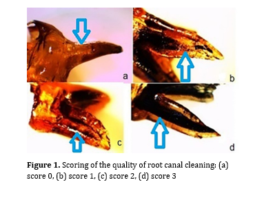
After dehydration, the teeth were immersed in methyl salicylate (Merck, Darmstadt, Germany) for final clearing. After clearing, assessment of the removal of Indian ink from the apical, middle, and cervical thirds of the root canals was performed under a stereomicroscope (Dewinter, Italy) at x10 magnification by one examiner who was blinded to the group allocation of the teeth, and scored as follows (Figure 1):
The minimum sample size was calculated to be 15 canals in each group based on the results of a previous study comparing rotary and hand files [16], with a 95% confidence level (α = 0.05), 80% study power (β = 0.2), and an effect size (d) of 0.5. The sample size was determined using the following formula:
Forty primary molars (including 20 maxillary and 20 mandibular primary molars) were evaluated in this study. The teeth were randomly divided into four groups (each group included 10 teeth and 30 canals).
The teeth were immersed in 0.5% sodium hypochlorite (Golrang, Tehran, Iran) for one week for disinfection, and stored in saline until the experiment [16].
Root canal preparation:
Access cavity was prepared in teeth using a round diamond bur (Mani Inc., Tokyo, Japan). After rinsing the root canals with saline (Samen, Mashahd, Iran), a #10 K-file (Mani Inc., Tokyo, Japan) was introduced into the canal until its tip was visible at the apex. The working length was determined 1 mm short of the apex. After introducing the file into the root canal, 1-2 mL of Indian ink was injected into the canal using an insulin syringe until ink leaked out from the apex. Ink injection was then repeated [16].
Study groups:
The teeth were randomly divided into four groups:
Group 1 (n=30 canals): Root canals were instrumented with #20 M3 Immatural file (M3; United Dental, Shanghai, China) and then #25 with 4% taper to the working length at 350 rpm with 1.5 N/cm torque by the single-length technique.
Group 2 (n=30 canals): Root canals were instrumented with Neoniti A1 (Neolix Creative Dental Instruments, Chȃtres-La, Foret, France) #25 file with 6% taper and 21 mm length at a speed of 300-500 rpm and 1.5 N/cm torque by the single-length technique.
Group 3 (n=30 canals): The mesial canals of mandibular molars and buccal canals of maxillary molars were instrumented with hand K-files (Mani Inc, Tochigi, Japan) up to #25, and the palatal canals of maxillary molars and distal canals of mandibular molars were filed up to #30 by the standard technique.
Group 4 (n=30 canals): This group served as the control group. No instrumentation was performed in this group. The canals were only rinsed with saline [17, 18].
Instrumentation with rotary files was performed using an endo-motor (Endo-Mate DT, NSK, Nakanishi Inc., Tokyo, Japan). In groups 1 and 2, the files were used with light pecking motion until the working length was reached. After using each file, the root canals were rinsed with 1 mL of saline (Samen, Mashahd, Iran). All root canal preparations were performed by the same operator.
After instrumentation, the access cavity was temporarily sealed with CavitTM (3M, Germany), and the teeth were stored in saline.
Clearing and evaluation of cleaning efficacy:
Root canal clearing was performed according to Silva et al. [19] in order to reveal the three-dimensional structure of the root canal system and assess the quality of cleaning by the files.
For the clearing process, each tooth was immersed in 10% hydrochloric acid in capped glass containers for 3 days. The acid was refreshed daily until the teeth were completely decalcified. The teeth were then rinsed under running water for 8 hours. This was followed by sequential dehydration in 70% ethanol for 16 hours (refreshed every 8 hours), 90% ethanol for 3 hours, and 100% ethanol for 3 hours (all alcohols from Merck, Darmstadt, Germany).
After dehydration, the teeth were immersed in methyl salicylate (Merck, Darmstadt, Germany) for final clearing. After clearing, assessment of the removal of Indian ink from the apical, middle, and cervical thirds of the root canals was performed under a stereomicroscope (Dewinter, Italy) at x10 magnification by one examiner who was blinded to the group allocation of the teeth, and scored as follows (Figure 1):
- Score 0: Completely clean canal with no ink remaining (Figure 1a)
- Score 1: More than 50% of the ink removed, with some remaining ink at certain points (Figure 1b)
- Score 2: Less than 50% of the ink removed, with visible ink lines in the canal (Figure 1c).
- Score 3: No ink removal (Figure 1d) [19].
Figure 1. Scoring of the quality of root canal cleaning: (a) score 0, (b) score 1, (c) score 2, (d) score 3
Statistical analysis:
Data were analyzed using SPSS version 26. For comparison of the cleaning efficacy of the groups in different parts of the roots, the Kruskal-Wallis followed by the Mann-Whitney U test, was applied at 0.05 level of significance.
Results
Statistical analysis:
Data were analyzed using SPSS version 26. For comparison of the cleaning efficacy of the groups in different parts of the roots, the Kruskal-Wallis followed by the Mann-Whitney U test, was applied at 0.05 level of significance.
Results
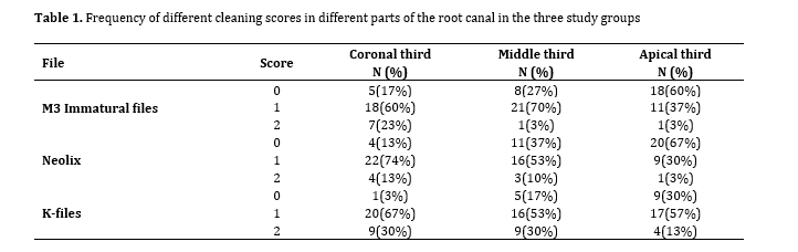
Table 1 presents the frequency of different cleaning scores. Since no cleaning was performed in the control group, all teeth in this group were scored 3.
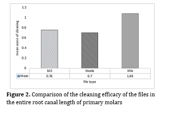
Comparison of the quality of cleaning revealed that the hand K-file group was significantly inferior to the M3 Immatural (P=0.001) and Neolix (P<0.001) groups; while no significant difference was noted in this respect between the two rotary systems (P=0.538; Figure 2).
Comparison of the quality of cleaning revealed that the hand K-file group was significantly inferior to the M3 Immatural (P=0.001) and Neolix (P<0.001) groups; while no significant difference was noted in this respect between the two rotary systems (P=0.538; Figure 2).
Figure 2. Comparison of the cleaning efficacy of the files in the entire root canal length of primary molars
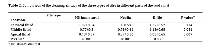
Table 2 compares the cleaning efficacy of the three types of files in different parts of the root. Comparison of the quality of cleaning in different parts of the root canals indicated no significant difference among the three groups in the coronal third (P=0.174). In the middle third, the difference was significant among the three groups (P=0.031), and hand K-files had a significantly lower performance than Neolix (P=0.024) and M3 Immatural (P=0.025) files but the two rotary systems were not significantly different in cleaning of the middle third of the root canals (P=0.711). Pairwise comparisons of the files in the apical third revealed significant differences between the hand K-files and the Neolix (P=0.004) and M3 Immatural file (P=0.015); but the two rotary systems were not significantly different in cleaning of the apical third of the root canals (P=0.611).
Table 2 compares the cleaning efficacy of the three types of files in different parts of the root. Comparison of the quality of cleaning in different parts of the root canals indicated no significant difference among the three groups in the coronal third (P=0.174). In the middle third, the difference was significant among the three groups (P=0.031), and hand K-files had a significantly lower performance than Neolix (P=0.024) and M3 Immatural (P=0.025) files but the two rotary systems were not significantly different in cleaning of the middle third of the root canals (P=0.711). Pairwise comparisons of the files in the apical third revealed significant differences between the hand K-files and the Neolix (P=0.004) and M3 Immatural file (P=0.015); but the two rotary systems were not significantly different in cleaning of the apical third of the root canals (P=0.611).
Table 1. Frequency of different cleaning scores in different parts of the root canal in the three study groups
Table 2. Comparison of the cleaning efficacy of the three types of files in different parts of the root canal
In general, the cleaning efficacy of all three files was higher in the apical third. In the hand K-file group, the difference between the coronal and middle thirds (P=0.475), or middle and apical thirds (P=0.086) was not significant; however, the cleaning efficacy was significantly higher in the apical third than the coronal third (P=0.008).
The difference in cleaning efficacy was not significant between the coronal and middle thirds in the M3 Immatural and Neolix rotary files (P=0.055 and P=0.072, respectively). However, in both the M3 Immatural file and Neolix groups, cleaning in the apical third was significantly superior to that in the coronal third (P<0.001 for both) and middle third (P=0.014 and P=0.019, respectively).
Discussion
This study assessed the cleaning efficacy of Neolix and M3 Immatural files in comparison with hand K-files. The results showed no significant difference among the three groups in the coronal third. However, in the middle and apical thirds, both rotary file systems demonstrated significantly superior cleaning compared to hand K-files, with no significant difference between the two rotary systems. Therefore, the null hypothesis of the study was rejected.
Pulpectomy is the treatment of choice for primary teeth with pulpal involvement. The success of pulpectomy in primary teeth relies on effective biomechanical instrumentation, using an appropriate obturating material with minimal voids, and achieving a strong, airtight seal [20]. The primary objective of pulpectomy is to establish an effective hermetic seal, which is influenced by several factors, including proper biomechanical preparation, type of obturating material, and the quality of obturation [21].
M3 Immatural is a multi-file rotary system designed for primary teeth. It has a constant taper of 4%, which decreases the risk of root perforation. In the present study, the cleaning efficacy of Neolix and M3 Immatural files in primary teeth was compared for the first time. The results indicated that the overall efficacy of both rotary systems was significantly higher than that of hand K-files. The present results were generally in line with those of Katge et al. [17]. They showed that DXL Pro and Prime Pedo file systems, which have been specifically designed for primary teeth, were superior to hand H-files in preparation of coronal and apical thirds of the root canals; however, the difference between the two files was not significant. In the present study, rotary files showed greater cleaning efficacy than hand K-files in the apical and middle thirds. Although both studies showed greater cleaning by rotary files, compared with hand files, fewer number of files used in the present study is an advantage, since it decreases the working time.
Jeevanandan and Govindaraju [22] in a clinical trial showed that the Kedo-S rotary file, which has been designed for primary teeth, yielded a higher obturation quality, and pulpectomy with this file had a higher success rate than K files in primary molars. Their results were in agreement with the present findings although they clinically evaluated the quality of obturation and treatment success. The files used in the present study had a lower and constant taper throughout their working length compared to files used in their study, which decreases the risk of procedural errors such as perforation. Conversely, Rathi et al. [13] reported that the Pro AF Baby Gold rotary system outperformed the Kedo-S system in terms of root canal cleaning efficacy. This difference may be attributed to variations in file design, metallurgical properties, and taper, which influence the cleaning efficiency.
While the findings of this study are consistent with those of Katge et al. [17] and Jeevanandan and Govindaraju [22], they differ from those of Katge et al. [3] and Nazari Moghaddam et al. [23]. The latter studies found no significant difference in cleaning efficacy between the rotary and hand files in the middle and apical thirds of primary molar root canals, with hand files being superior in the coronal third. Nazari Moghaddam et al. [23] found no significant difference between the efficacy of Flex Master rotary file with 4% taper and hand K-files in the middle and apical thirds of primary molar root canals. However, hand K-files were superior to rotary files only in the coronal third of the root canals. Katge et al. [3] found no significant difference between Mtwo rotary files and H-files in primary molars. This discrepancy may be attributed to several factors such as the irrigation protocol, file design, and number of instruments used, operator skills and technique, and clearing and evaluation techniques.
In the present study, only saline was used for irrigation to isolate the mechanical cleaning effect of the files, eliminating the influence of irrigants. This approach is considered a strength of the present study. In contrast, other studies used additional irrigants like sodium hypochlorite and EDTA, which may have contributed to differences in cleaning efficacy by enhancing debris removal, particularly in the coronal region [3, 23].
The current study used Neolix and M3 Immatural files, both of which have flexible structures with heat-treated properties, contributing to improved adaptation to the canal walls and debris removal. Katge et al. [3] used Mtwo rotary files, and their study involved a higher number of rotary instruments. Additionally, they used H-files instead of K-files for manual instrumentation, which may explain the observed differences.
The expertise of the operator in using hand files can influence the results. Hand K-files require a more labor-intensive technique, and their performance may vary depending on the clinician’s proficiency. The discrepancy in the results of previous studies may be due to variations in the operator technique, particularly in the use of hand files, where a more rigid cross-section could enhance coronal cleaning, as observed in the study by Nazari Moghaddam et al. [23].
The present study used the tissue clearing method with Indian ink, which provides a reliable visualization of root canal cleaning [14]. Similar methodologies were employed by Honardar et al. [24] and de Souza et al. [25], who confirmed that stereomicroscopy with the diaphanization process is a highly sensitive and cost-effective method for evaluation of cleaning efficacy. Score 3 in all canals in the control group indicated complete penetration of ink into the canals and the optimal efficacy of the clearing technique by using Indian ink. Based on the current results, the cleaning efficacy of all three file systems was higher in the apical third than the middle and coronal thirds of the root canals. Considering the fact that ink is injected into the canals by an insulin syringe through the canal orifice, the maximum concentration of ink is found in the coronal, and then the middle, and apical thirds, which may explain less cleaning of the root canals in the coronal third, as explained by Ramezanali et al. [16].
Clinical implications and limitations:
This study provides valuable insights into the mechanical cleaning efficacy of different instrumentation systems in primary molars. However, as an in vitro study, its findings should be interpreted with caution when applied to clinical settings. Factors such as irrigation dynamics, intracanal medicaments, and in vivo biological responses may influence the cleaning efficacy in actual patient scenarios. Future clinical trials are recommended to validate these results in real-world conditions.
Conclusion
The present results showed that the hand K-file group demonstrated significantly lower cleaning efficacy compared to both the M3 Immatural and Neolix rotary files, with no significant difference between the two rotary systems. Overall, all three file types performed better in the apical third of the root canal. Both M3 Immatural and Neolix rotary files showed superior cleaning efficacy in the apical third compared to the coronal and middle thirds.
Type of Study: Original article |
Subject:
Endodontics
References
1. Abanto J, Tsakos G, Olegário IC, Paiva SM, Mendes FM, Ardenghi TM, Bönecker M. Impact of pulpectomy versus tooth extraction in children's oral health-related quality of life: A randomized clinical trial. Community Dent Oral Epidemiol. 2024 Feb;52(1):13-23. [DOI:10.1111/cdoe.12895] [PMID]
2. Jeevanandan G. Kedo-S Paediatric Rotary Files for Root Canal Preparation in Primary Teeth - Case Report. J Clin Diagn Res. 2017 Mar;11(3):ZR03-5. [DOI:10.7860/JCDR/2017/25856.9508] [PMID] []
3. Katge F, Chimata VK, Poojari M, Shetty S, Rusawat B. Comparison of cleaning Efficacy and Instrumentation Time between Rotary and Manual Instrumentation Techniques in Primary Teeth: An in vitro Study. Int J Clin Pediatr Dent. 2016 Apr-Jun;9(2):124-7. [DOI:10.5005/jp-journals-10005-1347] [PMID] []
4. Maldupa I, Al-Yaseen W, Giese J, Ahmed Elagami R, Raggio DP. Recommended procedures for managing carious lesions in primary teeth with pulp involvement-a scoping review. BDJ Open. 2024 Sep 18;10(1):74. [DOI:10.1038/s41405-024-00259-8] [PMID] []
5. Kalra P, Rao A, Suman E, Shenoy R, Suprabha BS. Evaluation of conventional, protaper hand and protaper rotary instrumentation system for apical extrusion of debris, irrigants and bacteria- An in vitro randomized trial. J Clin Exp Dent. 2017 Feb 1;9(2):e254-8. [DOI:10.4317/jced.53340] [PMID] []
6. George S, Anandaraj S, Issac JS, John SA, Harris A. Rotary endodontics in primary teeth - A review. Saudi Dent J. 2016 Jan;28(1):12-7. [DOI:10.1016/j.sdentj.2015.08.004] [PMID] []
7. Barr ES, Kleier DJ, Barr NV. Use of nickel-titanium rotary files for root canal preparation in primary teeth. Pediatr Dent. 1999 Nov-Dec;21(7):453-4.
8. Pinheiro SL, Pessoa C, da Silva JN, Gonçalves RO, Duarte DA, da Silveira Bueno CE. Comparative Analysis of Protaper and Waveone Systems to Reduce Enterococcus Faecalis from Root Canal System in Primary Molars--An in Vitro Study. J Clin Pediatr Dent. 2016;40(2):124-8. [DOI:10.17796/1053-4628-40.2.124] [PMID]
9. Subramaniam P, Girish Babu KL, Tabrez TA. Effectiveness of Rotary Endodontic Instruments on Smear Layer Removal in Root Canals of Primary Teeth: A Scanning Electron Microscopy Study. J Clin Pediatr Dent. 2016;40(2):141-6. [DOI:10.17796/1053-4628-40.2.141] [PMID]
10. Pedullà E, Lo Savio F, La Rosa GRM, Miccoli G, Bruno E, Rapisarda S, et al. Cyclic fatigue resistance, torsional resistance, and metallurgical characteristics of M3 Rotary and M3 Pro Gold NiTi files. Restor Dent Endod. 2018 Apr 23;43(2):e25. [DOI:10.5395/rde.2018.43.e25] [PMID] []
11. Nazari Moghadam K, Farajian Zadeh N, Labbaf H, Kavosi A, Farajian Zadeh H. Negotiation, Centering Ability and Transportation of Three Glide Path Files in Second Mesiobuccal Canals of Maxillary Molars: A CBCT Assessment. Iran Endod J. 2019 Winter;14(1):47-51.
12. Dhingra A, Gupta R, Yadav V, Aggarwal N. Endodontic retreatment using single file neoniti system. Am J Oral Med Radiol. 2015;2(4):206-8.
13. Rathi N, Jain SA, Thosar N, Baliga S, Ahmed F, Mehta J. Comparative Evaluation of Cleaning Efficiency and Apical Extrusion of Debris Using Two Pediatric Rotary Endodontic Files: An In Vitro Study. Int J Clin Pediatr Dent. 2021 Mar-Apr;14(2):196-200. [DOI:10.5005/jp-journals-10005-1918] [PMID] []
14. Meng L, Song X, Wang J, Shi W, Gao L, Jiang X, Zhang W. Tissue clearing and its application in dental research. BMEMat. 2025 Mar;3(1):e12113. [DOI:10.1002/bmm2.12113]
15. Tomar AK, Pyasi SK, Dubey S, Sthapak U, Sahani S. To compare the efficacy of different file systems to remove filling material during root canal retreatment utilizing stereomicroscope: an in vitro study. IJADS. 2018;4(2):154-7.
16. Ramezanali F, Afkhami F, Soleimani A, Kharrazifard MJ, Rafiee F. Comparison of Cleaning Efficacy and Instrumentation Time in Primary Molars: Mtwo Rotary Instruments vs. Hand K-Files. Iran Endod J. 2015 Fall;10(4):240-3.
17. KATGE F, GHADGE S, POOJARI M, JAIN K, PATIL D. Comparative evaluation of cleaning efficacy of Prime Pedo™ and DXL-Pro™ Pedo rotary files with conventional H Files in root canals of primary teeth: an in vitro study. Journal of Clinical & Diagnostic Research. 2019 Jul 1;13(7). [DOI:10.7860/JCDR/2019/41425.12983]
18. Seraj B, Ramezani G, Ghadimi S, Mosharrafian SH, Motahhary P, Safari M. In-vitro comparison of instrumentation time and cleaning capacity between endodontic handpiece and manual preparation techniques in primary molar teeth. Minerva Stomatol. 2013 Jan-Feb;62(1-2):17-22.
19. Silva LA, Leonardo MR, Nelson-Filho P, Tanomaru JM. Comparison of rotary and manual instrumentation techniques on cleaning capacity and instrumentation time in deciduous molars. J Dent Child (Chic). 2004 Jan-Apr;71(1):45-7.
20. Grover R, Mehra M, Pandit IK, Srivastava N, Gugnani N, Gupta M. Clinical efficacy of various root canal obturating methods in primary teeth: a comparative study. Eur J Paediatr Dent. 2013 Jun;14(2):104-8.
21. Faghihian R, Amini K, Tahririan D. Rotary versus Manual Instrumentation for Root Canal Preparation in Primary Teeth: A Systematic Review and Meta-Analysis of Clinical Trials. Contemp Clin Dent. 2022 Jul-Sep;13(3):197-204. [DOI:10.4103/ccd.ccd_77_20] [PMID] []
22. Jeevanandan G, Govindaraju L. Clinical comparison of Kedo-S paediatric rotary files vs manual instrumentation for root canal preparation in primary molars: a double blinded randomised clinical trial. Eur Arch Paediatr Dent. 2018 Aug;19(4):273-8. [DOI:10.1007/s40368-018-0356-6] [PMID]
23. Nazari Moghaddam K, Mehran M, Farajian Zadeh H. Root canal cleaning efficacy of rotary and hand files instrumentation in primary molars. Iran Endod J. 2009 Spring;4(2):53-7.
24. Honardar K, Assadian H, Shahab S, Jafari Z, Kazemi A, Nazarimoghaddam K, et al. Cone-beam Computed Tomographic Assessment of Canal Centering Ability and Transportation after Preparation with Twisted File and Bio RaCe Instrumentation. J Dent (Tehran). 2014 Jul;11(4):440-6.
25. de Souza WA, Gonçalves PS, Rasquin LC, de Carvalho FB. Analysis of Cleaning Capacity of Three Instrumentation Techniques in Flattened Root Canals. Revista Bahiana de Odontologia. 2015 Apr;6(1):5-13. [DOI:10.17267/2238-2720revbahianaodonto.v6i1.597]
Send email to the article author
| Rights and permissions | |
 |
This work is licensed under a Creative Commons Attribution-NonCommercial 4.0 International License. |