

BibTeX | RIS | EndNote | Medlars | ProCite | Reference Manager | RefWorks
Send citation to:
URL: http://jrdms.dentaliau.ac.ir/article-1-88-en.html


2- Associate Professor, Restorative Dept, Member of Dental Material Research Center, Dental Branch of Tehran, Islamic Azad University, Tehran, Iran.
3- Assistant professor, Oral & Maxillofacial Pathology Dept, Dental Branch of Tehran, Islamic Azad University, Tehran, Iran.
4- Dentist
Abstract
Background and Aim: Oral mucosal lesions are an important issue in dentistry with various prevalence rates reported among different populations. There have been few studies on the frequency of oral mucosal lesions in Iranian population. This study aimed to determine the frequency of oral mucosal lesions and their related factors.
Materials and Methods: The descriptive study was conducted based on the existing data in archives of two referral centers, including Pathology Departments of Cancer Institute of Imam Khomeini and Boo-Ali hospitals in Tehran from 2000 to 2010. Age, sex, location of the lesions and microscopic diagnosis were retrieved from the files and the data were analyzed by SPSS13 using Chi-square test.
Results: Among 59273 files, 976 patients (1.56%) had oral mucosal lesions, and the most prevalent one was epithelial lesions (89.4%). The most prevalent mucosal lesion was in epithelial group (89.4%) and Squamous Cell Carcinoma (53%) was the most prevalent epithelial lesion. The most common location of oral mucosal lesions was lip (27.8%). Mean age of the patients was 44 ± 3 years. There was no correlation between sex, age and oral mucosal lesions (P<0.9).
Conclusion: The most prevalent mucosal lesion was Squamous Cell Carcinoma, which is a malignant tumor with epithelial origin and its early diagnosis is necessary.
Keywords: oral mucosa, cancer, oral, neoplasm, soft tissue
Introduction
Oral mucosal lesions have various prevalence rates in different populations. Complete oral examination is of great assistance in differential diagnosis of these lesions (1). If no local information is available about such lesions and they are not diagnosed early, the consequences of oral mucosal lesions would damage the patient as well as the society in both emotional and economic terms (2).
Scattered studies have been conducted on the frequency of oral mucosal lesions and their related factors among Iranian patients (3). Since early diagnosis is one way to deal with the disease, its early diagnosis prior to progression would increase recovery chances (4, 5, 6). Several authors conducted epidemiological studies all over the world on oral mucosal lesions, some of them are clinical based without histopathologic confirmation and some are retrospective evaluation of biopsied lesions. The prevalence of these lesions ranges from 11.83 to 58.7% in different reports (3, 5-9).
Despite the vast published articles in prevalence of oral mucosal lesions, conducted in other countries, there are few surveys in Iran. Fordyce granules, fissured tongue, leukoedema and hairy tongue were frequently founded in 598 referred Iranian patients in a clinical based study, conducted by Jahanbani et al. (3). A 10-year retrospective study of biopsied oral soft tissue lesions, in an Iranian population revealed 18.4% benign soft tissue tumors, including 91.2% reactive and 8.8% neoplastic lesions. The most common lesion was pyogenic granuloma (29.6%)(10). In a retrospective study by Seyedmajidi et al. the most common fibrous lesion of the oral cavity was irritation fibroma and the most common soft hemorrhagic lesion was pyogenic granuloma (11).
Because of different reports and geographic differences in prevalence of oral mucosal lesions, we carried out a survey of oral mucosal lesions in an Iranian population.
Materials and Methods
This retrospective study carried out on biopsied specimens in archives of two referral pathology laboratories of Tehran- Iran, including pathology departments of Cancer Institute of Imam Khomeini and Boo-Ali hospitals from 2000 to 2010. The samples with incomplete clinical records were excluded from the study. Age, gender, location of the lesion and microscopic diagnosis were retrieved. All data analyzed by SPSS13 statistical software using Chi-square test. The lesions were classified based on tissue origin and etiologic factor (12).
Results
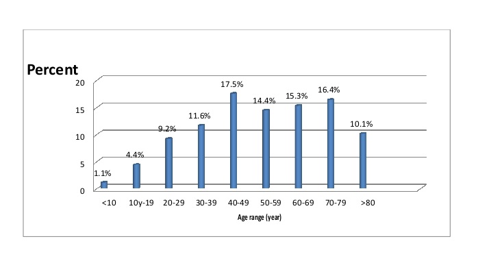
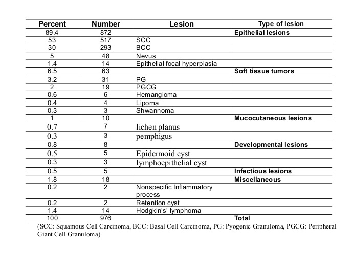
3.2% of 59273 recorded cases were oral lesions which 976(1.65%) of them were oral mucosal lesions. The most prevalent oral lesion was epithelial lesion (89.4%), followed by connective tissue lesions (6.5%). Among epithelial lesions, the most prevalent ones were Squamous Cell Carcinoma (53%), Basal Cell Carcinoma (30%) of lips, Melanocytic nevus (5%) and epithelial focal hyperplasia (1.4%), respectively. Pyogenic granuloma (3.2%), peripheral giant cell granuloma (2%), hemangioma (0.6%), lipoma (0.4%) and schwannoma (0.3%) were the most prevalent lesions among soft tissue lesions, respectively. Mucocutaneous lesions included lichen planus (0.7%) and pemphigus (0.3%). Developmental lesions included epidermoid cyst (0.5%) and lymphoepithelial cyst (0.3%). Other lesions consisted of inflammatory process and retention cysts each one 0.2% and Hodgkin’s lymphoma with 1.4 %( Table1). 535 lesions were found in males (54.8%) and 441 in females (45.2%). Mean age of the patients was 44±3 years old and most lesions (89.4%) were found in patients from 25 to 68 years with the peak of 5th decade of life (Fig 1).
Figure1- Distribution of the biopsied patients according to the age range (decade of life)
37.9% of the cases were below of the mean age (44±3 years old) and 66 (62.1%) above the mean age. The frequency of the biopsied oral mucosal lesions (types of the lesions) according to the age range (decade of life) are shown in table 2.
The most common locations of oral mucosal lesions were lips (27.8%) followed by tongue (25.6%), buccal vestibule (25.4%), gum (5.2%), palate (3.1%), respectively and 12.9% had non-specified location. There was no correlation between sex, age and oral mucosal lesions (P<0.9).
Table 1- Frequency of the biopsied oral mucosal lesions (the type of lesion) according to the age range (decade of life).
Discussion
This retrospective study was done among two referral biopsies centers of Tehran since 2000 to 2010. The results showed that oral lesions constituted 3.2% of the whole lesions, among them 976 cases (1.65%) were related to oral mucosal lesions. Although the results demonstrated that oral lesions had low percentage of the whole body lesions, the percentage of mucosal lesions, indicates the importance of these lesions. However, because of rare reports of the same studies in Iran, comparison is not feasible. Similar to the results obtained by Jahanbani et al. (3), Demko C.A., et al. (1) Pentenero M., et al. (13) most lesions in this study were of epithelial type. Nevertheless, these studies were clinical based in contrast to the present study which retrospectively evaluated biopsied cases and some differences in percentages of the lesions are related to study sample and the referral centers.
In the present study, the most common location of oral mucosal lesions was lips (27.8%) and tongue (25.6%), respectively. The most prevalent lesion in this study was SCC with the common location of lips, which is in line with the findings of other studies (12). Splieth CH., et al. (8) reported cheek mucosa, hard palate, alveolar bone and lips as the most prevalent locations of oral lesions. Al Khateeb (14) reported palate, tongue, upper lip and buccal mucosa as the most prevalent locations for benign neoplasmas and gum, buccal mucosa, lower lip and tongue as the most common locations of non-neoplastic lesions, respectively. Shulman et al. (9) reported that the most common locations were lips and tongue, respectively, which is similar to the findings of this study. The study of Splieth CH., et al. was conducted on normal individuals in clinic. Al-Khateeb only focused on benign lesions which can justify the differences in the results. They also analyzed benign lesions, which was more similar to the current study. This issue itself points to the importance of the research society, investigated lesion type as well as research method (clinical or pathological). Mehrotra et al. (15) reported tongue as the most common location, which was rather similar to this study in which tongue was the second most common location. In the study by Cebeci et al. (16), tongue lesions ranked third in terms of frequency. The most common site in our study could be resulted from the most common lesion we found here. In the other hand SCC is most commonly reported in the lips (related to UV exposure) and tongue.
In the present study, 535 lesions were found in males (54.8%) and 441 in females (45.2%) with M/F ratio of 1.2. Similarly, Jahanbani et al. (3) reported the frequency of oral mucosal lesions in males and females as 62.4% and 37.6%, respectively. Furthermore, Splieth et al. (8) stated the frequency of oral mucosal lesions in males as 12.20% and as 11.40% in females. Al Khateeb (14) founded the frequency of lesions in females and males as 60% and 40%, respectively; likewise, the frequencies were reported as 57.7% in females and 42.3% in males in the study by Al-Mobeeriek and Al-Dosari (17). The higher frequency of lesions in males in this study could be attributed to the most prevalent lesion in this research, i.e. SCC which has been mainly reported in males.
The patients in this study were in the mean age of 44±3 years old. Although most lesions (89.4%) were epithelial type and found in patients ranged from 25 to 68 years, the peak of prevalence was 5th decade of life. Looking at the fig 1 it can be concluded the most common biopsied lesion were found between 5th and 8th decades of life and decreasing of biopsied samples is revealed in 9th decade of life which is consistent with diminishing in life expectancy. In the survey of Al-Khateeb et al. (14), the mean age of the patients was 33 years old with the majority of the cases in the second to the fourth decades. Other studies were clinical based or focused merely on benign or malignant lesions.
SCC was the most prevalent lesion in this study. We also found positive correlation between age and oral mucosal lesion. More than half of the patients (62.1%) were older than the mean age. It can be concluded that the prevalence of SCC increases with age, which is similar to literature which states aging and higher exposure to risk factors increases the risk of cancers (12). The results of this study were similar to Jahanbani et al (3), Splieth et al (8), Al-alMobeeriek et al (17), Mumcu et al (18), Kovac-Kovacic et al (19) and Sixto-Requeijo et al (20) which showed the increase of oral lesion with aging clinically.
It should be considered that high frequency of SCC in this study may be a referral bias. Because Imam Cancer Institute is a referral center for cancer treatment, most of the biopsies are cancerous or precancerous lesions. This can shed some light on differences with other studies. For example, SCC constituted only 10% of biopsied oral lesions in the survey of Hoseinpour Jajarm and Mohtasham (21). However, it was the third most common lesion after inflammatory hyperplasia and lichen planus (21).
In a survey of benign oral masses in Jordanians, conducted by Al-Khateeb, only 4% of lesions were neoplastic and 96% non-neoplastic (14). Non-neoplastic lesions consisted of 43% traumatic lesions, 33% inflammatory/ infective lesions, 14% cystic lesions and 9% developmental lesions. Pyogenic granuloma constituted 19% and PGCG 6% of lesions (14) which is much higher than our results (Pyogenic granuloma 3.2%, peripheral giant cell granuloma 2%) which reflects importance of the referral centers studied. In other words, complete comparison seems impossible.
Finally we showed most of the lesions in this study were malignant which is in contrast with other clinical based studies. It could be related to the design of our study which includes just biopsied samples. It means some lesions are diagnosed clinically and biopsy is not necessary for all lesions. In fact biopsy is taken more frequently for undiagnosed clinical lesions and unfortunately some of them never are sent to laboratories especially when the surgeon thinks it is a benign lesion. Then it can be justified the differences between clinical or histopathological based studies about frequency of oral mucosal lesions besides the variation might be existed among different community, geography and race the authors study among.
Conclusion
The most prevalent mucosal lesion in this study was squamous cell carcinoma, which is a malignant tumor. However, it seems that this result may be related to referral centers studied in this survey. The most prevalent locations of mucosal lesions were lips and tongue. Incidence of mucosal lesions increased with age and no correlation was observed between mucosal lesions and sex. Although this study reflects some facts of oral mucosal lesions in Iran, further investigations are needed to clarify the true frequencies of theses lesions among Iranians.
| Rights and permissions | |
 |
This work is licensed under a Creative Commons Attribution-NonCommercial 4.0 International License. |