Journal of Research in Dental
and Maxillofacial Sciences
Volume 9, Issue 4 (12-2024)
J Res Dent Maxillofac Sci 2024, 9(4): 271-281 |
Back to browse issues page


Download citation:
BibTeX | RIS | EndNote | Medlars | ProCite | Reference Manager | RefWorks
Send citation to:


BibTeX | RIS | EndNote | Medlars | ProCite | Reference Manager | RefWorks
Send citation to:
Jaberi Ansari Z, Valian A, Afrasiabi A, Amdjadi P. Spectrophotometric Evaluation of the Effect of Bleaching on Color Changes of Stained Hybrid Ceramics. J Res Dent Maxillofac Sci 2024; 9 (4) :271-281
URL: http://jrdms.dentaliau.ac.ir/article-1-667-en.html
URL: http://jrdms.dentaliau.ac.ir/article-1-667-en.html
Spectrophotometric Evaluation of the Effect of Bleaching on Color Changes of Stained Hybrid Ceramics


1- Department of Restorative Dentistry, School of Dentistry, Shahid Beheshti University of Medical Sciences, Tehran, Iranof Medical Sciences, Tehran, Iran.
2- Department of Restorative Dentistry, School of Dentistry, Shahid Beheshti University of Medical Sciences, Tehran, Iran
3- Dental Research Center, School of Dentistry, Shahid Beheshti University of Medical Sciences, Tehran, Iran
4- Department of Dental Biomaterials, School of Dentistry, Shahid Beheshti University of Medical Sciences, Tehran, Iran ,amdjadiparisa@gmail.com
2- Department of Restorative Dentistry, School of Dentistry, Shahid Beheshti University of Medical Sciences, Tehran, Iran
3- Dental Research Center, School of Dentistry, Shahid Beheshti University of Medical Sciences, Tehran, Iran
4- Department of Dental Biomaterials, School of Dentistry, Shahid Beheshti University of Medical Sciences, Tehran, Iran ,
Full-Text [PDF 425 kb]
(950 Downloads)
| Abstract (HTML) (3053 Views)
Full-Text: (941 Views)
Abstract
Background and Aim: Hybrid ceramics are gaining popularity for cosmetic dental restorations due to their advantageous mechanical properties and optimal esthetic results. However, the potential effects of bleaching on their color stability are still a subject of interest. This study aimed to assess the effect of bleaching on stained hybrid ceramics in comparison with IPS e.max.
Materials and Methods: This in vitro study was conducted on 48 specimens fabricated from IPS e.max CAD, Vita Enamic, and Cerasmart (n=16) ceramics. The baseline color coordinates of the specimens were measured by a spectrophotometer. Eight specimens from each ceramic type underwent accelerated aging, and their color coordinates were measured again to calculate the color change (ΔE1). The remaining 8 specimens in each ceramic group were immersed in tea solution, and their ΔE was calculated (ΔE2). Subsequently, all specimens were exposed to 20% carbamide peroxide for 8 hours/day for 10 days, and ΔE3 and ΔE4 were calculated. Data were analyzed by two-way ANOVA and Tukey’s test (alpha=0.05).
Results: Type of ceramic, type of intervention (aging, immersion in tea solution, bleaching), and their interaction significantly affected the ΔE (P=0.0001). The maximum and minimum ΔE values were recorded for Cerasmart and IPS e.max ceramic, respectively.
Conclusion: Both accelerated aging and immersion in tea solution caused staining of hybrid ceramics. The minimum ΔE after bleaching occurred in aged ceramics. Bleaching of stained ceramics improved their color.
Keywords: Ceramics; Color; IPS e.max CAD LT; Spectrophotometry; Tooth Bleaching Agents
Introduction
Success of a restorative material is directly linked to its long-term clinical service, which is determined by its composition and the oral
environmental conditions. The oral environment can profoundly affect the esthetic and mechanical properties of dental restorations [1].
In the recent years, computer-aided design/computer-aided manufacturing (CAD/ CAM) esthetic restorative materials were developed [2]. Ceramic blocks have optimal mechanical properties such as high strength, stiffness, and excellent esthetics and biocompatibility. However, they are fragile and have low fracture toughness. Their milling is difficult and can also cause wear of the opposing natural teeth [3]. Composite resin blocks have low fragility and easier milling. Also, they do not cause significant wear of the opposing teeth [4]. However, they are prone to staining following long-term consumption of highly pigmented beverages like tea or coffee [5].
In an attempt to benefit from the advantages of both composite resins and ceramics, hybrid ceramics were introduced to the market. Vita Enamic is a hybrid ceramic with a dual structure. It is composed of a dominant porous network of sintered feldspathic ceramic, reinforced with a methacrylate polymer network. It has high flexural strength due to its fine ceramic structure and polymer network and has an elasticity close to that of dentin [6]. Also, it can provide a high bond strength following bonding to adhesives, requiring minimum tooth preparation. Moreover, it provides optimal marginal and internal fit [7]. Concerning color match, Vita Enamic has color properties resembling those of natural teeth [6].
Cerasmart by GC is a nano-hybrid ceramic that offers the ideal properties of both ceramic and composite resins. It has high elasticity and fracture resistance, optimal marginal adaptation, and high strength after bonding [8].
Discoloration is a drawback of hybrid ceramics, which occurs as a result of water sorption by their resin component. Dimethacrylate entraps a cross-linked network of unreacted monomers serving as a plasticizer. This network provides a structure for additional water sorption [9].
E.max CAD is a common glass ceramic used for CAD/CAM systems. It is composed of lithium disilicate and has a significantly higher flexural strength than other adhesive glass ceramics. The IPS e.max CAD blocks are produced in semi-crystalline form by the manufacturer for easier shaping and milling. The resultant glass ceramic restorations would have 1.5 µm particle size with 70% crystalline volume in a glass matrix. This ceramic has a reportedly high color stability [10].
Artificial aging involves a combination of high temperature, relative humidity, and UV irradiation [11]. In this process, UV irradiation at 340 nm wavelength (UVA spectrum) with 0.85 W/m2 intensity is used to simulate daylight. Materials used for esthetic restorations are exposed to sunlight. Thus, artificial aging can help predict their durability and color stability by simulating the oral environment [12].
Tooth bleaching is a highly popular dental procedure due to its optimal efficacy and non-invasiveness, which can be performed in dental office or at home. The effects of bleaching agents on discolored teeth or stained composite restorations have been widely investigated [13, 14]. However, the effects of bleaching agents on novel restorative materials such as hybrid ceramics have not been well studied. Thus, this study aimed to assess the effect of bleaching on stained hybrid ceramics in comparison with IPS e.max, which is a routinely used ceramic for cosmetic prosthetic restorations. The null hypotheses were that aging and immersion in tea solution would not cause staining of hybrid ceramics, and the color change (ΔE) of the three ceramic types would not be significantly different after aging, immersion in tea solution, or bleaching.
Materials and Methods
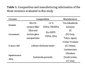
This in vitro, experimental study was conducted on 48 ceramic specimens fabricated from IPS e.max CAD, Vita Enamic, and Cerasmart ceramics (n=16 from each ceramic type). Table 1 presents the composition and manufacturing information of the three ceramics evaluated in this study. The sample size was calculated to be 48 considering alpha=0.05, beta=0.2, and study power of 0.8.
Interventions:
Sixteen CAD/CAM blocks of each ceramic were sectioned by a low-speed diamond saw (Buehler, IL, USA) under running water to obtain ceramic specimens measuring 12 × 6 × 2 mm. The IPS e.max specimens were sintered in a furnace according to the manufacturer’s instructions. The hybrid ceramics and IPS e.max specimens were then polished with a 600-grit diamond bur (D&Z, Wiesbaden, Germany) and were then polished with polishing mullets (EVE Diapol Polishing, Pforzheim, Germany) in three steps using the blue, pink, and gray colors of the polishers in an orderly manner [10]. Each mullet was used for 30 seconds, and the specimens were rinsed under running water after each step. Eight prepared specimens from each group were then mounted on a glass slab using cyanoacrylate glue (Faserverbundwerkstoffe GmbH, Waldenbuch, Germany) and were coded 1 to 8. Their CIE L*a*b* color coordinates were then measured by a spectrophotometer (Ci64; X-Rite, Grandville, MI, USA) against a gray background, and E1 was calculated. Next, the specimens underwent accelerated aging using a xenon lamp (Xenotest; Beta LM, Atlas, Germany), heat, and moisture. For this purpose, the specimens were first subjected to 340 nm xenon lamp irradiation with 0.85 W/m2 power density without water spray for 150 minutes under 60% humidity at 60°C and were then subjected to xenon lamp irradiation again with the same power density under 0% humidity at 25°C with water spray for 30 minutes. Finally, they were subjected to xenon lamp irradiation under 90% humidity at 45°C for 1440 minutes without water spray [11]. E2 of each specimen was then measured by the same spectrophotometer and ∆E1 was calculated. The remaining 8 specimens in each ceramic group were immersed in tea solution as explained by Sulieman et al [15]. For this purpose, a teabag (Lipton, UK) was immersed in 100 mL of 100°C boiling water for 5 minutes, and it was then allowed to cool down to room temperature (27°C). The specimens (n=8 from each group) were then immersed in the tea solution for 24 hours. They were then rinsed and placed in a freshly prepared tea solution. This was repeated three times. Eventually, the specimens were removed from the tea solution, rinsed with water for 30 seconds, stored at room temperature for 24 hours, and subjected to spectrophotometry again to calculate ∆E2.
Both subgroups of specimens (aged, and immersed in tea solution) were then subjected to bleaching such that 20% carbamide peroxide gel (Opalescence, Ultradent, South Jordan, UT, USA) was applied on the surface of the specimens for 8 hours daily and was then rinsed. This process was repeated for 10 days, and ∆E3 was then calculated for the aged group and ∆E4 was calculated for the specimens immersed in the tea solution. Eventually, ∆E5 was calculated to determine the color difference of aged and bleached specimens compared with the baseline color of the specimens (E1), and ∆E6 was calculated to determine the color differences of tea-stained and bleached specimens compared with the baseline color of the specimens (E1) [15].
Assessment of color stability:
The CIE L*a*b* color coordinates of the specimens were measured by a spectro-photometer immediately after aging and immersion in tea solution and also after bleaching. All measurements were made under standard D65 lighting conditions with 12-degree angle against a gray background (Checker Passport; X-Rite, Grandville, MI, USA). The 4-mm diaphragm of the device was adjusted at the center of each specimen, and the L*, a* and b* color coordinates were measured. ∆E was calculated using the equation:

Table 1. Composition and manufacturing information of the three ceramics evaluated in this study
Table 2. ∆E of different ceramics in different conditions
Table 3. Pairwise comparisons of the ∆E of ceramics after different interventions by the Tukey’s test
Table 4. ∆L of different ceramics after different interventions
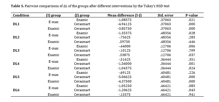
Table 5. Pairwise comparisons of ΔL of the groups after different interventions by the Tukey’s HSD test
Discussion
This study assessed the effect of bleaching on stained hybrid ceramics in comparison with IPS e.max, which is a routinely used ceramic for cosmetic prosthetic restorations. The results showed that IPS e.max experienced minimum discoloration while Cerasmart hybrid ceramic had maximum discoloration. The CIEL*a*b* color space was used in this study for the calculation of ∆E. Evidence shows that ∆E=1 is detectable by 50% of the observers under controlled conditions [16]. The clinically acceptable threshold for ∆E varies from 2.72 to 3.3 and even 3.7 [17]. In the present study, ∆E<1 was considered undetectable by the human eye; values between 1 and 3.3 were considered clinically acceptable, and values>3.3 were considered clinically unacceptable. The present results showed maximum ∆E following immersion in tea and minimum ∆E following bleaching of aged ceramics. Accelerated aging was performed for aging of specimens in the present study. This process does not affect external stains and focuses on internal discoloration of materials. Accelerated aging is widely used for assessment of color stability of dental materials such as composite resins, and resin cements [18]. It easily simulates the effects of long-term clinical service in the oral environment, and its validity has been previously confirmed [12, 19, 20]. Discoloration of dental restorative materials as the result of UV irradiation depends on chemical changes in the initiator system and the activator in the resin matrix. Destruction of residual amine and residual unreacted carbon-carbon double bonds result in formation of yellow compounds [21, 22]. It is not well understood that 1 year of clinical service equals how many hours of accelerated aging. However, the aging chamber manufacturer claims that 300 hours of aging corresponds to 1 year of clinical service [23]. A previous study demonstrated that 168 hours of UV irradiation caused detectable, but clinically acceptable, color change (∆E=2.07) in all composite resins [24]. In the present study, the maximum ∆E after aging was noted in Cerasmart (∆E=5.7), which was clinically unacceptable, followed by Vita Enamic (∆E=2.08). It is possible that chemical composition of materials and their resin content play a role in discoloration due to aging. Resin matrix, due to its polymer base, undergoes degradation and surface roughness following aging and clinical service, which adversely affects the color stability and stainability [14, 25]. Also, Cerasmart hybrid ceramic has higher translucency than Enamic despite having similar color shade, which may also explain higher discoloration of Cerasmart after aging.
Discoloration of dental ceramics in the oral environment directly depends on their appropriate polishing, non-critical cracks, and no reactivity of materials. Surface roughness adversely affects the biomechanical and esthetic properties of these restorations, and makes them susceptible to further aging [26-28]. In this study, the minimum ∆E (0.80) was recorded in IPS e.max CAD, which was not clinically perceivable. Slight discoloration of ceramics due to aging may be due to the breakdown of metal oxides, tints added to ceramics, or coloring agents used to achieve a desirable color match [29]. Breakdown of metal oxides as the result of UV irradiation leads to formation of peroxide compounds which may cause discoloration of ceramic materials [30].
Discoloration also occurs due to water sorption by the resin component. Thus, type of resin matrix plays an important role in color stability of restorative materials [31]. Higher resin matrix content of Cerasmart can explain its higher staining compared with Vita Enamic. According to the manufacturer, the resin component of Cerasmart is composed of DMA, UDMA, and bis-MMEP. Bis-MMEP is a polymer with a molecular weight almost twice that of TEGDMA. Materials containing high molecular weight polymers have higher water sorption due to lower cross-linking of molecular chains [32], which can be the case for Cerasmart hybrid ceramic. Vita Enamic contains 66wt% UDMA and 33wt% TEGDMA. Water sorption by TEGDMA is higher than that by bis-GMA [33-35]. Considering the high weight percentage of TEGDMA in Vita Enamic, it may allow the penetration of hydrophilic coloring agents into the resin matrix. However, UDMA is much more hydrophobic than bis-GMA, and therefore has higher color stability as well. It has been stated that dimethacrylate creates a cross-linked network of trapped unreacted monomers that serve as plasticizer and create a permeable structure for water sorption [36-38]. This may explain the greater discoloration of Vita Enamic and Cerasmart following immersion in tea solution. Immersion in tea solution causes adsorption of polar stains, resulting in superficial discoloration with no internal absorption [39].
The presence of higher amounts of organic matrix in the composition of composite resins compared with ceramics makes the composites more susceptible to chemical changes. Bleaching may cause discoloration of composite restorations, depending on the type of material. Some authors have reported that bleaching cannot clinically affect the color of tooth-colored restorations [13]; while, some others have reported discoloration of composite resins by bleaching [40, 41]. In the present study, the ΔE caused by bleaching of aged specimens was lower than other ΔE values. The minimum value was recorded for IPS e.max CAD (ΔE=0.38) and the maximum value was recorded for Vita Enamic (ΔE=0.89). Aging changes the internal structure of materials; thus, it causes internal staining. The ΔE of tea-stained and bleached materials was higher than the ΔE of aged and bleached materials. In other words, the bleaching agent had a greater effect on tea-stained ceramic specimens, probably due to the external nature of staining. The effect of bleaching on IPS e.max ceramic was lower than that on other hybrid ceramics, which may be attributed to its very low permeability to hydrogen peroxide. Polished ceramics are more susceptible to staining with external stains such as tea solution due to their rougher surface. The ΔE of IPS e.max CAD after bleaching can be due to the effect of bleaching on external stains. The ΔE of Cerasmart was higher than that of Vita Enamic after bleaching due to its higher polymer content and greater effect of bleaching agents on the resin component.
The ΔE5 value indicated the color difference of aged and bleached specimens with the control specimens, and was maximum in Cerasmart (almost equal to the value obtained after aging alone). This finding highlights the insignificant effect of the bleaching agent on aged Cerasmart ceramic; however, the ΔE5 of Vita Enamic and IPS e.max CAD was lower than the detectable threshold by the human eye (indicating their acceptable esthetics). The ΔE6 values of Cerasmart and Vita Enamic were almost equal, indicating that the color difference between tea-stained and bleached hybrid ceramics and the control specimens, although detectable by the human eye, was clinically acceptable. According to the present results, the null hypothesis of the study regarding no discoloration of hybrid ceramics due to aging or immersion in tea solution was rejected. The second part of the null hypothesis regarding no significant difference in ΔE of the three ceramic types was also rejected.
The ΔL indicates the change in lightness. In the present study, a direct correlation was noted between ΔE and ΔL after different interventions except for ΔL2 of Cerasmart which did not show such a correlation. The ΔE of ceramics following immersion in tea solution does not depend on the L* parameter; instead, it depends on Δb (which indicates yellowness-blueness) due to the effect of yellow tea stains on hybrid ceramics.
Kara et al. [9] evaluated the effect of bleaching on color stability of porcelains and Ceromer and showed that carbamide peroxide caused discoloration of both Ceromer and IPS Empress 2; their results were in agreement with ours, although they did not stain or age the specimens before the bleaching treatment. The present results were also in agreement with those of Canay and Çehreli [42], Kurtulmus-Yilmaz et al, [19] and Kwon et al, [43] on the effects of bleaching on composite resins and Culic et al, [41] on the effects of bleaching on hybrid ceramics. Mansour et al. [44] assessed the effects of bleaching on the conventional and hybrid ceramics and reported results in line with the present findings. However, they did not stain the specimens before bleaching. Catelan et al. [23] showed that aging with UV irradiation caused discoloration of composite resins. Their results were in accordance with ours. However, since we used hybrid ceramics which are a combination of composite resin and ceramic, the magnitude of discoloration was lower in the present study. Turgut et al. [20] assessed the effect of aging on IPS e.max ceramic laminates and showed their discoloration before and after cementation with resin cement. Their results were in line with ours although we assessed hybrid ceramics as well. Hamza et al. [11] assessed the effect of accelerated aging on color stability and surface roughness of ceramics. They compared three CAD/CAM ceramic blocks of translucent zirconia, Lava Ultimate, and zirconia veneered with VM9 feldspathic porcelain. They reported that aging had no significant effect on the color stability of ceramics and only caused surface roughening of Lava Ultimate. Their results were different from ours, which may be due to glazing of ceramics prior to aging in their study.
Further studies are required on the effects of bleaching on surface roughness of hybrid ceramics in the long-term. Also, the color stability of hybrid ceramics in other staining environments should be investigated.
Conclusion
The present results showed that both accelerated aging and immersion in tea solution caused staining of hybrid ceramics. The minimum ΔE following bleaching was noted in aged ceramics. Bleaching of stained ceramics improved their color.
Authors’ contributions
Conceptualization and Supervision: Z.J.A, Methodology and Formal analysis: A.V, Interpretation of data: Z.J.A, A.V, Writing the original draft: A.A, Writing the review and editing: P.A
Background and Aim: Hybrid ceramics are gaining popularity for cosmetic dental restorations due to their advantageous mechanical properties and optimal esthetic results. However, the potential effects of bleaching on their color stability are still a subject of interest. This study aimed to assess the effect of bleaching on stained hybrid ceramics in comparison with IPS e.max.
Materials and Methods: This in vitro study was conducted on 48 specimens fabricated from IPS e.max CAD, Vita Enamic, and Cerasmart (n=16) ceramics. The baseline color coordinates of the specimens were measured by a spectrophotometer. Eight specimens from each ceramic type underwent accelerated aging, and their color coordinates were measured again to calculate the color change (ΔE1). The remaining 8 specimens in each ceramic group were immersed in tea solution, and their ΔE was calculated (ΔE2). Subsequently, all specimens were exposed to 20% carbamide peroxide for 8 hours/day for 10 days, and ΔE3 and ΔE4 were calculated. Data were analyzed by two-way ANOVA and Tukey’s test (alpha=0.05).
Results: Type of ceramic, type of intervention (aging, immersion in tea solution, bleaching), and their interaction significantly affected the ΔE (P=0.0001). The maximum and minimum ΔE values were recorded for Cerasmart and IPS e.max ceramic, respectively.
Conclusion: Both accelerated aging and immersion in tea solution caused staining of hybrid ceramics. The minimum ΔE after bleaching occurred in aged ceramics. Bleaching of stained ceramics improved their color.
Keywords: Ceramics; Color; IPS e.max CAD LT; Spectrophotometry; Tooth Bleaching Agents
Introduction
Success of a restorative material is directly linked to its long-term clinical service, which is determined by its composition and the oral
environmental conditions. The oral environment can profoundly affect the esthetic and mechanical properties of dental restorations [1].
In the recent years, computer-aided design/computer-aided manufacturing (CAD/ CAM) esthetic restorative materials were developed [2]. Ceramic blocks have optimal mechanical properties such as high strength, stiffness, and excellent esthetics and biocompatibility. However, they are fragile and have low fracture toughness. Their milling is difficult and can also cause wear of the opposing natural teeth [3]. Composite resin blocks have low fragility and easier milling. Also, they do not cause significant wear of the opposing teeth [4]. However, they are prone to staining following long-term consumption of highly pigmented beverages like tea or coffee [5].
In an attempt to benefit from the advantages of both composite resins and ceramics, hybrid ceramics were introduced to the market. Vita Enamic is a hybrid ceramic with a dual structure. It is composed of a dominant porous network of sintered feldspathic ceramic, reinforced with a methacrylate polymer network. It has high flexural strength due to its fine ceramic structure and polymer network and has an elasticity close to that of dentin [6]. Also, it can provide a high bond strength following bonding to adhesives, requiring minimum tooth preparation. Moreover, it provides optimal marginal and internal fit [7]. Concerning color match, Vita Enamic has color properties resembling those of natural teeth [6].
Cerasmart by GC is a nano-hybrid ceramic that offers the ideal properties of both ceramic and composite resins. It has high elasticity and fracture resistance, optimal marginal adaptation, and high strength after bonding [8].
Discoloration is a drawback of hybrid ceramics, which occurs as a result of water sorption by their resin component. Dimethacrylate entraps a cross-linked network of unreacted monomers serving as a plasticizer. This network provides a structure for additional water sorption [9].
E.max CAD is a common glass ceramic used for CAD/CAM systems. It is composed of lithium disilicate and has a significantly higher flexural strength than other adhesive glass ceramics. The IPS e.max CAD blocks are produced in semi-crystalline form by the manufacturer for easier shaping and milling. The resultant glass ceramic restorations would have 1.5 µm particle size with 70% crystalline volume in a glass matrix. This ceramic has a reportedly high color stability [10].
Artificial aging involves a combination of high temperature, relative humidity, and UV irradiation [11]. In this process, UV irradiation at 340 nm wavelength (UVA spectrum) with 0.85 W/m2 intensity is used to simulate daylight. Materials used for esthetic restorations are exposed to sunlight. Thus, artificial aging can help predict their durability and color stability by simulating the oral environment [12].
Tooth bleaching is a highly popular dental procedure due to its optimal efficacy and non-invasiveness, which can be performed in dental office or at home. The effects of bleaching agents on discolored teeth or stained composite restorations have been widely investigated [13, 14]. However, the effects of bleaching agents on novel restorative materials such as hybrid ceramics have not been well studied. Thus, this study aimed to assess the effect of bleaching on stained hybrid ceramics in comparison with IPS e.max, which is a routinely used ceramic for cosmetic prosthetic restorations. The null hypotheses were that aging and immersion in tea solution would not cause staining of hybrid ceramics, and the color change (ΔE) of the three ceramic types would not be significantly different after aging, immersion in tea solution, or bleaching.
Materials and Methods
This in vitro, experimental study was conducted on 48 ceramic specimens fabricated from IPS e.max CAD, Vita Enamic, and Cerasmart ceramics (n=16 from each ceramic type). Table 1 presents the composition and manufacturing information of the three ceramics evaluated in this study. The sample size was calculated to be 48 considering alpha=0.05, beta=0.2, and study power of 0.8.
Interventions:
Sixteen CAD/CAM blocks of each ceramic were sectioned by a low-speed diamond saw (Buehler, IL, USA) under running water to obtain ceramic specimens measuring 12 × 6 × 2 mm. The IPS e.max specimens were sintered in a furnace according to the manufacturer’s instructions. The hybrid ceramics and IPS e.max specimens were then polished with a 600-grit diamond bur (D&Z, Wiesbaden, Germany) and were then polished with polishing mullets (EVE Diapol Polishing, Pforzheim, Germany) in three steps using the blue, pink, and gray colors of the polishers in an orderly manner [10]. Each mullet was used for 30 seconds, and the specimens were rinsed under running water after each step. Eight prepared specimens from each group were then mounted on a glass slab using cyanoacrylate glue (Faserverbundwerkstoffe GmbH, Waldenbuch, Germany) and were coded 1 to 8. Their CIE L*a*b* color coordinates were then measured by a spectrophotometer (Ci64; X-Rite, Grandville, MI, USA) against a gray background, and E1 was calculated. Next, the specimens underwent accelerated aging using a xenon lamp (Xenotest; Beta LM, Atlas, Germany), heat, and moisture. For this purpose, the specimens were first subjected to 340 nm xenon lamp irradiation with 0.85 W/m2 power density without water spray for 150 minutes under 60% humidity at 60°C and were then subjected to xenon lamp irradiation again with the same power density under 0% humidity at 25°C with water spray for 30 minutes. Finally, they were subjected to xenon lamp irradiation under 90% humidity at 45°C for 1440 minutes without water spray [11]. E2 of each specimen was then measured by the same spectrophotometer and ∆E1 was calculated. The remaining 8 specimens in each ceramic group were immersed in tea solution as explained by Sulieman et al [15]. For this purpose, a teabag (Lipton, UK) was immersed in 100 mL of 100°C boiling water for 5 minutes, and it was then allowed to cool down to room temperature (27°C). The specimens (n=8 from each group) were then immersed in the tea solution for 24 hours. They were then rinsed and placed in a freshly prepared tea solution. This was repeated three times. Eventually, the specimens were removed from the tea solution, rinsed with water for 30 seconds, stored at room temperature for 24 hours, and subjected to spectrophotometry again to calculate ∆E2.
Both subgroups of specimens (aged, and immersed in tea solution) were then subjected to bleaching such that 20% carbamide peroxide gel (Opalescence, Ultradent, South Jordan, UT, USA) was applied on the surface of the specimens for 8 hours daily and was then rinsed. This process was repeated for 10 days, and ∆E3 was then calculated for the aged group and ∆E4 was calculated for the specimens immersed in the tea solution. Eventually, ∆E5 was calculated to determine the color difference of aged and bleached specimens compared with the baseline color of the specimens (E1), and ∆E6 was calculated to determine the color differences of tea-stained and bleached specimens compared with the baseline color of the specimens (E1) [15].
Assessment of color stability:
The CIE L*a*b* color coordinates of the specimens were measured by a spectro-photometer immediately after aging and immersion in tea solution and also after bleaching. All measurements were made under standard D65 lighting conditions with 12-degree angle against a gray background (Checker Passport; X-Rite, Grandville, MI, USA). The 4-mm diaphragm of the device was adjusted at the center of each specimen, and the L*, a* and b* color coordinates were measured. ∆E was calculated using the equation:
Table 1. Composition and manufacturing information of the three ceramics evaluated in this study
Statistical analysis:
Normal distribution of data was evaluated using the Kolmogorov-Smirnov test. The equality of variances was assessed by the Levene’s test. Repeated measures two-way ANOVA was applied to assess the effect of type of ceramic and type of intervention (aging, staining, bleaching) on ∆E and ∆L. Since the interaction effect of type of ceramic and type of intervention was significant, within-group comparisons were performed by one-way ANOVA followed by pairwise comparisons by the Bonferroni test. Inter-group comparisons were performed by one-way ANOVA followed by the Tukey’s HSD test for pairwise comparisons. All statistical analyses were carried out by SPSS version 22 (SPSS Inc., IL, USA) at 0.05 level of significance.
Results
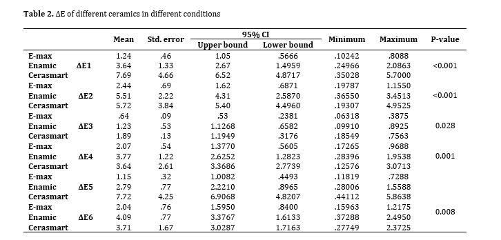
The Kolmogorov-Smirnov test confirmed normal distribution of ∆E data in all three groups; thus, parametric tests were applied for data analysis. Considering the presence of one within-group (aging, staining, bleaching) and one between-group (type of ceramic) factor, repeated measures two-way ANOVA was applied. Two-way ANOVA showed that type of ceramic (P<0.001) and type of intervention (P<0.001) significantly affected the ∆E. Their interaction effect was also significant (P<0.001). Thus, subgroup analysis was performed. Table 2 shows the ∆E of different ceramics after different interventions.
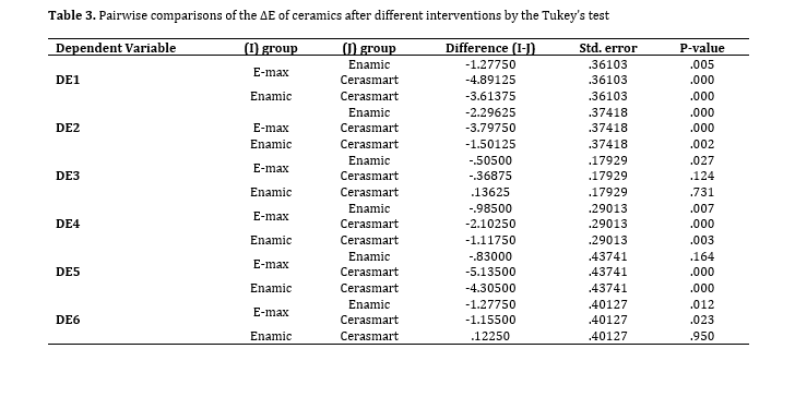
One-way ANOVA showed significant differences in ∆E among the three ceramic types after different interventions (P<0.05). Thus, pairwise comparisons were performed by the Tukey’s HSD test (Table 3). Regarding ∆E1 and ∆E2, IPS e.max showed minimum values compared with Enamic (P=0.009) and Cerasmart (P<0.001). Also, Enamic experienced significantly lower discoloration than Cerasmart (P<0.001). Concerning ∆E3, IPS e. max showed a significantly lower value than Enamic (P=0.027). The results regarding ∆E4 were similar to ∆E1. Regarding ∆E5, Cerasmart showed a significantly higher value than the other two ceramics (P<0.001). Concerning ∆E6, both Cerasmart and e.max showed significantly greater values than IPS e.max (P<0.05).
Subgroup analysis:
One-way ANOVA was applied to analyze the ∆E of each ceramic after different interventions. The results showed significant differences in all three ceramics after different interventions (P<0.001 for all three). Pairwise comparisons of different interventions within each ceramic group were performed by the Bonferroni test which revealed that:
In e.max ceramic, ∆E3 was significantly lower than ∆E1 (P=0.0027), and ∆E4 was significantly lower than ∆E2 (P=0.007). Other differences were not significant (P>0.05).
In Enamic, ∆E3 was significantly lower than ∆E1 (P=0.016) and ∆E2 (P=0.010). Also, ∆E5 was significantly lower than ∆E2 (P=0.025). Other differences were not significant (P>0.05).
In Cerasmart, ∆E3 was significantly lower than ∆E1 (P<0.001), ∆E4 (P<0.001), ∆E5 (P<0.001), and ∆E6 (P=0.013). Also, ∆E4 and ∆E6 were significantly lower than ∆E1 (P=0.004 and P=0.006, respectively) and ∆E2 (P=0.001 and P=0.004, respectively). ∆E5 was also significantly higher than ∆E4 (P=0.008) and ∆E6 (P=0.008).
Results of ∆L:
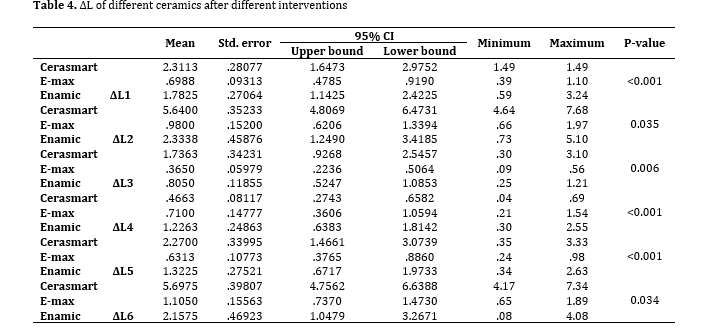
Table 4 shows the ∆L of different ceramics after different interventions. Two-way ANOVA showed that the effects of type of ceramic and different interventions, and their interaction effect were all significant on ∆L (P<0.001). Subgroup analysis revealed significant differences in ∆L of each ceramic after different interventions (P=0.05). Pairwise comparisons by the Tukey’s HSD test showed that ∆L1 of e.max was significantly lower than that of Enamic (P=0.021), and ∆L1 of Enamic was significantly lower than that of Cerasmart (P=0.0009). ∆L2 of e.max was significantly lower than that of Enamic (P=0.028). ∆L3 values of e.max (P=0.006) and Cerasmart (P=0.037) were significantly lower than that of Enamic, and ∆L4 of Cerasmart was significantly higher than that of e.max (P=0.001) and Enamic (P=0.024). The results for ∆L5 and ∆L6 were similar to those for ∆L4 (Table 5).
Normal distribution of data was evaluated using the Kolmogorov-Smirnov test. The equality of variances was assessed by the Levene’s test. Repeated measures two-way ANOVA was applied to assess the effect of type of ceramic and type of intervention (aging, staining, bleaching) on ∆E and ∆L. Since the interaction effect of type of ceramic and type of intervention was significant, within-group comparisons were performed by one-way ANOVA followed by pairwise comparisons by the Bonferroni test. Inter-group comparisons were performed by one-way ANOVA followed by the Tukey’s HSD test for pairwise comparisons. All statistical analyses were carried out by SPSS version 22 (SPSS Inc., IL, USA) at 0.05 level of significance.
Results
The Kolmogorov-Smirnov test confirmed normal distribution of ∆E data in all three groups; thus, parametric tests were applied for data analysis. Considering the presence of one within-group (aging, staining, bleaching) and one between-group (type of ceramic) factor, repeated measures two-way ANOVA was applied. Two-way ANOVA showed that type of ceramic (P<0.001) and type of intervention (P<0.001) significantly affected the ∆E. Their interaction effect was also significant (P<0.001). Thus, subgroup analysis was performed. Table 2 shows the ∆E of different ceramics after different interventions.
One-way ANOVA showed significant differences in ∆E among the three ceramic types after different interventions (P<0.05). Thus, pairwise comparisons were performed by the Tukey’s HSD test (Table 3). Regarding ∆E1 and ∆E2, IPS e.max showed minimum values compared with Enamic (P=0.009) and Cerasmart (P<0.001). Also, Enamic experienced significantly lower discoloration than Cerasmart (P<0.001). Concerning ∆E3, IPS e. max showed a significantly lower value than Enamic (P=0.027). The results regarding ∆E4 were similar to ∆E1. Regarding ∆E5, Cerasmart showed a significantly higher value than the other two ceramics (P<0.001). Concerning ∆E6, both Cerasmart and e.max showed significantly greater values than IPS e.max (P<0.05).
Subgroup analysis:
One-way ANOVA was applied to analyze the ∆E of each ceramic after different interventions. The results showed significant differences in all three ceramics after different interventions (P<0.001 for all three). Pairwise comparisons of different interventions within each ceramic group were performed by the Bonferroni test which revealed that:
In e.max ceramic, ∆E3 was significantly lower than ∆E1 (P=0.0027), and ∆E4 was significantly lower than ∆E2 (P=0.007). Other differences were not significant (P>0.05).
In Enamic, ∆E3 was significantly lower than ∆E1 (P=0.016) and ∆E2 (P=0.010). Also, ∆E5 was significantly lower than ∆E2 (P=0.025). Other differences were not significant (P>0.05).
In Cerasmart, ∆E3 was significantly lower than ∆E1 (P<0.001), ∆E4 (P<0.001), ∆E5 (P<0.001), and ∆E6 (P=0.013). Also, ∆E4 and ∆E6 were significantly lower than ∆E1 (P=0.004 and P=0.006, respectively) and ∆E2 (P=0.001 and P=0.004, respectively). ∆E5 was also significantly higher than ∆E4 (P=0.008) and ∆E6 (P=0.008).
Results of ∆L:
Table 4 shows the ∆L of different ceramics after different interventions. Two-way ANOVA showed that the effects of type of ceramic and different interventions, and their interaction effect were all significant on ∆L (P<0.001). Subgroup analysis revealed significant differences in ∆L of each ceramic after different interventions (P=0.05). Pairwise comparisons by the Tukey’s HSD test showed that ∆L1 of e.max was significantly lower than that of Enamic (P=0.021), and ∆L1 of Enamic was significantly lower than that of Cerasmart (P=0.0009). ∆L2 of e.max was significantly lower than that of Enamic (P=0.028). ∆L3 values of e.max (P=0.006) and Cerasmart (P=0.037) were significantly lower than that of Enamic, and ∆L4 of Cerasmart was significantly higher than that of e.max (P=0.001) and Enamic (P=0.024). The results for ∆L5 and ∆L6 were similar to those for ∆L4 (Table 5).
Table 2. ∆E of different ceramics in different conditions
Table 3. Pairwise comparisons of the ∆E of ceramics after different interventions by the Tukey’s test
Table 4. ∆L of different ceramics after different interventions
Table 5. Pairwise comparisons of ΔL of the groups after different interventions by the Tukey’s HSD test
Discussion
This study assessed the effect of bleaching on stained hybrid ceramics in comparison with IPS e.max, which is a routinely used ceramic for cosmetic prosthetic restorations. The results showed that IPS e.max experienced minimum discoloration while Cerasmart hybrid ceramic had maximum discoloration. The CIEL*a*b* color space was used in this study for the calculation of ∆E. Evidence shows that ∆E=1 is detectable by 50% of the observers under controlled conditions [16]. The clinically acceptable threshold for ∆E varies from 2.72 to 3.3 and even 3.7 [17]. In the present study, ∆E<1 was considered undetectable by the human eye; values between 1 and 3.3 were considered clinically acceptable, and values>3.3 were considered clinically unacceptable. The present results showed maximum ∆E following immersion in tea and minimum ∆E following bleaching of aged ceramics. Accelerated aging was performed for aging of specimens in the present study. This process does not affect external stains and focuses on internal discoloration of materials. Accelerated aging is widely used for assessment of color stability of dental materials such as composite resins, and resin cements [18]. It easily simulates the effects of long-term clinical service in the oral environment, and its validity has been previously confirmed [12, 19, 20]. Discoloration of dental restorative materials as the result of UV irradiation depends on chemical changes in the initiator system and the activator in the resin matrix. Destruction of residual amine and residual unreacted carbon-carbon double bonds result in formation of yellow compounds [21, 22]. It is not well understood that 1 year of clinical service equals how many hours of accelerated aging. However, the aging chamber manufacturer claims that 300 hours of aging corresponds to 1 year of clinical service [23]. A previous study demonstrated that 168 hours of UV irradiation caused detectable, but clinically acceptable, color change (∆E=2.07) in all composite resins [24]. In the present study, the maximum ∆E after aging was noted in Cerasmart (∆E=5.7), which was clinically unacceptable, followed by Vita Enamic (∆E=2.08). It is possible that chemical composition of materials and their resin content play a role in discoloration due to aging. Resin matrix, due to its polymer base, undergoes degradation and surface roughness following aging and clinical service, which adversely affects the color stability and stainability [14, 25]. Also, Cerasmart hybrid ceramic has higher translucency than Enamic despite having similar color shade, which may also explain higher discoloration of Cerasmart after aging.
Discoloration of dental ceramics in the oral environment directly depends on their appropriate polishing, non-critical cracks, and no reactivity of materials. Surface roughness adversely affects the biomechanical and esthetic properties of these restorations, and makes them susceptible to further aging [26-28]. In this study, the minimum ∆E (0.80) was recorded in IPS e.max CAD, which was not clinically perceivable. Slight discoloration of ceramics due to aging may be due to the breakdown of metal oxides, tints added to ceramics, or coloring agents used to achieve a desirable color match [29]. Breakdown of metal oxides as the result of UV irradiation leads to formation of peroxide compounds which may cause discoloration of ceramic materials [30].
Discoloration also occurs due to water sorption by the resin component. Thus, type of resin matrix plays an important role in color stability of restorative materials [31]. Higher resin matrix content of Cerasmart can explain its higher staining compared with Vita Enamic. According to the manufacturer, the resin component of Cerasmart is composed of DMA, UDMA, and bis-MMEP. Bis-MMEP is a polymer with a molecular weight almost twice that of TEGDMA. Materials containing high molecular weight polymers have higher water sorption due to lower cross-linking of molecular chains [32], which can be the case for Cerasmart hybrid ceramic. Vita Enamic contains 66wt% UDMA and 33wt% TEGDMA. Water sorption by TEGDMA is higher than that by bis-GMA [33-35]. Considering the high weight percentage of TEGDMA in Vita Enamic, it may allow the penetration of hydrophilic coloring agents into the resin matrix. However, UDMA is much more hydrophobic than bis-GMA, and therefore has higher color stability as well. It has been stated that dimethacrylate creates a cross-linked network of trapped unreacted monomers that serve as plasticizer and create a permeable structure for water sorption [36-38]. This may explain the greater discoloration of Vita Enamic and Cerasmart following immersion in tea solution. Immersion in tea solution causes adsorption of polar stains, resulting in superficial discoloration with no internal absorption [39].
The presence of higher amounts of organic matrix in the composition of composite resins compared with ceramics makes the composites more susceptible to chemical changes. Bleaching may cause discoloration of composite restorations, depending on the type of material. Some authors have reported that bleaching cannot clinically affect the color of tooth-colored restorations [13]; while, some others have reported discoloration of composite resins by bleaching [40, 41]. In the present study, the ΔE caused by bleaching of aged specimens was lower than other ΔE values. The minimum value was recorded for IPS e.max CAD (ΔE=0.38) and the maximum value was recorded for Vita Enamic (ΔE=0.89). Aging changes the internal structure of materials; thus, it causes internal staining. The ΔE of tea-stained and bleached materials was higher than the ΔE of aged and bleached materials. In other words, the bleaching agent had a greater effect on tea-stained ceramic specimens, probably due to the external nature of staining. The effect of bleaching on IPS e.max ceramic was lower than that on other hybrid ceramics, which may be attributed to its very low permeability to hydrogen peroxide. Polished ceramics are more susceptible to staining with external stains such as tea solution due to their rougher surface. The ΔE of IPS e.max CAD after bleaching can be due to the effect of bleaching on external stains. The ΔE of Cerasmart was higher than that of Vita Enamic after bleaching due to its higher polymer content and greater effect of bleaching agents on the resin component.
The ΔE5 value indicated the color difference of aged and bleached specimens with the control specimens, and was maximum in Cerasmart (almost equal to the value obtained after aging alone). This finding highlights the insignificant effect of the bleaching agent on aged Cerasmart ceramic; however, the ΔE5 of Vita Enamic and IPS e.max CAD was lower than the detectable threshold by the human eye (indicating their acceptable esthetics). The ΔE6 values of Cerasmart and Vita Enamic were almost equal, indicating that the color difference between tea-stained and bleached hybrid ceramics and the control specimens, although detectable by the human eye, was clinically acceptable. According to the present results, the null hypothesis of the study regarding no discoloration of hybrid ceramics due to aging or immersion in tea solution was rejected. The second part of the null hypothesis regarding no significant difference in ΔE of the three ceramic types was also rejected.
The ΔL indicates the change in lightness. In the present study, a direct correlation was noted between ΔE and ΔL after different interventions except for ΔL2 of Cerasmart which did not show such a correlation. The ΔE of ceramics following immersion in tea solution does not depend on the L* parameter; instead, it depends on Δb (which indicates yellowness-blueness) due to the effect of yellow tea stains on hybrid ceramics.
Kara et al. [9] evaluated the effect of bleaching on color stability of porcelains and Ceromer and showed that carbamide peroxide caused discoloration of both Ceromer and IPS Empress 2; their results were in agreement with ours, although they did not stain or age the specimens before the bleaching treatment. The present results were also in agreement with those of Canay and Çehreli [42], Kurtulmus-Yilmaz et al, [19] and Kwon et al, [43] on the effects of bleaching on composite resins and Culic et al, [41] on the effects of bleaching on hybrid ceramics. Mansour et al. [44] assessed the effects of bleaching on the conventional and hybrid ceramics and reported results in line with the present findings. However, they did not stain the specimens before bleaching. Catelan et al. [23] showed that aging with UV irradiation caused discoloration of composite resins. Their results were in accordance with ours. However, since we used hybrid ceramics which are a combination of composite resin and ceramic, the magnitude of discoloration was lower in the present study. Turgut et al. [20] assessed the effect of aging on IPS e.max ceramic laminates and showed their discoloration before and after cementation with resin cement. Their results were in line with ours although we assessed hybrid ceramics as well. Hamza et al. [11] assessed the effect of accelerated aging on color stability and surface roughness of ceramics. They compared three CAD/CAM ceramic blocks of translucent zirconia, Lava Ultimate, and zirconia veneered with VM9 feldspathic porcelain. They reported that aging had no significant effect on the color stability of ceramics and only caused surface roughening of Lava Ultimate. Their results were different from ours, which may be due to glazing of ceramics prior to aging in their study.
Further studies are required on the effects of bleaching on surface roughness of hybrid ceramics in the long-term. Also, the color stability of hybrid ceramics in other staining environments should be investigated.
Conclusion
The present results showed that both accelerated aging and immersion in tea solution caused staining of hybrid ceramics. The minimum ΔE following bleaching was noted in aged ceramics. Bleaching of stained ceramics improved their color.
Authors’ contributions
Conceptualization and Supervision: Z.J.A, Methodology and Formal analysis: A.V, Interpretation of data: Z.J.A, A.V, Writing the original draft: A.A, Writing the review and editing: P.A
Type of Study: Original article |
Subject:
Prosthodontics
References
1. Erdemir U, Yildiz E, Eren MM, Ozel S. Surface hardness evaluation of different composite resin materials: influence of sports and energy drinks immersion after a short-term period. J Appl Oral Sci. 2013 Mar-Apr;21(2):124-31. [DOI:10.1590/1678-7757201302185] [PMID] []
2. Soliman TA, Robaian A, Al-Gerny Y, Hussein EMR. Influence of surface treatment on repair bond strength of CAD/CAM long-term provisional restorative materials: an in vitro study. BMC Oral Health. 2023 May 30;23(1):342. [DOI:10.1186/s12903-023-03021-y] [PMID] []
3. Vaiani L, Boccaccio A, Uva AE, Palumbo G, Piccininni A, Guglielmi P, et al. Ceramic Materials for Biomedical Applications: An Overview on Properties and Fabrication Processes. J Funct Biomater. 2023 Mar 4;14(3):146. [DOI:10.3390/jfb14030146] [PMID] []
4. Ling L, Ma Y, Malyala R. A novel CAD/CAM resin composite block with high mechanical properties. Dent Mater. 2021 Jul;37(7):1150-5. [DOI:10.1016/j.dental.2021.03.006] [PMID]
5. Cinelli F, Scaminaci Russo D, Nieri M, Giachetti L. Stain Susceptibility of Composite Resins: Pigment Penetration Analysis. Materials (Basel). 2022 Jul 13;15(14):4874. [DOI:10.3390/ma15144874] [PMID] []
6. Steinbrenner H. Multichromatic and highly translucent hybrid ceramic Vita Enamic. Int J Comput Dent. 2018;21(3):239-50.
7. Azarbal A, Azarbal M, Engelmeier RL, Kunkel TC. Marginal Fit Comparison of CAD/CAM Crowns Milled from Two Different Materials. J Prosthodont. 2018 Jun;27(5):421-8. [DOI:10.1111/jopr.12683] [PMID]
8. Laborie M, Naveau A, Menard A. CAD-CAM resin-ceramic material wear: A systematic review. J Prosthet Dent. 2024 May;131(5):812-8. [DOI:10.1016/j.prosdent.2022.01.027] [PMID]
9. Kara HB, Aykent F, Ozturk B. The effect of bleaching agents on the color stability of ceromer and porcelain restorative materials in vitro. Oper Dent. 2013 Jan-Feb;38(1):E1-8. [DOI:10.2341/11-436-L] [PMID]
10. Ritter AV. Sturdevant's art & science of operative dentistry. 7th Edition Elsevier Health Sciences; 2017.
11. Hamza TA, Alameldin A, Elkouedi AY, Wee AG. Effect of artificial accelerated aging on surface roughness and color stability of different ceramic restorations. tomatological Dis Sci 2017;1:8-13. [DOI:10.20517/2573-0002.2016.05]
12. Haywood VB, Al Farawati F. Bleaching update and the future impact on prosthodontics. Br Dent J. 2019 May;226(10):753-60. [DOI:10.1038/s41415-019-0314-7] [PMID]
13. Hussain SK, Al-Abbasi SW, Refaat MM, Hussain AM. The effect of staining and bleaching on the color of two different types of composite restoration. J Clin Exp Dent. 2021 Dec 1;13(12):e1233-8. [DOI:10.4317/jced.58837] [PMID] []
14. Korać S, Ajanović M, Džanković A, Konjhodžić A, Hasić-Branković L, Gavranović-Glamoč A, et al. Color Stability of Dental Composites after Immersion in Beverages and Performed Whitening Procedures. Acta Stomatol Croat. 2022 Mar;56(1):22-32. [DOI:10.15644/asc56/1/3] [PMID] []
15. Sulieman M, Addy M, Macdonald E, Rees JS. The bleaching depth of a 35% hydrogen peroxide based in-office product: a study in vitro. J Dent. 2005 Jan;33(1):33-40. [DOI:10.1016/j.jdent.2004.07.008] [PMID]
16. Ragain JC Jr, Johnston WM. Minimum color differences for discriminating mismatch between composite and tooth color. J Esthet Restor Dent. 2001;13(1):41-8. [DOI:10.1111/j.1708-8240.2001.tb00250.x] [PMID]
17. Khashayar G, Bain PA, Salari S, Dozic A, Kleverlaan CJ, Feilzer AJ. Perceptibility and acceptability thresholds for colour differences in dentistry. J Dent. 2014 Jun;42(6):637-44. [DOI:10.1016/j.jdent.2013.11.017] [PMID]
18. Ching YC, AmpathUG, Ching Ky, Chuah CH, Sandu V, Singh R, et al. Effects of high temperature and ultraviolet radiation on polymer composites. 2019, Woodhead Publishing. p. 407-26. [DOI:10.1016/B978-0-08-102290-0.00018-0]
19. Kurtulmus-Yilmaz S, Cengiz E, Ulusoy N, Ozak ST, Yuksel E. The effect of home-bleaching application on the color and translucency of five resin composites. J Dent. 2013 Nov;41 Suppl 5:e70-5. [DOI:10.1016/j.jdent.2012.12.007] [PMID]
20. Turgut S, Bagis B. Colour stability of laminate veneers: an in vitro study. J Dent. 2011 Dec;39 Suppl 3:e57-64. [DOI:10.1016/j.jdent.2011.11.006] [PMID]
21. Saati K, Valizadeh S, Anaraki SN, Moosavi N. Effect of aging on color stability of amine-free resin cement through the ceramic laminate veneer. Dent Res J (Isfahan). 2021 Nov 22;18:99. [DOI:10.4103/1735-3327.330880] [PMID] []
22. Fidan M. Accelerated Aging Effects on Color Change, Translucency Parameter, and Surface Hardness of Resin Composites. Biomed Res Int. 2022 Aug 2;2022:6468281. [DOI:10.1155/2022/6468281] [PMID] []
23. Catelan A, Briso AL, Sundfeld RH, Goiato MC, dos Santos PH. Color stability of sealed composite resin restorative materials after ultraviolet artificial aging and immersion in staining solutions. J Prosthet Dent. 2011 Apr;105(4):236-41. [DOI:10.1016/S0022-3913(11)60038-3] [PMID]
24. Schulze KA, Marshall SJ, Gansky SA, Marshall GW. Color stability and hardness in dental composites after accelerated aging. Dent Mater. 2003 Nov;19(7):612-9. [DOI:10.1016/S0109-5641(03)00003-4] [PMID]
25. Raigrodski AJ. Contemporary materials and technologies for all-ceramic fixed partial dentures: a review of the literature. J Prosthet Dent. 2004 Dec;92(6):557-62. [DOI:10.1016/j.prosdent.2004.09.015] [PMID]
26. Porojan L, Vasiliu RD, Bîrdeanu MI, Porojan SD. Surface Characterization and Optical Properties of Reinforced Dental Glass-Ceramics Related to Artificial Aging. Molecules. 2020 Jul 28;25(15):3407. [DOI:10.3390/molecules25153407] [PMID] []
27. Bajraktarova-Valjakova E, Korunoska-Stevkovska V, Kapusevska B, Gigovski N, Bajraktarova-Misevska C, Grozdanov A. Contemporary Dental Ceramic Materials, A Review: Chemical Composition, Physical and Mechanical Properties, Indications for Use. Open Access Maced J Med Sci. 2018 Sep 24;6(9):1742-55. [DOI:10.3889/oamjms.2018.378] [PMID] []
28. Maciel LC, Silva CFB, de Jesus RH, Concílio LRDS, Kano SC, Xible AA. Influence of polishing systems on roughness and color change of two dental ceramics. J Adv Prosthodont. 2019 Aug;11(4):215-222. [DOI:10.4047/jap.2019.11.4.215] [PMID] []
29. Attin T, Hannig C, Wiegand A, Attin R. Effect of bleaching on restorative materials and restorations--a systematic review. Dent Mater. 2004 Nov;20(9):852-61. [DOI:10.1016/j.dental.2004.04.002] [PMID]
30. Arocha MA, Basilio J, Llopis J, Di Bella E, Roig M, Ardu S, et al. Colour stainability of indirect CAD-CAM processed composites vs. conventionally laboratory processed composites after immersion in staining solutions. J Dent. 2014 Jul;42(7):831-8. [DOI:10.1016/j.jdent.2014.04.002] [PMID]
31. O'Donnell JN, Langhorst SE, Fow MD, Antonucci JM, Skrtic D. Light-cured dimethacrylate-based resins and their composites: comparative study of mechanical strength, water sorption and ion release. J Bioact Compat Polym. 2008;23(3):207-26. [DOI:10.1177/0883911508089932] [PMID] []
32. Borges AL, Costa AK, Saavedra GS, Komori PC, Borges AB, Rode SM. Color stability of composites: effect of immersion media. Acta Odontol Latinoam. 2011;24(2):193-9.
33. Tuncer D, Karaman E, Firat E. Does the temperature of beverages affect the surface roughness, hardness, and color stability of a composite resin? Eur J Dent. 2013 Apr;7(2):165-71. [DOI:10.4103/1305-7456.110161] [PMID] []
34. Leite MLAES, Silva FDSDCME, Meireles SS, Duarte RM, Andrade AKM. The effect of drinks on color stability and surface roughness of nanocomposites. Eur J Dent. 2014 Jul;8(3):330-6. [DOI:10.4103/1305-7456.137640] [PMID] []
35. Acar O, Yilmaz B, Altintas SH, Chandrasekaran I, Johnston WM. Color stainability of CAD/CAM and nanocomposite resin materials. J Prosthet Dent. 2016 Jan;115(1):71-5. [DOI:10.1016/j.prosdent.2015.06.014] [PMID]
36. Barszczewska-Rybarek IM. A Guide through the Dental Dimethacrylate Polymer Network Structural Characterization and Interpretation of Physico-Mechanical Properties. Materials (Basel). 2019 Dec 5;12(24):4057. [DOI:10.3390/ma12244057] [PMID] []
37. Alqutaibi AY, Baik A, Almuzaini SA, Farghal AE, Alnazzawi AA, Borzangy S, et al. Polymeric Denture Base Materials: A Review. Polymers (Basel). 2023 Jul 31;15(15):3258. [DOI:10.3390/polym15153258] [PMID] []
38. Sarosi C, Moldovan M, Soanca A, Roman A, Gherman T, Trifoi A, et al. Effects of Monomer Composition of Urethane Methacrylate Based Resins on the C=C Degree of Conversion, Residual Monomer Content and Mechanical Properties. Polymers (Basel). 2021 Dec 16;13(24):4415. [DOI:10.3390/polym13244415] [PMID] []
39. Silva Costa SX, Becker AB, de Souza Rastelli AN, de Castro Monteiro Loffredo L, de Andrade MF, Bagnato VS. Effect of four bleaching regimens on color changes and microhardness of dental nanofilled composite. Int J Dent. 2009;2009:313845. [DOI:10.1155/2009/313845] [PMID] []
40. Epple M, Meyer F, Enax J. A Critical Review of Modern Concepts for Teeth Whitening. Dent J (Basel). 2019 Aug 1;7(3):79. [DOI:10.3390/dj7030079] [PMID] []
41. Culic B, Gasparik C, Mihai VA, Culic C, Dragos C, Silaghi-Dumitrescu L, et al. Evaluation of Bleaching on a CAD/CAM Hybrid Ceramic Material. 2017;62(1):61-71.
42. Canay S, Cehreli MC. The effect of current bleaching agents on the color of light-polymerized composites in vitro. J Prosthet Dent. 2003 May;89(5):474-8. [DOI:10.1016/S0022-3913(03)00168-9] [PMID]
43. Kwon YH, Shin DH, Yun DI, Heo YJ, Seol HJ, Kim HI. Effect of hydrogen peroxide on microhardness and color change of resin nanocomposites. Am J Dent. 2010 Feb;23(1):19-22.
44. Mansour CH, Kheiralla LS, Ghanem LA. Color Stability and Surface Roughness of CAD/CAM Ceramill Composite and Cerasmart Endocrowns. IOSR Journal of Dental and Medical Sciences. 2018 Sep;17(19):13-24.
Send email to the article author
| Rights and permissions | |
 |
This work is licensed under a Creative Commons Attribution-NonCommercial 4.0 International License. |