Journal of Research in Dental
and Maxillofacial Sciences
Volume 9, Issue 4 (12-2024)
J Res Dent Maxillofac Sci 2024, 9(4): 263-270 |
Back to browse issues page


Ethics code: IR.IAU.DENTAL.REC.1399.175
Download citation:
BibTeX | RIS | EndNote | Medlars | ProCite | Reference Manager | RefWorks
Send citation to:


BibTeX | RIS | EndNote | Medlars | ProCite | Reference Manager | RefWorks
Send citation to:
Sakhdari S, Ghasemi M, Shahrestani S. Age Estimation by Pulp/Tooth Area Ratio of Maxillary Canine Teeth on Cone-beam Computed Tomography Scans. J Res Dent Maxillofac Sci 2024; 9 (4) :263-270
URL: http://jrdms.dentaliau.ac.ir/article-1-578-en.html
URL: http://jrdms.dentaliau.ac.ir/article-1-578-en.html


1- Oral and Maxillofacial Radiology Department, Faculty of Dentistry, Tehran Medical Sciences, Islamic Azad University, Tehran, Iran
2- Periodontology Department, Faculty of Dentistry, Tehran Medical Sciences, Islamic Azad University, Tehran, Iran
3- Periodontology Department, Faculty of Dentistry, Shahed University, Tehran, Iran ,sahelshahrestani7@gmail.com
2- Periodontology Department, Faculty of Dentistry, Tehran Medical Sciences, Islamic Azad University, Tehran, Iran
3- Periodontology Department, Faculty of Dentistry, Shahed University, Tehran, Iran ,
Keywords: Age Determination by Teeth, Cone-Beam Computed Tomography, Cuspid, Dental Pulp, Dental Records, Forensic Dentistry
Full-Text [PDF 382 kb]
(1066 Downloads)
| Abstract (HTML) (2702 Views)
Full-Text: (834 Views)
Abstract
Background and Aim: This study aimed to assess the accuracy of age estimation by pulp/tooth area ratio (PTR) of maxillary canine teeth on cone-beam computed tomography (CBCT) scans.
Materials and Methods: This descriptive analytical study was conducted on 99 CBCT scans of patients between 18 to 71 years with fully erupted sound maxillary canine teeth. The CBCT scans were evaluated in five sections namely the mid-sagittal section, mid-coronal section, and axial planes [cementoenamel junction (CEJ) section, root section at one-quarter distance from the CEJ, and mid-root section] using OnDemand software. The tooth and pulp surface areas and PTR were calculated. The absolute error of age estimation was calculated by the regression test.
Results: In females, PTR of the CEJ section (-0.338) and one-quarter root section of the axial plane (-0.459; P=0.003 and P=0.000, respectively), and in males, PTR of the mid-root section of the axial plane (-0.346) and mid-sagittal section (-0.455; P=0.013 and P=0.002, respectively ) had significant correlations with age. The mean absolute error of age estimation in the present method, compared with the chronological age of individuals, was 7.227 years. In 39.5% of the cases, the obtained regression formula could estimate age with less than 5 years difference. No significant difference was noted in age estimation for males and females in this method.
Conclusion: It appears that PTR on CBCT images of maxillary canine teeth has a significant correlation with chronological age. The obtained formula in this study could estimate age with acceptable accuracy (less than 7.5 years).
Keywords: Age Determination by Teeth; Cone-Beam Computed Tomography; Cuspid; Dental Pulp; Dental Records; Forensic Dentistry
Introduction
Age estimation is highly important for forensic identification. It is also useful in crime investigations and identification of illegal
immigrants. According to Rai et al. [1], dental radiography was first used by Schuller in 1921 for forensic identification.
In 1995, the amount of secondary dentin deposition was quantified on dental radiographs, and its correlation with age was analyzed for the first time [2]. The most commonly applied methods for age estimation include the Gustafson and Johanson analyses, assessment of dentin translucency, and formation of cementum ring [3, 4]. Of these methods, assessment of secondary dentin deposition in the pulp chamber appears to be accurate. In some age determination methods, tooth cross-sections and dental radiographs are used to assess the size of pulp chamber. Panoramic and periapical radiographic modalities are often used for this purpose [5-7] especially when dental age is used to estimate the rate of development in young individuals [8, 9].
Previous methods based on two-dimensional (2D) radiographic modalities mainly had several shortcomings such as distortion and inaccurate magnification of images [10]. At present, CBCT can be used to eliminate many of the problems related to age estimation [11]. Several recent studies used CBCT to measure the tooth surface area. Uğur Aydın and Bayrak [12] found no significant difference between males and females in pulp/tooth surface area ratio (PTR) of maxillary central incisors while Rai et al. [1] found a direct correlation only between PTR in the axial plane and age.
To the best of the authors’ knowledge, PTR was not assessed in five sections using OnDemand software. Also, no previous study is available on pulp to tooth area measuring in five sections in the Iranian population. Due to the lack of studies and the information gap related to this issue, this study aimed to assess the accuracy of age estimation based on the PTR of maxillary canine teeth on CBCT scans using OnDemand software in an Iranian population.
Materials and Methods
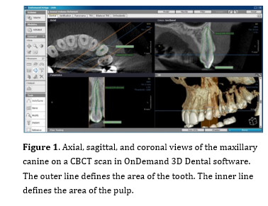
This analytical diagnostic study was conducted on 99 CBCT images of patients between 18 and 71 years, using the “confidence interval for one correlation” feature and considering α=0.05, R=0.5, and correlation coefficient of 0.3 between PTR and chronological age. The study sample was consisted of 51 females and 48 males. Ethical approval was granted by the Iran National Committee for Ethics in Biomedical Research (No: IR.IAU.DENTAL.REC.1399.175). The CBCT images used in this study had been obtained for diagnostic/therapeutic purposes. The patients’ age at the time of radiography was first calculated by subtracting their date of birth from the date of radiography. In this study, only sound fully erupted maxillary canine teeth were evaluated, and teeth with restorations, caries, wear, root resorption, cracks, periapical lesions, orthodontic, prosthodontic, and endodontic treatments, or developmental anomalies were excluded. All CBCT scans had been taken with Carestream9300 CBCT scanner (Kodak, France) with the exposure settings of 5 x 10 cm field of view, 10 mA, 90 kV, and 0.180 mm voxel size. The images had been obtained in standard conditions. The occlusal plane of the patients was parallel to the horizontal plane, and their mid-sagittal plane was perpendicular to the ground. The CBCT images were imported to OnDemand software version: 1.0.10.5385 (CyberMed, Korea), and five sections were obtained from each tooth in three planes, with a slice thickness of 1 mm and a slice interval of 0.1 mm. All CBCT images were assessed by one oral and maxillofacial radiologist with more than 10 years of experience in reading scans. The occlusal plane of the images was first parallelized to the horizontal plane, and in the axial plane, images were obtained at three sections: at the level of the cementoenamel junction (CEJ), at the mid-root, and at one-quarter distance from the CEJ. Next, in the axial plane, a curve was drawn over the maxillary arch. Using the reconstructed panoramic image, the mid-sagittal and mid-coronal sections were also made. Using the “Smart Pen” tool in the “Area” feature of OnDemand software, a minimum of 15 points were marked around the pulp, and a minimum of 30 points were marked around the tooth to outline the pulp and tooth. The tooth and pulp surface areas were calculated on each of the five sections (Figure 1). Next, the PTR was calculated and recorded for all five sections. The obtained values were used in regression formulae to assess their correlation with chronological age, gender, and standard error (1). Two weeks later, 10 CBCT images were randomly selected and measured again by the same observer to assess intra-examiner reliability.
Figure 1. Axial, sagittal, and coronal views of the maxillary canine on a CBCT scan in OnDemand 3D Dental software. The outer line defines the area of the tooth. The inner line defines the area of the pulp.
Results
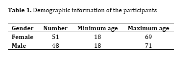
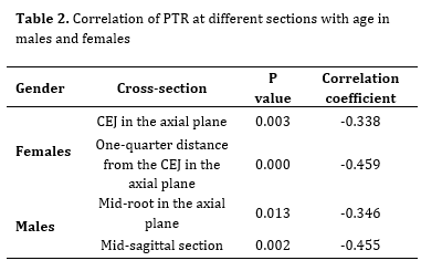
Table 1 presents the demographic information of the participants. In females, of the five sections evaluated, PTR at the CEJ (P=0.003) and one-quarter distance from the CEJ (P=0.000) sections had a significant correlation with age. In males, PTR at the axial mid-root (P=0.013) and mid-sagittal (longitudinal plane) sections (P=0.002) had a significant correlation with age (Table 2).
According to the PTR with significant correlations with age, the following formulae were designed for age estimation in males and females:
Estimated age in females = (X1 × -290/465) + (X2 × -232/598) + 68/309
X1: PTR at the cross-section at one-quarter distance from the CEJ
X2: PTR at the axial plane CEJ section
Estimated age in males = (X1 × -158/869) + (X2 × -414/823) + 82/481
X1: PTR at the mid-sagittal section
X2: PTR at the axial plane mid-root section
Table 1. Demographic information of the participants
Table 2. Correlation of PTR at different sections with age in males and females
Materials and Methods: This descriptive analytical study was conducted on 99 CBCT scans of patients between 18 to 71 years with fully erupted sound maxillary canine teeth. The CBCT scans were evaluated in five sections namely the mid-sagittal section, mid-coronal section, and axial planes [cementoenamel junction (CEJ) section, root section at one-quarter distance from the CEJ, and mid-root section] using OnDemand software. The tooth and pulp surface areas and PTR were calculated. The absolute error of age estimation was calculated by the regression test.
Results: In females, PTR of the CEJ section (-0.338) and one-quarter root section of the axial plane (-0.459; P=0.003 and P=0.000, respectively), and in males, PTR of the mid-root section of the axial plane (-0.346) and mid-sagittal section (-0.455; P=0.013 and P=0.002, respectively ) had significant correlations with age. The mean absolute error of age estimation in the present method, compared with the chronological age of individuals, was 7.227 years. In 39.5% of the cases, the obtained regression formula could estimate age with less than 5 years difference. No significant difference was noted in age estimation for males and females in this method.
Conclusion: It appears that PTR on CBCT images of maxillary canine teeth has a significant correlation with chronological age. The obtained formula in this study could estimate age with acceptable accuracy (less than 7.5 years).
Keywords: Age Determination by Teeth; Cone-Beam Computed Tomography; Cuspid; Dental Pulp; Dental Records; Forensic Dentistry
Introduction
Age estimation is highly important for forensic identification. It is also useful in crime investigations and identification of illegal
immigrants. According to Rai et al. [1], dental radiography was first used by Schuller in 1921 for forensic identification.
In 1995, the amount of secondary dentin deposition was quantified on dental radiographs, and its correlation with age was analyzed for the first time [2]. The most commonly applied methods for age estimation include the Gustafson and Johanson analyses, assessment of dentin translucency, and formation of cementum ring [3, 4]. Of these methods, assessment of secondary dentin deposition in the pulp chamber appears to be accurate. In some age determination methods, tooth cross-sections and dental radiographs are used to assess the size of pulp chamber. Panoramic and periapical radiographic modalities are often used for this purpose [5-7] especially when dental age is used to estimate the rate of development in young individuals [8, 9].
Previous methods based on two-dimensional (2D) radiographic modalities mainly had several shortcomings such as distortion and inaccurate magnification of images [10]. At present, CBCT can be used to eliminate many of the problems related to age estimation [11]. Several recent studies used CBCT to measure the tooth surface area. Uğur Aydın and Bayrak [12] found no significant difference between males and females in pulp/tooth surface area ratio (PTR) of maxillary central incisors while Rai et al. [1] found a direct correlation only between PTR in the axial plane and age.
To the best of the authors’ knowledge, PTR was not assessed in five sections using OnDemand software. Also, no previous study is available on pulp to tooth area measuring in five sections in the Iranian population. Due to the lack of studies and the information gap related to this issue, this study aimed to assess the accuracy of age estimation based on the PTR of maxillary canine teeth on CBCT scans using OnDemand software in an Iranian population.
Materials and Methods
This analytical diagnostic study was conducted on 99 CBCT images of patients between 18 and 71 years, using the “confidence interval for one correlation” feature and considering α=0.05, R=0.5, and correlation coefficient of 0.3 between PTR and chronological age. The study sample was consisted of 51 females and 48 males. Ethical approval was granted by the Iran National Committee for Ethics in Biomedical Research (No: IR.IAU.DENTAL.REC.1399.175). The CBCT images used in this study had been obtained for diagnostic/therapeutic purposes. The patients’ age at the time of radiography was first calculated by subtracting their date of birth from the date of radiography. In this study, only sound fully erupted maxillary canine teeth were evaluated, and teeth with restorations, caries, wear, root resorption, cracks, periapical lesions, orthodontic, prosthodontic, and endodontic treatments, or developmental anomalies were excluded. All CBCT scans had been taken with Carestream9300 CBCT scanner (Kodak, France) with the exposure settings of 5 x 10 cm field of view, 10 mA, 90 kV, and 0.180 mm voxel size. The images had been obtained in standard conditions. The occlusal plane of the patients was parallel to the horizontal plane, and their mid-sagittal plane was perpendicular to the ground. The CBCT images were imported to OnDemand software version: 1.0.10.5385 (CyberMed, Korea), and five sections were obtained from each tooth in three planes, with a slice thickness of 1 mm and a slice interval of 0.1 mm. All CBCT images were assessed by one oral and maxillofacial radiologist with more than 10 years of experience in reading scans. The occlusal plane of the images was first parallelized to the horizontal plane, and in the axial plane, images were obtained at three sections: at the level of the cementoenamel junction (CEJ), at the mid-root, and at one-quarter distance from the CEJ. Next, in the axial plane, a curve was drawn over the maxillary arch. Using the reconstructed panoramic image, the mid-sagittal and mid-coronal sections were also made. Using the “Smart Pen” tool in the “Area” feature of OnDemand software, a minimum of 15 points were marked around the pulp, and a minimum of 30 points were marked around the tooth to outline the pulp and tooth. The tooth and pulp surface areas were calculated on each of the five sections (Figure 1). Next, the PTR was calculated and recorded for all five sections. The obtained values were used in regression formulae to assess their correlation with chronological age, gender, and standard error (1). Two weeks later, 10 CBCT images were randomly selected and measured again by the same observer to assess intra-examiner reliability.
Figure 1. Axial, sagittal, and coronal views of the maxillary canine on a CBCT scan in OnDemand 3D Dental software. The outer line defines the area of the tooth. The inner line defines the area of the pulp.
Results
Table 1 presents the demographic information of the participants. In females, of the five sections evaluated, PTR at the CEJ (P=0.003) and one-quarter distance from the CEJ (P=0.000) sections had a significant correlation with age. In males, PTR at the axial mid-root (P=0.013) and mid-sagittal (longitudinal plane) sections (P=0.002) had a significant correlation with age (Table 2).
According to the PTR with significant correlations with age, the following formulae were designed for age estimation in males and females:
Estimated age in females = (X1 × -290/465) + (X2 × -232/598) + 68/309
X1: PTR at the cross-section at one-quarter distance from the CEJ
X2: PTR at the axial plane CEJ section
Estimated age in males = (X1 × -158/869) + (X2 × -414/823) + 82/481
X1: PTR at the mid-sagittal section
X2: PTR at the axial plane mid-root section
Table 1. Demographic information of the participants
Table 2. Correlation of PTR at different sections with age in males and females
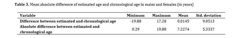
The mean absolute difference of estimated age from the chronological age of individuals was 7.227 years. In other words, a mean error of 7 years existed in age estimation compared with the chronological age of individuals (Table 3).
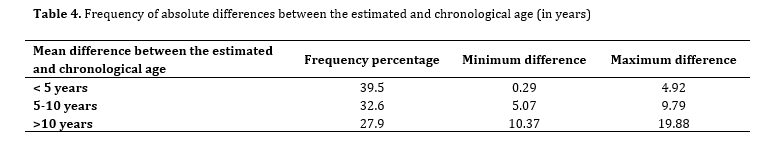
The method used in this study was capable of estimating age with a correlation coefficient of R=0.76. No significant difference was noted in age estimation for males and females in this method. Using the regression formula obtained in this study, in 39.5% of the cases, the estimated age had a difference less than 5 years with the chronological age. In 32.6%, the difference between the estimated and chronological age was 5 to 10 years. In 27.9%, the estimated age had a difference over 10 years with the chronological age (Table 4).
After 2 weeks, 10 CBCT scans were randomly selected and re-evaluated. In all cases, the intra-class correlation coefficient was found to be over 0.8, indicating high intra-examiner reliability.
Table 3. Mean absolute difference of estimated age and chronological age in males and females (in years)
Table 4. Frequency of absolute differences between the estimated and chronological age (in years)
It appears that PTR of maxillary canine teeth on CBCT scans has a significant correlation with chronological age of individuals. The formula designed in this study may be used for age estimation with acceptable accuracy.
Discussion
In forensic medicine, age estimation is highly important for forensic identification. The size of pulp chamber depends on the amount of secondary dentin deposition. Deposition of secondary dentin increases, and size of pulp chamber decreases with age [4]. The reduction in size of pulp chamber can be indirectly measured by radiography [13]. Previous studies used different methods for age estimation by CBCT, such as linear or surface area measurements [1]. Recently, researchers evaluated the pulp/tooth volume ratio [14]. Salemi et al. [15] discussed that PTR had a more significant correlation with age than linear measurements. CBCT has higher accuracy than 2D imaging modalities since it can provide different planes and sections, and enables 3D reconstruction of images [5]. Therefore, CBCT was used to calculate PTR in the present study. Selection of maxillary canine teeth for this study was because of the fact that maxillary canine teeth have the largest root size and longest survival in dental arch. They have been used for assessment of tooth dimensions and structure in many previous studies [16, 17]. Since previous investigations did not find a significant difference between teeth in the right and left quadrants [18], teeth from both quadrants were equally used in the present study. Rai et al. [1] evaluated the PTR in the sagittal, coronal, and axial planes. Accordingly, in the present study, five sections were evaluated in the three spatial planes for a more comprehensive assessment. The results showed that by assessment of PTR at the suggested cross-sections, age could be estimated in both males and females with a mean absolute error of 7.22 years. In 39.5% of the cases, the difference between the estimated and chronological age was less than 5 years. The difference between the estimated and chronological age by using the formula obtained in the present study was less than 10 years, which is acceptable in forensic medicine [3, 16, 19].
Also, in a study by Bansal et al. [20], five cross-sections of maxillary central incisor and canine teeth were examined by AutoCAD software. In their study, as in the present study, axial cross-sections at the CEJ of canine teeth were better for age estimation; although the estimated average age difference was 8.25 years, which was higher than the average age calculated in the present study. In the current study, of four sections that had a significant correlation with age and were used for designing of the age estimation formula for both males and females, three sections were in the axial plane. Haghanifar et al. [21] evaluated the PTR of maxillary and mandibular central incisors and canine teeth and reported similar results (7.45 for maxillary central incisors), although the number of examined teeth in the abovementioned study was higher than that in the present study. They assessed the axial CEJ section and the mid-sagittal section of the aforementioned teeth. They showed that maxillary central incisors had optimal potential for age estimation; however, in both males and females, the axial section had a stronger correlation with age than the sagittal section. Similar to the study by Haghanifar et al. [21], Trottier et al. [22] examined the PTR in 12 anterior teeth of the maxilla and mandible, and concluded that the correlation between PTR and chronological age was the strongest in the axial plane and in maxillary teeth. Axial sections, due to their distance from the site of occlusal load application, show lower deposition of secondary dentin compared with other sections, and thus, changes occurring in this section are mainly attributed to age rather than secondary dentin deposition due to other factors [23]. In a study by Alqarni et al, [24] maxillary central incisors and canine teeth were examined in two axial and sagittal sections. In their study, central incisors and axial sections were superior to the sagittal sections for age estimation; although, contrary to the present study and that of Haghanifar et al. [21], Alqarni et al. [24] found a weak correlation between the PTR and chronological age.
In the present study, the surface area of the teeth and pulp chamber was calculated, and the results were used to create a formula for age estimation. Salemi et al. [15] evaluated maxillary canine teeth by the Kvaal’s method, and found that tooth area ratios are superior to tooth length or width ratios for age estimation. Similar to the present study, they evaluated maxillary canine teeth due to their longer survival compared with other teeth, less wear, and having a distinct pulp chamber. They found no significant difference in age estimation between males and females, which was in accordance with the present results [15].
The method adopted in the present study was similar to that of Rai et al. [1] and the same tooth type and the same sections were evaluated for calculation of PTR. However, Rai et al. [1] used AutoCAD software in their study. Unlike the present study, only the CEJ section in the axial plane had a significant correlation with age, and age was estimated with 14.7 years difference from the chronological age, which was higher than the difference between the estimated and chronological age in the present study. Controversy in the results of the two studies may be due to their smaller sample size or use of different software programs. No significant correlation was found between PTR in the coronal plane and age in males or females, neither in the present study nor in the study by Rai et al. [1].
Ugur Aydin and Bayrak [12] used the PTR of maxillary central incisors on CBCT images for age estimation. They used inVivo 5 software and evaluated the teeth only in the sagittal plane. They found a significant correlation between PTR in the sagittal plane and chronological age of individuals. Bhogte et al. [25] also estimated the PTR in mid-axial, coronal and sagittal sections of maxillary central incisors and, contrary to the study by Ugur Aydin and Bayrak [12], they concluded that the mid‑sagittal sections had the least correlation for age prediction, although only 50 teeth were investigated. However, in the present study, only the sagittal plane in males had a significant correlation with age. Differences in tooth type and software programs may explain the variations in the results. However, PTR was not significantly different between males and females in both studies.
Afify et al. [16] evaluated maxillary and mandibular canine teeth, and indicated a significant correlation between PTR and age in both males and females. Moreover, they reported that maxillary canine teeth were superior to mandibular canine teeth for age estimation. Similar to other studies, they found no significant difference in statistical analyses between males and females. In both males and females, both the mid-sagittal and axial sections had significant correlations with age, but the mid-sagittal section showed a stronger correlation. This finding was different from the results of Rai et al, [1] the present results, and those of Haghanifar et al, [21] Trottier et al, [22] and Bhogte et al, [25] who demonstrated a stronger correlation between the PTR in the axial section and age, compared with the sagittal section. The present study found a mean difference of about 7 years between the estimated and chronological age. Similarly, Afify et al. [16] estimated age with the same level of error by designing a formula based on PTR in the axial plane. However, the formula estimated age with ±4.7-8.1 years difference with the chronological age in their study.
Recently, age estimation by 3D measurement of tooth volume has gained increasing popularity, and has been the topic of many investigations. However, specific software programs should be necessarily used for the techniques based on volumetric measurements [5, 26]. Computed tomography has also been applied for dental volumetric measurements [27, 28]. Nonetheless, CBCT largely replaced computed tomography for maxillofacial imaging due to its numerous advantages [29].
A higher correlation coefficient might have been obtained between the estimation method and the estimated age of patients in the present study if a larger sample size had been used. Finding eligible sound canine teeth was difficult, which led to a relatively small sample size.
Since studies on age estimation in different populations and racial groups have used different instruments and techniques for this purpose, some differences exist in the reported results. Thus, it appears that age estimation formulae that are based on tooth structure are exclusive to the respective racial and ethnic groups.
Conclusion
It appears that PTR of maxillary canine teeth on CBCT scans has a significant correlation with chronological age of individuals. The formula designed in this study may be used for age estimation with acceptable accuracy.
Acknowledgement
The authors appreciate the assistance of the staff of the Radiology Department of Islamic Azad University of Tehran, who helped in collection of CBCT scans, and also Dr. Mohammad Javad Kharazifard for his help in statistical analysis.
Type of Study: Original article |
Subject:
Radiology
References
1. Rai A, Acharya AB, Naikmasur VG. Age estimation by pulp-to-tooth area ratio using cone-beam computed tomography: A preliminary analysis. J Forensic Dent Sci. 2016 Sep-Dec;8(3):150-4. [DOI:10.4103/0975-1475.195118] [PMID] []
2. Zaher JF, Fawzy IA, Habib SR, Ali MM. Age estimation from pulp/tooth area ratio in maxillary incisors among Egyptians using dental radiographic images. J Forensic Leg Med. 2011 Feb;18(2):62-5. [DOI:10.1016/j.jflm.2010.12.004] [PMID]
3. Solheim T, Sundnes PK. Dental age estimation of Norwegian adults--a comparison of different methods. Forensic Sci Int. 1980 Jul-Aug;16(1):7-17. [DOI:10.1016/0379-0738(80)90174-7] [PMID]
4. Kvaal S, Solheim T. A non-destructive dental method for age estimation. J Forensic Odontostomatol. 1994 Jun;12(1):6-11.
5. Jagannathan N, Neelakantan P, Thiruvengadam C, Ramani P, Premkumar P, Natesan A, Herald JS, Luder HU. Age estimation in an Indian population using pulp/tooth volume ratio of mandibular canines obtained from cone beam computed tomography. J Forensic Odontostomatol. 2011 Jul 1;29(1):1-6.
6. Marroquin TY, Karkhanis S, Kvaal SI, Vasudavan S, Kruger E, Tennant M. Age estimation in adults by dental imaging assessment systematic review. Forensic Sci Int. 2017 Jun;275:203-11. [DOI:10.1016/j.forsciint.2017.03.007] [PMID]
7. Sakhdari S, Mehralizadeh S, Zolfaghari M, Madadi M. Age Estimation from Pulp/Tooth Area Ratio Using Digital Panoramic Radiography. J Iran Dent Assoc 2015;27(1) :19-23
8. Paryab M, Varaghi P, Mosharafian S, Kharrazi fard M J. Timing of Permanent Tooth Development in an Iranian Subpopulation. J Res Dent Maxillofac Sci 2023;8(3(:187-95. [DOI:10.61186/jrdms.8.3.187]
9. Nouri A, Sheikhi M, Haji Seyed Javadi S K. Relationship between the Estimated Dental Age and Chronological Age Using the Demirjian Method in an Iranian Population. J Res Dent Maxillofac Sci 2023;8 (4):243-8 [DOI:10.61186/jrdms.8.4.243]
10. Biuki N, Razi T, Faramarzi M. Relationship between pulp-tooth volume ratios and chronological age in different anterior teeth on CBCT. J Clin Exp Dent. 2017 May 1;9(5):e688-93. [DOI:10.4317/jced.53654] [PMID] []
11. Marroquin Penaloza TY, Karkhanis S, Kvaal SI, Nurul F, Kanagasingam S, Franklin D, et al. Application of the Kvaal method for adult dental age estimation using Cone Beam Computed Tomography (CBCT). J Forensic Leg Med. 2016 Nov;44:178-82. [DOI:10.1016/j.jflm.2016.10.013] [PMID]
12. Uğur Aydın Z, Bayrak S. Relationship Between Pulp Tooth Area Ratio and Chronological Age Using Cone-beam Computed Tomography Images. J Forensic Sci. 2019 Jul;64(4):1096-9. [DOI:10.1111/1556-4029.13986] [PMID]
13. Cameriere R, Ferrante L, Belcastro MG, Bonfiglioli B, Rastelli E, Cingolani M. Age estimation by pulp/tooth ratio in canines by peri-apical X-rays. J Forensic Sci. 2007 Jan;52(1):166-70. [DOI:10.1111/j.1556-4029.2006.00336.x] [PMID]
14. Star H, Thevissen P, Jacobs R, Fieuws S, Solheim T, Willems G. Human dental age estimation by calculation of pulp-tooth volume ratios yielded on clinically acquired cone beam computed tomography images of monoradicular teeth. J Forensic Sci. 2011 Jan;56 Suppl 1:S77-82. [DOI:10.1111/j.1556-4029.2010.01633.x] [PMID]
15. Salemi F, Farhadian M, Sabzkouhi BA, Saati S, Nafisi N. Age estimation by pulp to tooth area ratio in canine teeth using cone-beam computed tomography. Egypt J Forensic Sci. 2020;10(1):2. [DOI:10.1186/s41935-019-0176-9]
16. Afify M, Salem W, Mahmoud N. Age Estimation from Pulp/Tooth Area Ratio of Canines using Cone-Beam Computed Tomography Image Analysis: Study of an Egyptian Sample. J Forensic Res. 2019;10(434):2. [DOI:10.1186/s41935-019-0176-9]
17. Du C, Zhu Y, Hong L. Age-related changes in pulp cavity of incisors as a determinant for forensic age identification. J Forensic Sci. 2011 Jan;56 Suppl 1:S72-6. [DOI:10.1111/j.1556-4029.2010.01577.x] [PMID]
18. Asif MK, Nambiar P, Mani SA, Ibrahim NB, Khan IM, Sukumaran P. Dental age estimation employing CBCT scans enhanced with Mimics software: Comparison of two different approaches using pulp/tooth volumetric analysis. J Forensic Leg Med. 2018 Feb;54:53-61. [DOI:10.1016/j.jflm.2017.12.010] [PMID]
19. Asif MK, Nambiar P, Mani SA, Ibrahim NB, Khan IM, Lokman NB. Dental age estimation in Malaysian adults based on volumetric analysis of pulp/tooth ratio using CBCT data. Leg Med (Tokyo). 2019 Feb;36:50-58. [DOI:10.1016/j.legalmed.2018.10.005] [PMID]
20. Bansal V, Konidena A, Nagi R, Kataria APS, Yumnam N, Farooq F. Correlation of pulp-to-tooth area ratio with age and gender using CBCT of maxillary central incisor and canine: A comparative study. JIAOMR. 2022;34(1):87-94. [DOI:10.4103/jiaomr.jiaomr_67_21]
21. Haghanifar S, Ghobadi F, Vahdani N, Bijani A. Age estimation by pulp/tooth area ratio in anterior teeth using cone-beam computed tomography: comparison of four teeth. J Appl Oral Sci. 2019 Aug 12;27:e20180722. [DOI:10.1590/1678-7757-2018-0722] [PMID] []
22. Trottier M, Keenan S, Fairgrieve SI. Individual age estimation using pulp-to-tooth area ratio in single-rooted teeth. Can Soc Forensic Sci. 2023:1-14. [DOI:10.1080/00085030.2023.2276562]
23. Nemsi H, Haj Salem N, Bouanene I, Ben Jomaa S, Belhadj M, Mosrati MA, Aissaoui A, Ben Amor F, Chadly A. Age assessment in canine and premolar by cervical axial sections of cone-beam computed tomography. Leg Med (Tokyo). 2017 Sep;28:31-6. [DOI:10.1016/j.legalmed.2017.07.004] [PMID]
24. Alqarni A, Ajmal M, Hakami RM, Alassmi AA, Chalikkandy SN, Arem S. Relationship between Pulp-Tooth Area Ratio and Chronological Age among Saudi Arabian Adults: A Cone Beam Computed Tomography Image Analysis. Appl Sci. 2023;13(13):7945. [DOI:10.3390/app13137945]
25. Bhogte SA, Jois HS, Anusha AV, Pattnaik A, Rana M. Estimation of Age by Calculating Pulp-to-Tooth Area Ratio using CBCT in Maxillary Central Incisors: A Retrospective Cross-Sectional Preliminary Analysis on the Hyderabad Population. JIAOMR. 2023;35(2):246-9. [DOI:10.4103/jiaomr.jiaomr_59_23]
26. Gulsahi A, Kulah CK, Bakirarar B, Gulen O, Kamburoglu K. Age estimation based on pulp/tooth volume ratio measured on cone-beam CT images. Dentomaxillofac Radiol. 2018 Jan;47(1):20170239. [DOI:10.1259/dmfr.20170239] [PMID] []
27. Pinchi V, Pradella F, Buti J, Baldinotti C, Focardi M, Norelli GA. A new age estimation procedure based on the 3D CBCT study of the pulp cavity and hard tissues of the teeth for forensic purposes: A pilot study. J Forensic Leg Med. 2015 Nov;36:150-7. [DOI:10.1016/j.jflm.2015.09.015] [PMID]
28. Queiroz CL, Silva RF, Silva RHA. Computed tomography use on age estimation in forensic dentistry: A review. J Forensic Sci Criminol. 2016;4(1):1-6. [DOI:10.15744/2348-9804.4.105]
29. Sue M, Oda T, Sasaki Y, Ogura I. Age-related changes in the pulp chamber of maxillary and mandibular molars on cone-beam computed tomography images. Oral Radiol. 2018 Sep;34(3):219-23. [DOI:10.1007/s11282-017-0300-1] [PMID]
Send email to the article author
| Rights and permissions | |
 |
This work is licensed under a Creative Commons Attribution-NonCommercial 4.0 International License. |