Journal of Research in Dental
and Maxillofacial Sciences
Volume 9, Issue 1 (3-2024)
J Res Dent Maxillofac Sci 2024, 9(1): 57-60 |
Back to browse issues page


Download citation:
BibTeX | RIS | EndNote | Medlars | ProCite | Reference Manager | RefWorks
Send citation to:


BibTeX | RIS | EndNote | Medlars | ProCite | Reference Manager | RefWorks
Send citation to:
Sinha D, Sinha I. Autogenous Bone Grafting for Treatment of a Complex Odontoma: A Case Report. J Res Dent Maxillofac Sci 2024; 9 (1) :57-60
URL: http://jrdms.dentaliau.ac.ir/article-1-508-en.html
URL: http://jrdms.dentaliau.ac.ir/article-1-508-en.html


1- Department of Oral and Maxillofacial Surgery, SRM Kattankulathur Dental College and Hospital, Chengalpattu District, Tamil Nadu, India , devanshusinha456@gmail.com
2- Department of Orthodontics and Dentofacial Orthopedics, A.B. Shetty Memorial Institute of Dental Sciences, Deralakatte, Mangalore, Karnataka, India.
2- Department of Orthodontics and Dentofacial Orthopedics, A.B. Shetty Memorial Institute of Dental Sciences, Deralakatte, Mangalore, Karnataka, India.
Full-Text [PDF 354 kb]
(1089 Downloads)
| Abstract (HTML) (2363 Views)
Full-Text: (735 Views)
Abstract
Background and Aim: Complex odontomas are commonly found in the anterior maxilla and mandible, and are typically asymptomatic unless they cause tooth displacement or other complications. Management of odontomas depends on the size and location of the lesion, and may include surgical excision and bone grafting.
Case Presentation: This case report presents a rare case of complex odontoma in a 20-year-old female patient complaining of a maxillary malposed tooth along with a swelling for the past 5 years. The patient underwent excision of the lesion along with extraction of the affected tooth and autogenous bone grafting of the defect site. Histopathological analysis confirmed the provisional diagnosis of odontoma.
Conclusion: This case report highlights the importance of proper diagnosis and management of odontomas to prevent potential complications.
Key Words: Odontoma; Autografts; Maxilla; Mandible
Introduction
Odontoma is a developmental abnormality of the oral cavity that arises from the proliferation of odontogenic epithelium and mesenchyme, resulting in formation of tooth-like structures. It is the most common odontogenic tumor, accounting for approximately 67% of all odontogenic tumors, and is often detected incidentally in routine radiographic examination [1]. The incidence of odontoma is estimated to be 1 per 1000 to 2000 individuals, with a slight predilection for males and a peak incidence in the second decade of life [2]. Although odontomas can occur in any location of the oral cavity, they are most commonly found in the posterior maxilla, followed by the anterior maxilla and mandible [3]. Odontomas are classified into two main types: compound odontomas, which consist of multiple small tooth-like structures, and complex odontomas, which consist of a disorganized mass of dental tissues that does not resemble normal teeth [4]. Management of odontomas depends on the type, size, and location of the lesion. Small, asymptomatic lesions may require no treatment and can be monitored with regular radiographic examinations. Larger or symptomatic lesions may require surgical excision, often with the use of autogenous bone grafts to fill the resultant defect [5,6]. In this case report, we present a patient with a complex odontoma in the anterior maxilla, which was managed by surgical excision and autogenous bone grafting. By reporting this case, the authors wish to raise awareness about this common developmental abnormality and its management.
Case Presentation
A 20-year-old female patient was referred to the Department of Oral and Maxillofacial Surgery complaining of a maxillary malposed tooth along with a swelling for the past 5 years. The swelling was asymptomatic in nature, with no history of associated pain. The patient did not have any underlying condition or syndromic history. The patient was informed about the procedure and signed written informed consent before treatment.
On examination, the patient had a normal facial profile with no gross asymmetry. She had normal mouth opening and normal temporomandibular movement. The patient had a partially erupted left central incisor that only its incisal third was visible. A swelling was present over the apical portion of it, which was bony hard on palpation. There was no sign of infection in or around the tooth region.
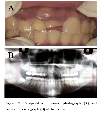
Panoramic radiograph revealed a radiopaque mass that resembled a bulbous tooth measuring approximately 3 x 2 cm in size, which led to provisional diagnosis of odontoma (Figure 1).
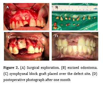
Due to the size of the lesion and its proximity to the nasal floor, an excisional biopsy was performed under general anesthesia. A mucoperiosteal flap was reflected, peripheral osteotomy was performed, and the entire mass was excised, which created a defect at the operated site. A block graft was harvested from the left parasymphyseal region and placed at the defect site, and was eventually secured with a titanium screw (Figure 2).
Figure 1. Preoperative intraoral photograph (A) and panoramic radiograph (B) of the patient
Figure 2. (A) Surgical exploration, (B) excised odontoma, (C) symphyseal block graft placed over the defect site, (D) postoperative photograph after one month
The histopathology report confirmed the provisional diagnosis of odontoma. The patient had an uneventful postoperative recovery, and was followed up for a period of 6 months. There was no evidence of recurrence, and the surgical site healed satisfactorily.
This case report highlights the importance of considering odontoma as a differential diagnosis in patients presenting with a malposed tooth and a swelling in the oral cavity. It also highlights the significance of meticulous surgical planning and execution to prevent any complications during the surgical procedure.
Discussion
Odontomas are the most common odontogenic tumors that arise from the epithelial and mesenchymal remnants of tooth buds. They are classified as either compound or complex depending on their histological characteristics. Compound odontomas are typically composed of multiple small tooth-like structures, while complex odontomas consist of a disorganized mass of dental tissues. Odontomas are generally asymptomatic and are often discovered incidentally on routine radiographs or during routine dental examinations. However, they may cause impaction or displacement of the adjacent teeth, root resorption, and in rare cases, facial asymmetry and/or pain.
The treatment of odontomas usually involves surgical removal of the lesion with an adequate safety margin. In cases where the odontoma is located in a critical area or involves vital structures, a conservative approach may be adopted. The use of autogenous bone grafts is a popular technique for reconstruction of osseous defects following odontoma excision. Autogenous bone grafts have several advantages, including their osteogenic potential, low antigenicity, and lack of disease transmission.
Several studies have reported a high success rate for autogenous bone grafts in reconstruction of osseous defects following odontoma excision. For instance, Kadri et al. [7] reported 97.6% success rate for autogenous bone grafting in 42 cases of odontoma excision. Chang et al. [8] conducted a study on 81 Taiwanese patients with odontomas. Odontomas were more common in the first two decades of life, and had higher frequency in the maxilla and in the anterior region. Compound odontomas were predominated. Odontomas were often associated with impacted teeth, particularly maxillary central incisors. Histologically, odontomas displayed diverse components and occasional dentigerous cysts. Surgical excision showed no recurrence during 1-15 years of follow-up. Maltagliati et al. [9] found that complex odontomas, like the one reported in the present case report, are more commonly found in the anterior maxilla and mandible, and are typically asymptomatic unless they cause tooth displacement or other complications.
Tomizawa et al. [10] conducted a retrospective study on 39 odontoma cases in Japanese children (1979-2002). Delayed tooth eruption was among the chief complaints of patients. Most cases had tooth eruption issues in the maxillary anterior region. The treatment involved surgical removal, with rare recurrence emphasizing the importance of periodic observations in very young children with early-stage odontomas. Kämmerer et al. [11] assessed the management of retained teeth in odontomas. Among 45 patients, initial symptoms included delayed eruption, pain, and swellings. Odontomas were incidentally discovered in 28 cases. Extractions were more common in older patients, and among the non-extracted teeth, orthodontic alignment was proven to be successful in some cases. No relapse was observed in any case, emphasizing the value of panoramic radiography for their early detection. Their study underscored that odontoma removal can facilitate normal tooth eruption, often avoiding extractions, and orthodontic alignment is a reasonable therapeutic option. Nguyen and Van Huynh [12] conducted a retrospective study on 90 odontoma cases in Vietnam. Complex odontoma patients were older, and 67% of them showed intraoral bone expansion. Complex odontomas had larger dimensions and caused fewer problems in dentition than compact odontomas, which were associated with more complications in the adjacent teeth. Odontoma development varied with age, subtype, and gender. Their study emphasized on the significance of clinical and radiographic features for early diagnosis and tailored treatment, especially for younger patients.
Sun et al. [13] reviewed multiple odontomas by assessing 12 cases reported in the literature. They reported that multiple odontomas can occur in two to four jaw quadrants, presenting as compound or complex type. They highlighted the diverse manifestations of multiple odontomas in terms of localization and histological types.
Conclusion
Odontomas are benign tumors that commonly occur in the oral cavity. They are usually asymptomatic and are often discovered incidentally on routine radiographs or during routine dental examinations. Surgical excision with an adequate safety margin is the treatment of choice, and autogenous bone grafting is a popular technique for reconstruction of osseous defects following odontoma excision, with a high success rate reported in the literature. Regular dental checkups and radiographic examinations are essential for early detection and management of odontomas.
Background and Aim: Complex odontomas are commonly found in the anterior maxilla and mandible, and are typically asymptomatic unless they cause tooth displacement or other complications. Management of odontomas depends on the size and location of the lesion, and may include surgical excision and bone grafting.
Case Presentation: This case report presents a rare case of complex odontoma in a 20-year-old female patient complaining of a maxillary malposed tooth along with a swelling for the past 5 years. The patient underwent excision of the lesion along with extraction of the affected tooth and autogenous bone grafting of the defect site. Histopathological analysis confirmed the provisional diagnosis of odontoma.
Conclusion: This case report highlights the importance of proper diagnosis and management of odontomas to prevent potential complications.
Key Words: Odontoma; Autografts; Maxilla; Mandible
Introduction
Odontoma is a developmental abnormality of the oral cavity that arises from the proliferation of odontogenic epithelium and mesenchyme, resulting in formation of tooth-like structures. It is the most common odontogenic tumor, accounting for approximately 67% of all odontogenic tumors, and is often detected incidentally in routine radiographic examination [1]. The incidence of odontoma is estimated to be 1 per 1000 to 2000 individuals, with a slight predilection for males and a peak incidence in the second decade of life [2]. Although odontomas can occur in any location of the oral cavity, they are most commonly found in the posterior maxilla, followed by the anterior maxilla and mandible [3]. Odontomas are classified into two main types: compound odontomas, which consist of multiple small tooth-like structures, and complex odontomas, which consist of a disorganized mass of dental tissues that does not resemble normal teeth [4]. Management of odontomas depends on the type, size, and location of the lesion. Small, asymptomatic lesions may require no treatment and can be monitored with regular radiographic examinations. Larger or symptomatic lesions may require surgical excision, often with the use of autogenous bone grafts to fill the resultant defect [5,6]. In this case report, we present a patient with a complex odontoma in the anterior maxilla, which was managed by surgical excision and autogenous bone grafting. By reporting this case, the authors wish to raise awareness about this common developmental abnormality and its management.
Case Presentation
A 20-year-old female patient was referred to the Department of Oral and Maxillofacial Surgery complaining of a maxillary malposed tooth along with a swelling for the past 5 years. The swelling was asymptomatic in nature, with no history of associated pain. The patient did not have any underlying condition or syndromic history. The patient was informed about the procedure and signed written informed consent before treatment.
On examination, the patient had a normal facial profile with no gross asymmetry. She had normal mouth opening and normal temporomandibular movement. The patient had a partially erupted left central incisor that only its incisal third was visible. A swelling was present over the apical portion of it, which was bony hard on palpation. There was no sign of infection in or around the tooth region.
Panoramic radiograph revealed a radiopaque mass that resembled a bulbous tooth measuring approximately 3 x 2 cm in size, which led to provisional diagnosis of odontoma (Figure 1).
Due to the size of the lesion and its proximity to the nasal floor, an excisional biopsy was performed under general anesthesia. A mucoperiosteal flap was reflected, peripheral osteotomy was performed, and the entire mass was excised, which created a defect at the operated site. A block graft was harvested from the left parasymphyseal region and placed at the defect site, and was eventually secured with a titanium screw (Figure 2).
Figure 1. Preoperative intraoral photograph (A) and panoramic radiograph (B) of the patient
Figure 2. (A) Surgical exploration, (B) excised odontoma, (C) symphyseal block graft placed over the defect site, (D) postoperative photograph after one month
The histopathology report confirmed the provisional diagnosis of odontoma. The patient had an uneventful postoperative recovery, and was followed up for a period of 6 months. There was no evidence of recurrence, and the surgical site healed satisfactorily.
This case report highlights the importance of considering odontoma as a differential diagnosis in patients presenting with a malposed tooth and a swelling in the oral cavity. It also highlights the significance of meticulous surgical planning and execution to prevent any complications during the surgical procedure.
Discussion
Odontomas are the most common odontogenic tumors that arise from the epithelial and mesenchymal remnants of tooth buds. They are classified as either compound or complex depending on their histological characteristics. Compound odontomas are typically composed of multiple small tooth-like structures, while complex odontomas consist of a disorganized mass of dental tissues. Odontomas are generally asymptomatic and are often discovered incidentally on routine radiographs or during routine dental examinations. However, they may cause impaction or displacement of the adjacent teeth, root resorption, and in rare cases, facial asymmetry and/or pain.
The treatment of odontomas usually involves surgical removal of the lesion with an adequate safety margin. In cases where the odontoma is located in a critical area or involves vital structures, a conservative approach may be adopted. The use of autogenous bone grafts is a popular technique for reconstruction of osseous defects following odontoma excision. Autogenous bone grafts have several advantages, including their osteogenic potential, low antigenicity, and lack of disease transmission.
Several studies have reported a high success rate for autogenous bone grafts in reconstruction of osseous defects following odontoma excision. For instance, Kadri et al. [7] reported 97.6% success rate for autogenous bone grafting in 42 cases of odontoma excision. Chang et al. [8] conducted a study on 81 Taiwanese patients with odontomas. Odontomas were more common in the first two decades of life, and had higher frequency in the maxilla and in the anterior region. Compound odontomas were predominated. Odontomas were often associated with impacted teeth, particularly maxillary central incisors. Histologically, odontomas displayed diverse components and occasional dentigerous cysts. Surgical excision showed no recurrence during 1-15 years of follow-up. Maltagliati et al. [9] found that complex odontomas, like the one reported in the present case report, are more commonly found in the anterior maxilla and mandible, and are typically asymptomatic unless they cause tooth displacement or other complications.
Tomizawa et al. [10] conducted a retrospective study on 39 odontoma cases in Japanese children (1979-2002). Delayed tooth eruption was among the chief complaints of patients. Most cases had tooth eruption issues in the maxillary anterior region. The treatment involved surgical removal, with rare recurrence emphasizing the importance of periodic observations in very young children with early-stage odontomas. Kämmerer et al. [11] assessed the management of retained teeth in odontomas. Among 45 patients, initial symptoms included delayed eruption, pain, and swellings. Odontomas were incidentally discovered in 28 cases. Extractions were more common in older patients, and among the non-extracted teeth, orthodontic alignment was proven to be successful in some cases. No relapse was observed in any case, emphasizing the value of panoramic radiography for their early detection. Their study underscored that odontoma removal can facilitate normal tooth eruption, often avoiding extractions, and orthodontic alignment is a reasonable therapeutic option. Nguyen and Van Huynh [12] conducted a retrospective study on 90 odontoma cases in Vietnam. Complex odontoma patients were older, and 67% of them showed intraoral bone expansion. Complex odontomas had larger dimensions and caused fewer problems in dentition than compact odontomas, which were associated with more complications in the adjacent teeth. Odontoma development varied with age, subtype, and gender. Their study emphasized on the significance of clinical and radiographic features for early diagnosis and tailored treatment, especially for younger patients.
Sun et al. [13] reviewed multiple odontomas by assessing 12 cases reported in the literature. They reported that multiple odontomas can occur in two to four jaw quadrants, presenting as compound or complex type. They highlighted the diverse manifestations of multiple odontomas in terms of localization and histological types.
Conclusion
Odontomas are benign tumors that commonly occur in the oral cavity. They are usually asymptomatic and are often discovered incidentally on routine radiographs or during routine dental examinations. Surgical excision with an adequate safety margin is the treatment of choice, and autogenous bone grafting is a popular technique for reconstruction of osseous defects following odontoma excision, with a high success rate reported in the literature. Regular dental checkups and radiographic examinations are essential for early detection and management of odontomas.
Type of Study: Case report |
Subject:
Oral & maxillofacial surgery
References
1. Reichart PA, Philipsen HP. Odontogenic Tumors and Allied Lesions. London, UK: Quintessence Publishing Co; 2004.
2. Neville BW, Dam DD, Allen CM, Bouquot JE. Oral and maxillofacial pathology. 3rd ed. Philadelphia: WB. Saunders; 2016.
3. Slootweg PJ. An analysis of the interrelationship of the mixed odontogenic tumors--ameloblastic fibroma, ameloblastic fibro-odontoma, and the odontomas. Oral Surg Oral Med Oral Pathol.1981 Mar;51(3):266-76.
4. Shear M, Speight P. Cysts of the Oral and Maxillofacial Regions. 4th ed. Wiley; 2007. [DOI:10.1002/9780470759769]
5. de Oliveira BH, Campos V, Marçal S. Compound odontoma--diagnosis and treatment: three case reports. Pediatr Dent. 2001 Mar-Apr;23(2):151-7.
6. Etemadi A, Bitaraf T, Amini A, Goudarzi M, Nadafpour N. Bacterial Accumulation on Triclosan-Coated and Silk Sutures After Dental Implant Surgery. J Res Dent Maxillofac Sci 2019; 4 (3) :1-4. [DOI:10.29252/jrdms.4.3.1]
7. Kadri M, Grand A, Mondoloni M, Walter P, Boussouni S, Rochefort J. A Giant Posterior Maxillary Intra-Sinusal Complex Odontoma: A Clinical Case. Cureus. 2023 Jul 27;15(7):e42546. [DOI:10.7759/cureus.42546]
8. Chang JY, Wang JT, Wang YP, Liu BY, Sun A, Chiang CP. Odontoma: a clinicopathologic study of 81 cases. J Formos Med Assoc. 2003 Dec;102(12):876-82.
9. Maltagliati A, Ugolini A, Crippa R, Farronato M, Paglia M, Blasi S, Angiero F. Complex odontoma at the upper right maxilla: Surgical management and histomorphological profile. Eur J Paediatr Dent. 2020 Sep;21(3):199-202.
10. Tomizawa M, Otsuka Y, Noda T. Clinical observations of odontomas in Japanese children: 39 cases including one recurrent case. Int J Paediatr Dent. 2005 Jan;15(1):37-43. [DOI:10.1111/j.1365-263X.2005.00607.x] [PMID]
11. Kämmerer PW, Schneider D, Schiegnitz E, Schneider S, Walter C, Frerich B, Kunkel M. Clinical parameter of odontoma with special emphasis on treatment of impacted teeth-a retrospective multicentre study and literature review. Clin Oral Investig. 2016 Sep;20(7):1827-35. [DOI:10.1007/s00784-015-1673-3] [PMID]
12. Nguyen DK, Van Huynh D. Clinical and radiological characteristics of odontomas: A retrospective study of 90 cases. Imaging Sci Dent. 2023 Jun;53(2):117-26. [DOI:10.5624/isd.20220184] [PMID] []
13. Sun L, Sun Z, Ma X. Multiple complex odontoma of the maxilla and the mandible. Oral Surg Oral Med Oral Pathol Oral Radiol. 2015 Jul;120(1): e11-6. [DOI:10.1016/j.oooo.2015.02.488] [PMID]
Send email to the article author
| Rights and permissions | |
 |
This work is licensed under a Creative Commons Attribution-NonCommercial 4.0 International License. |