

Volume 8, Issue 1 (1-2023)
J Res Dent Maxillofac Sci 2023, 8(1): 28-37 |
Back to browse issues page


Ethics code: IR.IAU.DENTAL.REC.1400.161
Download citation:
BibTeX | RIS | EndNote | Medlars | ProCite | Reference Manager | RefWorks
Send citation to:


BibTeX | RIS | EndNote | Medlars | ProCite | Reference Manager | RefWorks
Send citation to:
Beikian Ghavidel P, Moshari A A, Toursavadkouhi S. Effectiveness of D-RaCe, SP1 and R-Endo Rotary Systems for Root Canal Filling Material Removal from the Human Mandibular Molars: A Micro-CT Study. J Res Dent Maxillofac Sci 2023; 8 (1) :28-37
URL: http://jrdms.dentaliau.ac.ir/article-1-478-en.html
URL: http://jrdms.dentaliau.ac.ir/article-1-478-en.html


1- Endodontic Department, Dental School, Islamic Azad University of Medical sciences, Tehran, Iran , s_savadkouhi@yahoo.com
2- Endodontic Department, Dental School, Islamic Azad University of Medical sciences, Tehran, Iran
3- Dental Material Research Center, Endodontic Department, Dental School, Islamic Azad University of Medical sciences, Tehran, Iran
2- Endodontic Department, Dental School, Islamic Azad University of Medical sciences, Tehran, Iran
3- Dental Material Research Center, Endodontic Department, Dental School, Islamic Azad University of Medical sciences, Tehran, Iran
Full-Text [PDF 828 kb]
(429 Downloads)
| Abstract (HTML) (950 Views)
Introduction
Inability to efficiently clean the root canal from filling materials in retreatment results in incomplete removal of microorganisms and inadequate cleaning, and subsequent reduction in retreatment success rate [1]. Root canal therapy has a success rate of approximately 90% [2]. Nonetheless, treatment failure occurs due to a number of reasons, such as poor adherence to the standard protocols, inadequate instrumentation of the root canal system, residual microorganisms remaining in the canal, poor quality of obturation, under-extension or over-extension of root filling material, and coronal leakage [3].
Although no retreatment method is capable of complete removal of root filling materials from the root canal, maximum removal of root filling material is imperative in endodontic retreatment to ensure a high success rate [1].
Several endodontic instruments have been suggested for more efficient elimination of root filling materials from the root canals such as hand files, ultrasonic instruments, laser, rotary instruments including Gates-Glidden drills and nickel-titanium (NiTi) rotary files, and solvents [1], among others. D-RaCe, SP1, and R-Endo are among the commonly used retreatment rotary systems designed specifically for intra-canal filling material removal.
Several methods have been suggested for evaluation of the efficacy of endodontic instruments in removal of root filling materials in root canal retreatment such as assessment by stereomicroscope, cone-beam computed tomography, scanning electron microscopy, and micro-computed tomography (micro-CT) [4]. The main advantage of micro-CT is its high accuracy for detection and quantification of residual root filling materials without physical manipulation of root canals [5].
Considering the gap of information regarding the comparative retreatment efficacy of D-RaCe, SP1, and R-Endo in removal of root filling materials, this study aimed to compare the retreatment efficacy of D-RaCe, SP1, and R-Endo in removal of gutta-percha from the mesiobuccal canal of mandibular molars using micro-CT. The null hypothesis was that no significant difference would be found in retreatment efficacy of D-RaCe, SP1, and R-Endo in removal of gutta-percha from the mesiobuccal canal of mandibular molars.
Materials and Methods
This experimental in vitro study was carried out on 30 mandibular molar teeth that had been extracted as the result of hopeless periodontal prognosis or as part of prosthetic treatment plan. The study protocol gained approval from the ethics committee of Islamic Azad University, School of Dentistry (IR.IAU.DENTAL.REC.1400.161).
Sample size:
The sample size was calculated to be 10 in each group based on previous investigations and assuming α=0.05, β=0.2, and study power of 80% to find a maximum difference 30% larger than the standard deviation [6,7].
Eligibility criteria:
The inclusion criteria were mandibular molars with completely formed roots, no internal/external root resorption, no calcification, no fracture, and no previous endodontic treatment. Also, the mesiobuccal canals had to have 20 to 30-degree curvature as measured according to the Schneider’s method [6]. The teeth underwent periapical radiography in mesiodistal and buccolingual directions to ensure their eligibility for study inclusion [6].
Methodology:
The tooth crowns were cut by a diamond disc (KG Sorensen, Cotia, SP, Brazil) at low speed under water coolant to standardize the root length at 16 mm in all teeth. Access cavity was created by a round diamond bur (Bosphorus, Istanbul, Turkey). To determine the working length, a #15 K-file (Mani Inc., Tochigi, Japan) was inserted into the canal until its tip was seen at the apex. Working length was determined 0.5 mm shorter than the apical foramen [8]. After access cavity preparation, the root canals were rinsed with 10 mL of 2.5% sodium hypochlorite. For this purpose, a #15 manual K-file was first introduced into the canal to find the canal path.
The root canals were then instrumented with Denco-D-Super files (Shenzhen Denco medical Ltd., China) at a speed of 150 to 350 rpm and 2 N/cm torque as instructed by the manufacturer. This system has 6 file sizes, including 3 shaping and 3 finishing files. SX is the orifice shaper with 19 mm length and 3.5% to 19% variable taper from the tip to the shank. S1 file (purple) is size 10 with 25 mm length, S2 file (white) is size 15 with 25 mm length, F1 file (yellow) is size 20 with 25 mm length, F2 file (red) is size 25 with 25 mm length, and F3 file (blue) is size 30 with 25 mm length, that were used in an orderly manner as instructed by the manufacturer. In the process of instrumentation, the root canals were frequently irrigated with 10 mL of 2.5% sodium hypochlorite (Microvem, Istanbul, Turkey) with a 30-gauge needle (aviTip, Ultradent Products, South Jordan, UT, USA). Next, the canals were rinsed with 17% EDTA (Imicryl, Konya, Turkey) for 1 minute for smear layer removal. The root canals were finally irrigated with 10 mL of 2.5% sodium hypochlorite followed by a final rinse with saline. They were then dried with paper points (Diadent, South Korea) and obturated with gutta-percha (Diadent, South Korea) and AH Plus sealer (Dentsply, DeTrey, Konstanz, Germany) by the lateral compaction technique. The root filling length was 12 mm. Excess gutta-percha was removed by a hot instrument, and the root filling was packed with an endodontic plugger. The teeth were then temporarily restored with Cavit (3M ESPE, Germany) with 4 mm thickness to ensure optimal coronal seal [3]. The teeth were then incubated at 37°C for 3 weeks to allow complete setting of sealer. Next, the teeth underwent micro-CT (LOTUS-InVivo) with 19 µm pixel size, 80 kV voltage, 100 µAm amperage, and 28-minute time for each root. To reconstruct 2D images in the axial plane, cross-sectional slices with 50 µm slice thickness were reconstructed perpendicular to the longitudinal axis of the tooth from the apex towards the coronal region. Accordingly, 2D images were reconstructed using LOTUS-InVivo-ACQ software. Empty spaces and filled areas were reconstructed according to their difference in density first two-dimensionally, and then three-dimensionally using the software program, and their volume was quantified and reported in cubic millimeters (mm3) [9].
Next, the teeth were randomly divided into three groups (n=10) for retreatment with D-RaCe, SP1, and R-Endo rotary systems using a table of random numbers. The teeth then underwent retreatment by the same operator. For this purpose, root filling materials were removed from the coronal part of the canal using #2 and #3 Gates-Glidden drills.
Afterwards, 1 drop of chloroform solvent was added to the canal [10]. Depending on group allocation of the teeth, they underwent retreatment with D-RaCe NiTi rotary system (FKG, La Chaux-de Fonds, Switzerland), SP1 (Dentaire, SP1-RTRA, China), or R-Endo (Micro-Mega, France) as instructed by the manufacturer.
D-RaCe group: D-RaCe retreatment system has two files of DR1 and DR2. DR1 operates at 1000 rpm with 1 to 1.5 N/cm torque and is used for the coronal third while DR2 operates at 600 rpm with 1 to 1.5 N/cm torque and is used for the middle and apical thirds of the canals.
SP1 group: SP1 files with size 30 are used for the coronal third, those with size 25 are used for the middle third, and files with size 20 are used for the apical third of the canal with a speed of 350 rpm and 2 N/cm torque.
R-Endo group: This system includes three files of R1, R2, and R3. R1 is used for the coronal third, R2 for the middle third, and R3 for the apical third, with a suggested speed of 300 to 400 rpm and 1.5 N/cm torque. The files are used with gentle apical pressure.
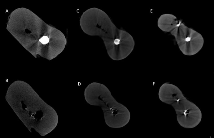
All files were discarded after use in 5 canals [11]. In the process of endodontic retreatment, the root canals were rinsed with 10 mL of 2.5% sodium hypochlorite with a 30-gauge needle (aviTip, Ultradent Products, South Jordan, UT, USA) after using each file. Also, after completion of instrumentation, the canals were rinsed with 17% EDTA for 1 minute followed by 10 mL of 2.5% sodium hypochlorite, and a final rinse with saline. They were then dried with paper points. The criteria for accomplishment of retreatment included reaching the primary working length, observing smooth canal walls, and absence of any root filling material on the files. The teeth underwent micro-CT again to quantify the residual root filling material in the canals in each group. The volume of residual gutta-percha and sealer in the canal was divided by the total volume of root filling material and multiplied by 100 to calculate the volumetric percentage of cleaning efficiency of the rotary systems. Figures 1-3 show the micrographs of representative specimens from the three groups before and after retreatment.
Figure 1. Micro-CT view of the canal before and after retreatment with SP1 files: (A) apical third before retreatment, (B) apical third after retreatment; (C) middle third before retreatment; (D) middle third after retreatment, (E) coronal third before retreatment; (F) coronal third after retreatment
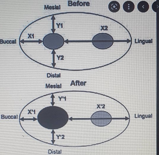
Apical transportation was also calculated by LOTUS InVivo-REC software as a secondary outcome [9]. Canal transportation was only calculated in the apical third by using a digital caliper for the measurements on the scans with the formula below:
Canal transportation = (M1-M2) – (D1-D2) Where M1 is the shortest distance between the mesial wall of the root and mesial wall of the uninstrumented canal
Figure 2. Micro-CT view of the canal before and after retreatment with D-RaCe files: (A) apical third before retreatment, (B) apical third after retreatment; (C) middle third before retreatment; (D) middle third after retreatment, (E) coronal third before retreatment; (F) coronal third after retreatments
Figure 3. Micro-CT view of the canal before and after retreatment with R-Endo files: (A) apical third before retreatment, (B) apical third after retreatment; (C) middle third before retreatment; (D) middle third after retreatment, (E) coronal third before retreatment; (F) coronal third after retreatment
M2 is the shortest distance between the mesial wall of the root and mesial wall of the instrumented canal D1 is the shortest distance between the distal wall of the root and distal wall of the uninstrumented canal D2 is the shortest distance between the distal wall of the root and distal wall of the instrumented canal [12] (Figure 4).
Statistical analysis:
Data were statistically analyzed by SPSS 25.
Figure 4. Schematic view of measurement of apical transportation
The percentage of residual root filling material in the coronal, middle, and apical thirds of the canals in the three groups was calculated and reported. Repeated measures ANOVA and two-way ANOVA were used to analyze the effect of file type and root canal region (coronal, middle or apical third) on the percentage of residual gutta-percha.
Pairwise comparisons were performed by the Tukey’s test. One-way ANOVA was also applied to compare the three groups regarding canal transportation. Level of significance was set at 0.05.
Results
Table 1 indicates the measures of central dispersion for the percentage of cleaning efficiency of the three systems. Two-way ANOVA showed significant effects of type of retreatment rotary system (P=0.05) and root canal region (coronal, middle or apical third) (P<0.001) on the percentage of cleaning efficiency of the files; however, their interaction effect was not significant (P=0.48).
Pairwise comparisons of the groups by the Tukey’s test showed significantly superior cleaning efficiency of SP1 to R-Endo (P=0.04). No other significant differences were noted (P>0.05, Table 2). Also, pairwise comparisons of different regions of the root canals by the Tukey’s test revealed significant differences between the middle and coronal (P<0.001), and also apical and coronal (P=0.002) thirds (Table 3) such that the cleaning efficiency of the files was significantly lower in the coronal third.
Table 4 presents the measures of central dispersion for canal transportation in the three groups. According to one-way ANOVA, the three groups had no significant difference in this regard (P=0.78).
Table 1. MIC and MBC of P. granatum extract and CHX against the microorganism
Table 2. Pairwise comparisons of the rotary systems regarding their percentage of cleaning efficiency
Table 3. Pairwise comparisons of three parts of the root regarding cleaning efficacy of retreatment files
Table 4. Measures of central dispersion for canal transportation in the three groups (n=10)
Discussion
This study compared the retreatment efficacy of D-RaCe, SP1, and R-Endo in removal of gutta-percha from the mesiobuccal canal of mandibular molars using micro-CT. The results showed that none of the tested systems could completely remove the root filling materials from the root canal system, which was in agreement with previous findings [13,14]. Due to anatomical complexities of the root canals, and inability of endodontic instruments to access all parts of the root canal, complete elimination of root filling materials is not possible [6,15]. The results also showed a significant difference in cleaning efficiency of the three systems, such that D-RaCe had the highest cleaning efficiency in the apical third and SP1 had the highest cleaning efficiency in the middle and coronal thirds. R-Endo had the lowest efficiency in the entire root canal system. In total, the difference between SP1 and R-Endo was significant. Thus, the null hypothesis of the study was rejected. However, the difference in canal transportation was not significant among the three groups.
The retreatment efficacy of the files in removal of gutta-percha depends on file design, cutting efficiency, and mechanical and physical properties of the files [16]. It has been reported that continuous rotation of the files in the root canal system results in more efficient cutting through the root filling materials, and their more effective removal [17]. Different properties of the files such as their cross-sectional design, size, and taper can explain their differences in removal of root filling materials. The D-RaCe rotary system has a triangular and plain cross-section along with intermittent cutting edges, which increase its cutting efficiency and removal of gutta-percha from the root canal system. These files also have an active cutting tip [8].
Parameters such as cross-sectional design of the files, their cutting efficiency, and kinematics affect removal of root filling materials, such that a larger cross-section enhances the removal of root filling materials and debris. On the other hand, a smaller contact area between the instrument and the root canal wall decreases the risk of file fracture and provides a space for coronal extrusion of gutta-percha. The type of metal alloy of the files can also increase their efficacy [8]. SP1 rotary files are made of CM wire alloy, and have a titanium oxide coating. Also, these files have two active cutting points, and are activated in the canal with full-rotation movements; these parameters are probably responsible for higher efficacy of these files in removal of root filling materials from the root canal [18].
All three rotary systems used in the current study have NiTi alloys; also, R-Endo and SP1 files have a square-shaped cross-section, which should confer further resistance to these files compared with rectangular cross-section [10]. SP1 and D-RaCe files used for the coronal third better prepared the path for cleaning of middle and apical thirds, due to their greater taper while this was not the case for R-Endo files. These files have not been previously assessed in the literature to compare our results with.
Rödig et al. [19] confirmed excellent cleaning efficacy of D-RaCe files for endodontic retreatment, which was in line with the present findings, and can be attributed to the intermittent cutting edges of these files and their smooth surface created by electrochemical coating, resulting in their excellent sharpness and optimal cleaning of the canal. Özlek and Gündüz [8] compared ProTaper Universal Retreatment, EdgeFile XR, ProTaper NEXT, and EdgeFile® X3 NiTi rotary systems for elimination of root filling materials using micro-CT and showed their optimal efficacy and safe application, although none of them could completely remove the root filling materials from the root canal system.
Alberto Rubino et al. [5] evaluated the removal of root filling materials from oval-shaped canals by ProTaper Universal and Mani NRT-GPR systems by using micro-CT. They concluded that ProTaper Universal was more effective than Mani NRT-GPR in removal of filling materials from the root canals. Silva et al. [15]
evaluated the retreatment cleaning efficiency of XP-Endo Shaper and XP-Endo Shaper R using micro-CT, and found a significant difference between the two systems, which was in accord with the present findings. However, Martins et al. [13] compared ProTaper Next and Reciproc with micro-CT and detected no significant difference between them regarding the mean volume of residual filling materials. Their results were in contrast to the present results. Application of different instruments, methodologies, tooth types, and canal curvatures can explain the variations in the reported literature.
In the current study, the cleaning efficiency of the files was significantly different in different parts of the root canals, such that the cleaning efficiency in the coronal third was lower than that in the middle and apical thirds, while the cleaning efficiency was the same in the middle and apical thirds. Faus-Matoses et al. [7]
reported that the residual amount of gutta-percha in the middle and coronal thirds was significantly higher than that in the apical third which was not in accordance with the present findings regarding the middle third. In their study, the root canals were primarily shaped with PTG F2 (#25/0.08), and #40/0.06 file was used in the process of retreatment.
However, in the present study, primary instrumentation of the canals was conducted by Denco-D-Super files. It has been recommended that in the process of retreatment, the root canals must be reshaped with a larger apical size file than the file used for primary shaping [20]. Therefore, taper size of the files used in retreatment in the current study probably resulted in lower cleaning efficiency in the coronal third.
In the present study, the highest cleaning efficiency belonged to SP1 file in the middle third of the canal with only 8.5% of gutta-percha remaining in the canal. The lowest efficiency belonged to R-Endo in the coronal third, with 63% residual gutta-percha. Extrusion of root filling materials from the apical third and anatomical complexity of the mesial root canals of mandibular molars [21]
particularly in the apical third further complicate the process of retreatment, and result in higher volume of residual root filling material [22].
Rödig et al. [23] showed 14.2% to 19.3% residual gutta-percha in use of ProTaper for retreatment, which was lower than the values in the present study. Another study reported 3.7% to 11.3% residual gutta-percha in retreatment of curved canals with Hedstrom, Reciproc, ProTaper, and D-RaCe files [24] which was also in contrast to the present results. Different amounts of residual gutta-percha can also be due to the obturation technique, such that warm vertical condensation techniques often result in increased density of gutta-percha and fewer voids, compared with lateral compaction technique, and decrease the efficacy of the files.
This study had some limitations. Root canals with 25 to 30-degree curvature were evaluated in this study. Thus, the results cannot be generalized to straight canals or those with a curvature higher or lower than this range [25,26]. Moreover, the present study was conducted in vitro on mandibular molars. Thus, the results cannot be directly generalized to the clinical setting and other teeth. The effect of experience and expertise of the operator on the results cannot be ignored either.
Similar studies are required on other teeth with different levels of anatomical complexity and also in the clinical setting to obtain more reliable results.
Conclusion
Although none of the systems had 100% cleaning efficiency, the cleaning efficiency of D-RaCe and SP1 was superior to that of R-Endo for retreatment of mesiobuccal canal of mandibular molars.
Full-Text: (410 Views)
| Abstract
Background and Aim: The present study compared the retreatment efficacy of D-RaCe, SP1, and R-Endo rotary files in gutta-percha removal from the mesiobuccal canal of mandibular molars by micro-computed tomography (micro-CT). Materials and Methods: In this experimental in vitro study, 30 mandibular molars were prepared with Denco-D-Super files and filled using gutta-percha and AH Plus sealer by the lateral compaction technique. Root canals then underwent micro-CT, and the amount of root filling material was measured on images using LOTUS InVivo-REC software. The teeth were randomly assigned to three groups (n=10) for retreatment with D-RaCe, SP1, and R-Endo systems. They underwent micro-CT again after retreatment, and the volume of residual root filling material in the root canals was calculated. The percentage of cleaning efficiency of each system in removal of gutta-percha from the mesiobuccal canal was calculated and compared among the three groups by two-way ANOVA and Tukey’s test (alpha=0.05). Results: File type (P=0.05) and root canal region (apical, middle, or coronal third) (P<0.001) had significant effects on the percentage of cleaning efficiency. SP1 was significantly superior to R-Endo regarding cleaning efficiency (P=0.04). D-RaCe had the highest cleaning efficiency in the apical third (85.12%) while SP1 had the highest cleaning efficiency in the middle (91.57%) and coronal (68.22%) thirds. R-Endo had the lowest cleaning efficiency in the entire root canal system. Conclusion: Although none of the systems had 100% cleaning efficiency, the cleaning efficiency of D-RaCe and SP1 was superior to R-Endo for retreatment of mesiobuccal canal of mandibular molars. Key Words: Mandible; Molar; Retreatment; Root Canal Therapy; X-Ray Microtomography |
Introduction
Inability to efficiently clean the root canal from filling materials in retreatment results in incomplete removal of microorganisms and inadequate cleaning, and subsequent reduction in retreatment success rate [1]. Root canal therapy has a success rate of approximately 90% [2]. Nonetheless, treatment failure occurs due to a number of reasons, such as poor adherence to the standard protocols, inadequate instrumentation of the root canal system, residual microorganisms remaining in the canal, poor quality of obturation, under-extension or over-extension of root filling material, and coronal leakage [3].
Although no retreatment method is capable of complete removal of root filling materials from the root canal, maximum removal of root filling material is imperative in endodontic retreatment to ensure a high success rate [1].
Several endodontic instruments have been suggested for more efficient elimination of root filling materials from the root canals such as hand files, ultrasonic instruments, laser, rotary instruments including Gates-Glidden drills and nickel-titanium (NiTi) rotary files, and solvents [1], among others. D-RaCe, SP1, and R-Endo are among the commonly used retreatment rotary systems designed specifically for intra-canal filling material removal.
Several methods have been suggested for evaluation of the efficacy of endodontic instruments in removal of root filling materials in root canal retreatment such as assessment by stereomicroscope, cone-beam computed tomography, scanning electron microscopy, and micro-computed tomography (micro-CT) [4]. The main advantage of micro-CT is its high accuracy for detection and quantification of residual root filling materials without physical manipulation of root canals [5].
Considering the gap of information regarding the comparative retreatment efficacy of D-RaCe, SP1, and R-Endo in removal of root filling materials, this study aimed to compare the retreatment efficacy of D-RaCe, SP1, and R-Endo in removal of gutta-percha from the mesiobuccal canal of mandibular molars using micro-CT. The null hypothesis was that no significant difference would be found in retreatment efficacy of D-RaCe, SP1, and R-Endo in removal of gutta-percha from the mesiobuccal canal of mandibular molars.
Materials and Methods
This experimental in vitro study was carried out on 30 mandibular molar teeth that had been extracted as the result of hopeless periodontal prognosis or as part of prosthetic treatment plan. The study protocol gained approval from the ethics committee of Islamic Azad University, School of Dentistry (IR.IAU.DENTAL.REC.1400.161).
Sample size:
The sample size was calculated to be 10 in each group based on previous investigations and assuming α=0.05, β=0.2, and study power of 80% to find a maximum difference 30% larger than the standard deviation [6,7].
Eligibility criteria:
The inclusion criteria were mandibular molars with completely formed roots, no internal/external root resorption, no calcification, no fracture, and no previous endodontic treatment. Also, the mesiobuccal canals had to have 20 to 30-degree curvature as measured according to the Schneider’s method [6]. The teeth underwent periapical radiography in mesiodistal and buccolingual directions to ensure their eligibility for study inclusion [6].
Methodology:
The tooth crowns were cut by a diamond disc (KG Sorensen, Cotia, SP, Brazil) at low speed under water coolant to standardize the root length at 16 mm in all teeth. Access cavity was created by a round diamond bur (Bosphorus, Istanbul, Turkey). To determine the working length, a #15 K-file (Mani Inc., Tochigi, Japan) was inserted into the canal until its tip was seen at the apex. Working length was determined 0.5 mm shorter than the apical foramen [8]. After access cavity preparation, the root canals were rinsed with 10 mL of 2.5% sodium hypochlorite. For this purpose, a #15 manual K-file was first introduced into the canal to find the canal path.
The root canals were then instrumented with Denco-D-Super files (Shenzhen Denco medical Ltd., China) at a speed of 150 to 350 rpm and 2 N/cm torque as instructed by the manufacturer. This system has 6 file sizes, including 3 shaping and 3 finishing files. SX is the orifice shaper with 19 mm length and 3.5% to 19% variable taper from the tip to the shank. S1 file (purple) is size 10 with 25 mm length, S2 file (white) is size 15 with 25 mm length, F1 file (yellow) is size 20 with 25 mm length, F2 file (red) is size 25 with 25 mm length, and F3 file (blue) is size 30 with 25 mm length, that were used in an orderly manner as instructed by the manufacturer. In the process of instrumentation, the root canals were frequently irrigated with 10 mL of 2.5% sodium hypochlorite (Microvem, Istanbul, Turkey) with a 30-gauge needle (aviTip, Ultradent Products, South Jordan, UT, USA). Next, the canals were rinsed with 17% EDTA (Imicryl, Konya, Turkey) for 1 minute for smear layer removal. The root canals were finally irrigated with 10 mL of 2.5% sodium hypochlorite followed by a final rinse with saline. They were then dried with paper points (Diadent, South Korea) and obturated with gutta-percha (Diadent, South Korea) and AH Plus sealer (Dentsply, DeTrey, Konstanz, Germany) by the lateral compaction technique. The root filling length was 12 mm. Excess gutta-percha was removed by a hot instrument, and the root filling was packed with an endodontic plugger. The teeth were then temporarily restored with Cavit (3M ESPE, Germany) with 4 mm thickness to ensure optimal coronal seal [3]. The teeth were then incubated at 37°C for 3 weeks to allow complete setting of sealer. Next, the teeth underwent micro-CT (LOTUS-InVivo) with 19 µm pixel size, 80 kV voltage, 100 µAm amperage, and 28-minute time for each root. To reconstruct 2D images in the axial plane, cross-sectional slices with 50 µm slice thickness were reconstructed perpendicular to the longitudinal axis of the tooth from the apex towards the coronal region. Accordingly, 2D images were reconstructed using LOTUS-InVivo-ACQ software. Empty spaces and filled areas were reconstructed according to their difference in density first two-dimensionally, and then three-dimensionally using the software program, and their volume was quantified and reported in cubic millimeters (mm3) [9].
Next, the teeth were randomly divided into three groups (n=10) for retreatment with D-RaCe, SP1, and R-Endo rotary systems using a table of random numbers. The teeth then underwent retreatment by the same operator. For this purpose, root filling materials were removed from the coronal part of the canal using #2 and #3 Gates-Glidden drills.
Afterwards, 1 drop of chloroform solvent was added to the canal [10]. Depending on group allocation of the teeth, they underwent retreatment with D-RaCe NiTi rotary system (FKG, La Chaux-de Fonds, Switzerland), SP1 (Dentaire, SP1-RTRA, China), or R-Endo (Micro-Mega, France) as instructed by the manufacturer.
D-RaCe group: D-RaCe retreatment system has two files of DR1 and DR2. DR1 operates at 1000 rpm with 1 to 1.5 N/cm torque and is used for the coronal third while DR2 operates at 600 rpm with 1 to 1.5 N/cm torque and is used for the middle and apical thirds of the canals.
SP1 group: SP1 files with size 30 are used for the coronal third, those with size 25 are used for the middle third, and files with size 20 are used for the apical third of the canal with a speed of 350 rpm and 2 N/cm torque.
R-Endo group: This system includes three files of R1, R2, and R3. R1 is used for the coronal third, R2 for the middle third, and R3 for the apical third, with a suggested speed of 300 to 400 rpm and 1.5 N/cm torque. The files are used with gentle apical pressure.
All files were discarded after use in 5 canals [11]. In the process of endodontic retreatment, the root canals were rinsed with 10 mL of 2.5% sodium hypochlorite with a 30-gauge needle (aviTip, Ultradent Products, South Jordan, UT, USA) after using each file. Also, after completion of instrumentation, the canals were rinsed with 17% EDTA for 1 minute followed by 10 mL of 2.5% sodium hypochlorite, and a final rinse with saline. They were then dried with paper points. The criteria for accomplishment of retreatment included reaching the primary working length, observing smooth canal walls, and absence of any root filling material on the files. The teeth underwent micro-CT again to quantify the residual root filling material in the canals in each group. The volume of residual gutta-percha and sealer in the canal was divided by the total volume of root filling material and multiplied by 100 to calculate the volumetric percentage of cleaning efficiency of the rotary systems. Figures 1-3 show the micrographs of representative specimens from the three groups before and after retreatment.
Figure 1. Micro-CT view of the canal before and after retreatment with SP1 files: (A) apical third before retreatment, (B) apical third after retreatment; (C) middle third before retreatment; (D) middle third after retreatment, (E) coronal third before retreatment; (F) coronal third after retreatment
Apical transportation was also calculated by LOTUS InVivo-REC software as a secondary outcome [9]. Canal transportation was only calculated in the apical third by using a digital caliper for the measurements on the scans with the formula below:
Canal transportation = (M1-M2) – (D1-D2) Where M1 is the shortest distance between the mesial wall of the root and mesial wall of the uninstrumented canal
Figure 2. Micro-CT view of the canal before and after retreatment with D-RaCe files: (A) apical third before retreatment, (B) apical third after retreatment; (C) middle third before retreatment; (D) middle third after retreatment, (E) coronal third before retreatment; (F) coronal third after retreatments
Figure 3. Micro-CT view of the canal before and after retreatment with R-Endo files: (A) apical third before retreatment, (B) apical third after retreatment; (C) middle third before retreatment; (D) middle third after retreatment, (E) coronal third before retreatment; (F) coronal third after retreatment
M2 is the shortest distance between the mesial wall of the root and mesial wall of the instrumented canal D1 is the shortest distance between the distal wall of the root and distal wall of the uninstrumented canal D2 is the shortest distance between the distal wall of the root and distal wall of the instrumented canal [12] (Figure 4).
Statistical analysis:
Data were statistically analyzed by SPSS 25.
Figure 4. Schematic view of measurement of apical transportation
The percentage of residual root filling material in the coronal, middle, and apical thirds of the canals in the three groups was calculated and reported. Repeated measures ANOVA and two-way ANOVA were used to analyze the effect of file type and root canal region (coronal, middle or apical third) on the percentage of residual gutta-percha.
Pairwise comparisons were performed by the Tukey’s test. One-way ANOVA was also applied to compare the three groups regarding canal transportation. Level of significance was set at 0.05.
Results
Table 1 indicates the measures of central dispersion for the percentage of cleaning efficiency of the three systems. Two-way ANOVA showed significant effects of type of retreatment rotary system (P=0.05) and root canal region (coronal, middle or apical third) (P<0.001) on the percentage of cleaning efficiency of the files; however, their interaction effect was not significant (P=0.48).
Pairwise comparisons of the groups by the Tukey’s test showed significantly superior cleaning efficiency of SP1 to R-Endo (P=0.04). No other significant differences were noted (P>0.05, Table 2). Also, pairwise comparisons of different regions of the root canals by the Tukey’s test revealed significant differences between the middle and coronal (P<0.001), and also apical and coronal (P=0.002) thirds (Table 3) such that the cleaning efficiency of the files was significantly lower in the coronal third.
Table 4 presents the measures of central dispersion for canal transportation in the three groups. According to one-way ANOVA, the three groups had no significant difference in this regard (P=0.78).
Table 1. MIC and MBC of P. granatum extract and CHX against the microorganism
Table 2. Pairwise comparisons of the rotary systems regarding their percentage of cleaning efficiency
Table 3. Pairwise comparisons of three parts of the root regarding cleaning efficacy of retreatment files
Table 4. Measures of central dispersion for canal transportation in the three groups (n=10)
Discussion
This study compared the retreatment efficacy of D-RaCe, SP1, and R-Endo in removal of gutta-percha from the mesiobuccal canal of mandibular molars using micro-CT. The results showed that none of the tested systems could completely remove the root filling materials from the root canal system, which was in agreement with previous findings [13,14]. Due to anatomical complexities of the root canals, and inability of endodontic instruments to access all parts of the root canal, complete elimination of root filling materials is not possible [6,15]. The results also showed a significant difference in cleaning efficiency of the three systems, such that D-RaCe had the highest cleaning efficiency in the apical third and SP1 had the highest cleaning efficiency in the middle and coronal thirds. R-Endo had the lowest efficiency in the entire root canal system. In total, the difference between SP1 and R-Endo was significant. Thus, the null hypothesis of the study was rejected. However, the difference in canal transportation was not significant among the three groups.
The retreatment efficacy of the files in removal of gutta-percha depends on file design, cutting efficiency, and mechanical and physical properties of the files [16]. It has been reported that continuous rotation of the files in the root canal system results in more efficient cutting through the root filling materials, and their more effective removal [17]. Different properties of the files such as their cross-sectional design, size, and taper can explain their differences in removal of root filling materials. The D-RaCe rotary system has a triangular and plain cross-section along with intermittent cutting edges, which increase its cutting efficiency and removal of gutta-percha from the root canal system. These files also have an active cutting tip [8].
Parameters such as cross-sectional design of the files, their cutting efficiency, and kinematics affect removal of root filling materials, such that a larger cross-section enhances the removal of root filling materials and debris. On the other hand, a smaller contact area between the instrument and the root canal wall decreases the risk of file fracture and provides a space for coronal extrusion of gutta-percha. The type of metal alloy of the files can also increase their efficacy [8]. SP1 rotary files are made of CM wire alloy, and have a titanium oxide coating. Also, these files have two active cutting points, and are activated in the canal with full-rotation movements; these parameters are probably responsible for higher efficacy of these files in removal of root filling materials from the root canal [18].
All three rotary systems used in the current study have NiTi alloys; also, R-Endo and SP1 files have a square-shaped cross-section, which should confer further resistance to these files compared with rectangular cross-section [10]. SP1 and D-RaCe files used for the coronal third better prepared the path for cleaning of middle and apical thirds, due to their greater taper while this was not the case for R-Endo files. These files have not been previously assessed in the literature to compare our results with.
Rödig et al. [19] confirmed excellent cleaning efficacy of D-RaCe files for endodontic retreatment, which was in line with the present findings, and can be attributed to the intermittent cutting edges of these files and their smooth surface created by electrochemical coating, resulting in their excellent sharpness and optimal cleaning of the canal. Özlek and Gündüz [8] compared ProTaper Universal Retreatment, EdgeFile XR, ProTaper NEXT, and EdgeFile® X3 NiTi rotary systems for elimination of root filling materials using micro-CT and showed their optimal efficacy and safe application, although none of them could completely remove the root filling materials from the root canal system.
Alberto Rubino et al. [5] evaluated the removal of root filling materials from oval-shaped canals by ProTaper Universal and Mani NRT-GPR systems by using micro-CT. They concluded that ProTaper Universal was more effective than Mani NRT-GPR in removal of filling materials from the root canals. Silva et al. [15]
evaluated the retreatment cleaning efficiency of XP-Endo Shaper and XP-Endo Shaper R using micro-CT, and found a significant difference between the two systems, which was in accord with the present findings. However, Martins et al. [13] compared ProTaper Next and Reciproc with micro-CT and detected no significant difference between them regarding the mean volume of residual filling materials. Their results were in contrast to the present results. Application of different instruments, methodologies, tooth types, and canal curvatures can explain the variations in the reported literature.
In the current study, the cleaning efficiency of the files was significantly different in different parts of the root canals, such that the cleaning efficiency in the coronal third was lower than that in the middle and apical thirds, while the cleaning efficiency was the same in the middle and apical thirds. Faus-Matoses et al. [7]
reported that the residual amount of gutta-percha in the middle and coronal thirds was significantly higher than that in the apical third which was not in accordance with the present findings regarding the middle third. In their study, the root canals were primarily shaped with PTG F2 (#25/0.08), and #40/0.06 file was used in the process of retreatment.
However, in the present study, primary instrumentation of the canals was conducted by Denco-D-Super files. It has been recommended that in the process of retreatment, the root canals must be reshaped with a larger apical size file than the file used for primary shaping [20]. Therefore, taper size of the files used in retreatment in the current study probably resulted in lower cleaning efficiency in the coronal third.
In the present study, the highest cleaning efficiency belonged to SP1 file in the middle third of the canal with only 8.5% of gutta-percha remaining in the canal. The lowest efficiency belonged to R-Endo in the coronal third, with 63% residual gutta-percha. Extrusion of root filling materials from the apical third and anatomical complexity of the mesial root canals of mandibular molars [21]
particularly in the apical third further complicate the process of retreatment, and result in higher volume of residual root filling material [22].
Rödig et al. [23] showed 14.2% to 19.3% residual gutta-percha in use of ProTaper for retreatment, which was lower than the values in the present study. Another study reported 3.7% to 11.3% residual gutta-percha in retreatment of curved canals with Hedstrom, Reciproc, ProTaper, and D-RaCe files [24] which was also in contrast to the present results. Different amounts of residual gutta-percha can also be due to the obturation technique, such that warm vertical condensation techniques often result in increased density of gutta-percha and fewer voids, compared with lateral compaction technique, and decrease the efficacy of the files.
This study had some limitations. Root canals with 25 to 30-degree curvature were evaluated in this study. Thus, the results cannot be generalized to straight canals or those with a curvature higher or lower than this range [25,26]. Moreover, the present study was conducted in vitro on mandibular molars. Thus, the results cannot be directly generalized to the clinical setting and other teeth. The effect of experience and expertise of the operator on the results cannot be ignored either.
Similar studies are required on other teeth with different levels of anatomical complexity and also in the clinical setting to obtain more reliable results.
Conclusion
Although none of the systems had 100% cleaning efficiency, the cleaning efficiency of D-RaCe and SP1 was superior to that of R-Endo for retreatment of mesiobuccal canal of mandibular molars.
Type of Study: Original article |
Subject:
Endodontics
References
1. Simsek N, Keles A, Ahmetoglu F, Ocak MS, Yologlu S. Comparison of different retreatment techniques and root canal sealers: a scanning electron microscopic study. Braz Oral Res. 2014;28:S1806-83242014000100221. [DOI:10.1590/1807-3107BOR-2014.vol28.0006] [PMID]
2. Olcay K, Ataoglu H, Belli S. Evaluation of Related Factors in the Failure of Endodontically Treated Teeth: A Cross-sectional Study. J Endod. 2018 Jan;44(1):38-45. [DOI:10.1016/j.joen.2017.08.029] [PMID]
3. Tabassum S, Khan FR. Failure of endodontic treatment: The usual suspects. Eur J Dent. 2016 Jan-Mar;10(1):144-7. [DOI:10.4103/1305-7456.175682] [PMID] [PMCID]
4. Velozo C, Albuquerque D. Microcomputed Tomography Studies of the Effectiveness of XP-endo Shaper in Root Canal Preparation: A Review of the Literature. Scientific World Journal. 2019 Aug 19;2019:3570870. [DOI:10.1155/2019/3570870] [PMID] [PMCID]
5. Alberto Rubino G, de Miranda Candeiro GT, Gonzales Freire L, Faga Iglecias E, de Mello Lemos É, Luiz Caldeira C, Gavini G. Micro-CT Evaluation of Gutta-Percha Removal by Two Retreatment Systems. Iran Endod J. 2018 Spring; 13 (2):221-7.
6. Schneider SW. A comparison of canal preparations in straight and curved root canals. Oral Surg Oral Med Oral Pathol. 1971 Aug;32(2):271-5. [DOI:10.1016/0030-4220(71)90230-1] [PMID]
7. Faus-Matoses V, Pasarín-Linares C, Faus-Matoses I, Foschi F, Sauro S, Faus-Llácer VJ. Comparison of Obturation Removal Efficiency from Straight Root Canals with ProTaper Gold or Reciproc Blue: A Micro-Computed Tomography Study. J Clin Med. 2020 Apr 18;9(4):1164. [DOI:10.3390/jcm9041164] [PMID] [PMCID]
8. Özlek E, Gündüz H. Effectiveness of different rotary file systems in removing the root canal filling material: A micro-computed tomography study. J Dent Res Dent Clin Dent Prospects. 2021 Fall;15(4):273-8. [DOI:10.34172/joddd.2021.045] [PMID] [PMCID]
9. Kalantar Motamedi MR, Mortaheb A, Zare Jahromi M, Gilbert BE. Micro-CT Evaluation of Four Root Canal Obturation Techniques. Scanning. 2021 Feb 25;2021: 6632822. [DOI:10.1155/2021/6632822] [PMID] [PMCID]
10. Silva EJ, Orlowsky NB, Herrera DR, Machado R, Krebs RL, Coutinho-Filho Tde S. Effectiveness of rotatory and reciprocating movements in root canal filling material removal. Braz Oral Res. 2015;29:1-6. [DOI:10.1590/1807-3107BOR-2015.vol29.0008] [PMID]
11. Azevedo MAD, Silva TGD, Fernandes Â, Piasecki L, Fariniuk LF, Silva Neto UXD. Endodontic Retreatment Using a Single Instrument from four Nickel-Titanium Systems - A Micro-CT Study. Braz Dent J. 2020 Nov-Dec;31(6):605-10. [DOI:10.1590/0103-6440202003463] [PMID]
12. Vorster M, van der Vyver PJ, Paleker F. Canal Transportation and Centering Ability of WaveOne Gold in Combination with and without Different Glide Path Techniques. J Endod. 2018 Sep;44(9):1430-5. [DOI:10.1016/j.joen.2018.06.003] [PMID]
13. Martins MP, Duarte MA, Cavenago BC, Kato AS, da Silveira Bueno CE. Effectiveness of the ProTaper Next and Reciproc Systems in Removing Root Canal Filling Material with Sonic or Ultrasonic Irrigation: A Micro-computed Tomographic Study. J Endod. 2017 Mar;43(3):467-1. [DOI:10.1016/j.joen.2016.10.040] [PMID]
14. Yılmaz F, Koç C, Kamburoğlu K, Ocak M, Geneci F, Uzuner MB, Çelik HH. Evaluation of 3 Different Retreatment Techniques in Maxillary Molar Teeth by Using Micro-computed Tomography. J Endod. 2018 Mar;44(3): 480-4. [DOI:10.1016/j.joen.2017.09.006] [PMID]
15. Silva EJNL, Belladonna FG, Zuolo AS, Rodrigues E, Ehrhardt IC, Souza EM, De-Deus G. Effectiveness of XP-endo Finisher and XP-endo Finisher R in removing root filling remnants: a micro-CT study. Int Endod J. 2018 Jan;51(1):86-91. [DOI:10.1111/iej.12788] [PMID]
16. Vasconcelos RA, Arias A, Peters OA. Lateral and axial cutting efficiency of instruments manufactured with conventional nickel-titanium and novel gold metallurgy. Int Endod J. 2018 May;51(5):577-83. [DOI:10.1111/iej.12822] [PMID]
17. Crozeta BM, Silva-Sousa YT, Leoni GB, Mazzi-Chaves JF, Fantinato T, Baratto-Filho F, Sousa-Neto MD. Micro-Computed Tomography Study of Filling Material Removal from Oval-shaped Canals by Using Rotary, Reciprocating, and Adaptive Motion Systems. J Endod. 2016 May;42(5):793-7. [DOI:10.1016/j.joen.2016.02.005] [PMID]
18. Maserat V, Aghaei Soltani F, Kazemi R, Saeedi Robat M, Labafchi A. Comparison of Three Rotary File Systems of Superfile Denco, Sp1 Gold, and ProTaper Universal in terms of Debris Extrusion. J Dent Mater Tech 2022;11(1):62-7.
19. Rödig T, Hausdörfer T, Konietschke F, Dullin C, Hahn W, Hülsmann M. Efficacy of D-RaCe and ProTaper Universal Retreatment NiTi instruments and hand files in removing gutta-percha from curved root canals - a micro-computed tomography study. Int Endod J. 2012 Jun;45(6):580-9. [DOI:10.1111/j.1365-2591.2012.02014.x] [PMID]
20. Siqueira JF Jr, Rôças IN, Ricucci D, Hülsmann M. Causes and management of post-treatment apical periodontitis. Br Dent J. 2014 Mar;216(6):305-12. [DOI:10.1038/sj.bdj.2014.200] [PMID]
21. Schirrmeister JF, Wrbas KT, Schneider FH, Altenburger MJ, Hellwig E. Effectiveness of a hand file and three nickel-titanium rotary instruments for removing gutta-percha in curved root canals during retreatment. Oral Surg Oral Med Oral Pathol Oral Radiol Endod. 2006 Apr; 101 (4):542-7. [DOI:10.1016/j.tripleo.2005.03.003] [PMID]
22. Endal U, Shen Y, Knut A, Gao Y, Haapasalo M. A high-resolution computed tomographic study of changes in root canal isthmus area by instrumentation and root filling. J Endod. 2011 Feb;37(2):223-7. [DOI:10.1016/j.joen.2010.10.012] [PMID]
23. Rödig T, Wagner J, Wiegand A, Rizk M. Efficacy of the ProTaper retreatment system in removing Thermafil, GuttaCore or vertically compacted gutta-percha from curved root canals assessed by micro-CT. Int Endod J. 2018 Jul; 51(7):808-15. [DOI:10.1111/iej.12893] [PMID]
24. Rödig T, Reicherts P, Konietschke F, Dullin C, Hahn W, Hülsmann M. Efficacy of reciprocating and rotary NiTi instruments for retreatment of curved root canals assessed by micro-CT. Int Endod J. 2014 Oct;47(10):942-8. [DOI:10.1111/iej.12239] [PMID]
25. Rodrigues CT, Duarte MA, de Almeida MM, de Andrade FB, Bernardineli N. Efficacy of CM-Wire, M-Wire, and Nickel-Titanium Instruments for Removing Filling Material from Curved Root Canals: A Micro-Computed Tomography Study. J Endod. 2016 Nov;42(11):1651-5. [DOI:10.1016/j.joen.2016.08.012] [PMID]
26. Nevares G, de Albuquerque DS, Freire LG, Romeiro K, Fogel HM, Dos Santos M, Cunha RS. Efficacy of ProTaper NEXT Compared with Reciproc in Removing Obturation Material from Severely Curved Root Canals: A Micro-Computed Tomography Study. J Endod. 2016 May; 42 (5):803-8. [DOI:10.1016/j.joen.2016.02.010] [PMID]
Send email to the article author
| Rights and permissions | |
 |
This work is licensed under a Creative Commons Attribution-NonCommercial 4.0 International License. |
