

BibTeX | RIS | EndNote | Medlars | ProCite | Reference Manager | RefWorks
Send citation to:
URL: http://jrdms.dentaliau.ac.ir/article-1-467-en.html


2- International Campus, Tehran University of Medical Sciences, Teh-ran, Iran
3- Department of Pediatric Dentistry, Tehran University of Medical Sciences, Tehran, Iran
4- Tehran University of Medical Sciences, Tehran, Iran
| Abstract Background and Aim: Studies conducted on the Iranian population have mainly focused on tooth eruption or estimation of chronological age of children. This study aimed to assess the time of onset and completion of calcification of permanent teeth in an Iranian subpopulation. Materials and Methods: This descriptive, cross-sectional study was performed on 778 panoramic radiographs of subjects presenting to a public clinic in Tehran. Date of birth and exact date of taking the radiograph were recorded. The Demirjian’s method was used for determination of dental developmental stage. The median age for different calcification stages was separately determined for the maxillary and mandibular teeth. Time of each calcification stage in the maxilla and mandible in males and females was compared using the Wilcoxon signed rank test and independent sample t-test. Results: The chronological age of subjects was between 3 to 18 years. The median age for different developmental stages of teeth was determined. All developmental stages began sooner in the mandible (P<0.05). In comparison of males and females, no significant difference was noted in development of maxillary central incisors and first molars and mandibular central and lateral incisors and first molars (P>0.05). Completion of crown and root of other teeth specially the canines occurred faster and terminated sooner in females than males (P<0.05). Conclusion: The present results showed that the development of teeth in female children occurred sooner and in a different order. This difference was more significant in development of root of last erupted teeth during late mixed dentition period. Key Words: Dentition; Permanent; Radiography; Tooth Calcification |
Introduction
Human growth and development may have variable trends. As a result, two individuals at the same chronological age may have different trends of physiological, dental, and skeletal growth and development, secondary sex characteristics, weight and height. Thus, physiological age is used for growth assessments [1].
Of the parameters used for assessment of human growth and development, dental development is less affected by endocrine disorders or environmental factors [1-4]. Thus, knowledge about the normal age of dental development can help estimate the biological development. Also, this information can help dentists in diagnosis and treatment planning. According to the literature, dental age is related to skeletal age and can be used for estimation of proper time for initiation of orthodontic treatment [5]. Moreover, knowledge about dental development can help the dentists to provide necessary instructions regarding the nutritional regimen at the time of dental development and predictions in situations such as trauma, systemic diseases, and therapeutic interventions. Dental developmental stage is correlated to the chronological age and is therefore, used in forensic medicine.
The time of onset and completion of dental calcification has been previously investigated. A histological study by Logan and Kronfeld provided comprehensive information in this respect [6]. Other studies were conducted on this topic using oral radiographs [7-17]. A previous study by Moorrees et al. [18] evaluated primary teeth in this respect.
Studies on the Iranian population have mostly assessed the age of dental calcification for estimation of chronological age [19, 20] or have been performed on tooth eruption [21]. Tooth eruption is only a small part of the continuous process of dental development, and is influenced by several environmental factors such as the crowding of dental arch, and infection and ankylosis of primary teeth. Thus, information about other stages of dental development such as time of calcification of root and crown may be more reliable than data regarding tooth eruption for assessment of physiological development [22].
But tooth calcification data are not available separately for different teeth to assess the difference in pattern of development of different teeth or their order of development. ImaniMoghaddam et al. [23] studied the developmental stages of mandibular teeth in a group of children in Mashhad and compared the stages between the left and right side of the mandible. This study aimed to assess the time of initiation and completion of calcification of permanent teeth in an Iranian subpopulation.
Materials and Methods
This descriptive, cross-sectional study was approved by the ethics committee of the Tehran University of Medical Sciences with the ethical code IR.TUMS.REC.1394.286. Panoramic radiographs of subjects between 3 to 18 years presenting to a public clinic in Tehran during one year were evaluated, and 778 radiographs that met our inclusion criteria were selected. The inclusion criteria were as follows: (I) No systemic disease, premature birth or abnormality, (II) having at least 7 permanent teeth or tooth buds in the left quadrants of the maxilla and mandible, (III) No history of factors affecting dental calcification such as trauma or inflammation at the site, (IV) no tooth extraction, (V) Good quality of radiographs for diagnostic purposes, (VI) accurate birth date and date of radiography had to be available to calculate the age of person at the time of radiography.
A form was filled out for each patient including the name, birth date and exact date of taking the radiograph. The chronological age of individuals at the time of radiography was determined by calculating the difference between their birth date and date of radiography. For further accuracy, age was recorded in months.
All radiographs had been taken by the same technician using the same device (2002 CC; Planmeca, Finland). To increase the accuracy of observations, the observer was instructed on how to use the Demirjian’s method and was calibrated. Only 20 radiographs were assessed per day. Radiographs were observed on a 15-inch monitor in a room with adequate lighting. For classification of the dental developmental stage, the Demirjian’s method [24] was used (Figure 1). In this method, dental calcification is divided into eight stages. To assess the reproducibility of observations by the observer (intra-observer agreement), 122 radiographs were viewed again after one week and the results were analyzed using the Pearson’s correlation coefficient, which was calculated to be 0.915.
Figure 1. Classification of mineralization stage of permanent teeth according to the Demirjian’s method
After observing all radiographs, data were analyzed using SPSS version 24 (SPSS Inc., IL, USA). The median age and age range for each dental calcification stage were separately determined for the left maxillary and mandibular teeth. Also, the time of each calcification stage for each tooth was compared in males and females and in the maxilla and mandible using the Wilcoxon signed rank test and independent sample t-test.
Results
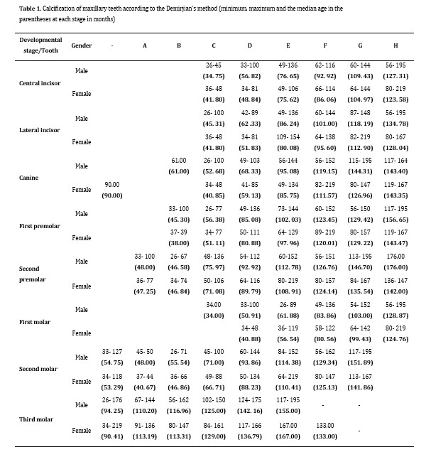
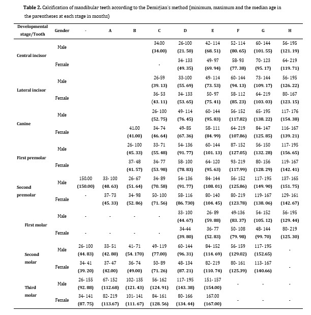
779 radiographs were included in this study. A total of 410 radiographs belonged to males in the age range of 26-195 months and 368 belonged to females in the age range of 34-219 months. Table 1 shows the maximum and minimum and the median (age with the highest frequency) age at each calcification stage for the maxillary teeth. Table 2 shows the maximum and minimum and the median (age with the highest frequency) age at each calcification stage for the mandibular teeth. Early stages of calcification (stages 0, A and B) of incisors started before the age of 3. Also, last stages of root completion (stages G and H) of second and third molars occurred after the age evaluated in this study. Thus, we did not have these stages in our findings.
As shown in Tables 1 and 2, the order of development of the crown of maxillary and mandibular teeth in both males and females was as follows: First molar, central incisor, lateral incisor, canine, first premolar, second premolar, second molar, and third molar teeth.
The order of root development of permanent maxillary and mandibular teeth in females was as follows: First molar, central incisor, lateral incisor, canine, first premolar, second premolar, second molar, and third molar. This order was slightly different in males and was as follows: First molar, central and lateral incisors, first premolar, canine, second premolar, second molar, and third molar.
The age of dental development in the maxilla and mandible was compared for each developmental stage using the Wilcoxon signed rank test. The results showed that tooth development in all stages occurred sooner in the mandible, which was significant in all anterior teeth and first and second premolars (P=0.017).
The age of dental development in males and females was analyzed separately for each stage using independent sample t-test. The results showed that development in all stages occurred sooner in females. In details, statistically significant differences were found in the stages G and H (root completion) for central incisors and lateral incisors and first molars of the mandible and for central incisors and first molars of the maxilla, and in the stages D to H (initiation of root formation to root completion) for canines, first premolars, second premolars, and second molars of the mandible and lateral incisors, canines, first premolars, second premolars and second molars of the maxilla (P<0.05).
Table 1. Calcification of maxillary teeth according to the Demirjian’s method (minimum, maximum and the median age in the parentheses at each stage in months)
Table 2. Calcification of mandibular teeth according to the Demirjian’s method (minimum, maximum and the median age in the parentheses at each stage in months)
In premolars and canines, stages of root completion occurred significantly sooner and faster in females.
Dental chart:
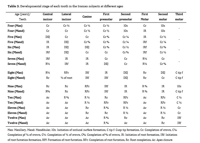
Table 3 shows the final results with regard to developmental stage of each tooth in different ages.
Clinical viewpoints:
No clinically significant difference was noted for central incisors of both jaws.
Development of permanent first molars started sooner in females but eventually continued with the same trend in boys and girls. Root completion of permanent first molars occurred at the age of 10-10.5 years which is important in cases with carious lesions who need pulp therapy. In some cases, we have to deal with a severe carious first molar which has a weak prognosis. The best age for extraction of these teeth is the time of completion of second molar furcation. The furcation of second molar (stage E) of both jaws formed at the age of 9-9.5 years.
Table 3. Developmental stage of each tooth in the Iranian subjects at different ages
In second premolars, which have the highest frequency of missing, the initiation of crown calcification (stage 1) is clear in most children at 4 years of age but can be found up to 8 years in males. It was remarkable that although the developmental stage 1 of canines and second molars started sooner (especially in females) and was visible on radiographs at a younger age, other developmental stages of these teeth were completed at older ages in comparison with first incisors and premolars.
The bud stage of third molar (stage 0) could be seen in the mandible at age 7- 7.5 years and in maxilla at age 7.5-8 years, and occurred significantly sooner in females. The oldest age showing stage 0 of calcification of third molars was 12.9 years.
Discussion
Growth and development of human organs and body parts are variable in the same person and among different individuals [1]. Thus, knowledge in this respect is imperative for making therapeutic decisions. Dental development is important for clinicians since it affects growth and development of alveolar bone and facial skeleton, and plays an important role in obtaining a balanced occlusion. This study assessed the time of initiation and completion of calcification of permanent teeth using panoramic radiographs. Also, the values were compared between the maxilla and mandible, and males and females.
Assessment of dental calcification based on radiographs is important for monitoring of dental development. Several methods have been suggested by Nolla [7] in 1960, Moorrees et al. [8] in 1963 and Demirjian and Goldstein [24] in 1976 for classification of dental calcification. The Demirjian’s method was used in the current study, which has a simple staging system and high reproducibility [23, 25].
Panoramic radiographs were used for this purpose in our study since they visualize the tooth buds of all permanent teeth in one radiograph and have a suitable reproducibility [11]. Since dental development initiates before birth and radiographs are not often requested in the first couple of years after birth, some primary stages of development, occurring in the first couple of years after birth, cannot be evaluated. The age of occurrence of primary stages of development has not been determined in previous studies either.
The present results showed that completion of crown and root of anterior teeth occurred at the same age as reported by Karadayı et al. [15] at a younger age compared to studies by AlQahtani et al. [16] and Moorrees et al. [8] (central and lateral incisors), and at an older age compared to a study by Liversidge and Speechly [10]. Compared to the study by Nolla [7], completion of crown occurred at the same age but root completion occurred at a younger age in our population.
Assessment of the data obtained for the posterior teeth in the present study showed that completion of crown of first and second premolars and permanent first molars occurred at the same age as reported by Nolla [7], Garn et al. [14], Karadayı et al. [15] and AlQahtani et al, [16] at a lower age compared to the study by Nanda and Chawla [9], and at an older age compared to studies by Logan and Kronfeld [6], Moorrees et al. [8] Liversidge and Speechly [10], and Garn et al. [14]. The age of completion of crown of second molars in our study was the same as that reported by Logan and Kronfeld [6], Nolla [7], Garn et al. [14], Karadayı et al. [15] and AlQahtani et al, [16] and lower than that reported by Nanda and Chawla [9] in Indian children. Regarding third molars, the first signs of tooth buds were seen at an older age in our study compared to studies by Logan and Kronfeld [6], Garn et al, [14] and AlQahtani et al [16] but completion of crown occurred at a younger age in our study compared to others. Completion of root of posterior teeth in our study occurred at the same age as in studies by Garn et al, [14] and Karadayı et al, [15] at a younger age compared to studies by Logan and Kronfeld [6], Nolla [7], Nanda and Chawla [9], Garn et al. [14] and AlQahtani et al, [16] and at an older age compared to the study by Liversidge and Speechly [10]. Regarding the order of root development, first molars ranked first in our study versus central incisors in the study by Nolla [7]. However, order of root development in other teeth was the same in both studies.
In both studies, root of maxillary canines developed with a greater delay than the mandibular canines such that their development terminated after the development of first premolars.
In the study by ImaniMoghaddam et al. [23], stages of root development were assessed and the root completion of teeth occurred at an older age compared to the present study.
Formation of root furcation was similar to the results found in this study.
Such a difference in the results may be attributed to differences in methodology such as type of radiography and method of classification of tooth calcification, racial differences, sample size, and age range of participants; and as reported by Nanda and Chawla [9], it may be related to the effects of lifestyle, nutrition, health and more importantly race on dental development.
Different classification systems for assessment of dental development have been used in previous studies. El-Yazeed et al. [11] used the Nolla’s method [7], Liversidge and Speechly [10], and Esan and Schepartz [17] used the Demirjian’s method, Nanda and Chawla [9] used a 15-stage classification system and Moorrees et al. [8] used a 14-stage classification system. Moorrees et al, [8] and Nanda and Chawla [9] used periapical and lateral radiographs, which do not allow assessment of all teeth. Nolla [7] used a series of patient radiographs.
Comparison of development of teeth in the maxilla and mandible revealed that most mandibular teeth in both males and females developed at a younger age compared to the maxillary teeth, which was in agreement with the results of Nolla [7], Nanda and Chawla [9] and El-Yazeed et al [11]. This difference for canine teeth was significant in females such that maxillary canines had a much slower pace of root development concomitant with the development of second premolars while mandibular canines developed sooner than first premolars.
Comparison of males and females in this respect showed that crown and root development of the maxillary lateral incisors and canines, premolars and second molars of both jaws occurred sooner in females compared to males. Males and females were significantly different in later developmental stages of teeth including root completion of incisors and crown and root completion of canines and posterior teeth which occur after the age of 9. In studies by Nolla [7], Moorrees et al. [8], Liversidge and Speechly [10], El-Yazeed et al, [11] and Karadayı et al, [15] at least one stage of dental development occurred sooner in females compared to males. Also, duration of completion of root length in females was shorter than that in males, which was in line with the results of Nolla [7].
However, cross-sectional studies with a small sample size in each developmental stage and non-random selection of samples do not allow accurate comparisons between males and females. At least 100 females and 100 males are required for each stage of dental development for accurate comparisons.
In general, considering the dental development in Iranian children, particularly for posterior teeth, it appears that completion of crowns occurs at an older age while completion of roots occurs at a younger age in Iranian children compared to previous studies, which may be related to racial differences. Faster development of mandibular teeth compared to the maxillary teeth and in females compared to males in our study was in agreement with the results of studies conducted on different populations.
Regarding the key points used for diagnosis and treatment planning, detection of signs of development of permanent third molars at the age of 8-9 years and initiation of furcation formation in second molars at the age of 9-10 years can be named, which should be taken into account in extraction treatment planning for first molars with severe caries. Also, assessment of the age range of formation of bud of second premolars revealed that initiation of calcification of this tooth bud may occur as late as 8 years of age.
Our study had a cross-sectional design, which was similar to many previous studies but different from the design of Nolla’s study [7], which was longitudinal. One limitation of cross-sectional studies is that tooth length is compared with adjacent teeth on radiographs; whereas, several radiographs taken with short intervals are required to determine the duration of completion of each dental calcification stage. Studies with larger sample size and longitudinal design are required to more accurately determine the age range of dental development in the Iranian children and provide the relevant tabulations.
Conclusion
The time of tooth calcification in this study was different in some stages from the others due to racial and methodological differences. The results showed that dental development occurred sooner in the mandible and in females. This difference in dental development was more significant between the maxillary and mandibular canine teeth, and premolars and second molars to some extent.
| Rights and permissions | |
 |
This work is licensed under a Creative Commons Attribution-NonCommercial 4.0 International License. |