

Volume 8, Issue 1 (1-2023)
J Res Dent Maxillofac Sci 2023, 8(1): 38-42 |
Back to browse issues page


Ethics code: do not have ethic code
Download citation:
BibTeX | RIS | EndNote | Medlars | ProCite | Reference Manager | RefWorks
Send citation to:


BibTeX | RIS | EndNote | Medlars | ProCite | Reference Manager | RefWorks
Send citation to:
Ruhollahi M, Talaeipour A, Tour Savadkouh S, Roghanizad N. Centering Ability of F6 SkyTaper and RaCe Rotary Files: A Cone-Beam Computed Tomography Study. J Res Dent Maxillofac Sci 2023; 8 (1) :38-42
URL: http://jrdms.dentaliau.ac.ir/article-1-376-en.html
URL: http://jrdms.dentaliau.ac.ir/article-1-376-en.html


1- Endodontics Department, Faculty of Dentistry, Tehran Medical Sciences, Islamic Azad University, Tehran, Iran
2- Oral and Maxillofacial Radiology Department, Faculty of Dentistry, Tehran Medical Sciences, Islamic Azad University, Tehran, Iran
3- Endodontics Department, Faculty of Dentistry, Tehran Medical Sciences, Islamic Azad University, Tehran, Iran , nr686@yahoo.com
2- Oral and Maxillofacial Radiology Department, Faculty of Dentistry, Tehran Medical Sciences, Islamic Azad University, Tehran, Iran
3- Endodontics Department, Faculty of Dentistry, Tehran Medical Sciences, Islamic Azad University, Tehran, Iran , nr686@yahoo.com
Full-Text [PDF 728 kb]
(377 Downloads)
| Abstract (HTML) (738 Views)
Introduction
The purpose of mechanical preparation of the root canal system is to remove all the infectious and necrotic debris. Mechanical preparation improves irrigation and obturation [1]. To achieve the best therapeutic results, the taper, shape, and position of the canal before and after canal preparation should be consistent. Maintaining this consistency is particularly difficult in curved canals.
Endodontic files have a tendency to straighten the curved canals, leading to problems such as ledge formation, zipping and perforation [2]. Numerous techniques and instruments have been introduced to solve this problem [3].
Nickel-titanium (NiTi) rotary files have been highly successful in this regard due to their tremendous flexibility. Biomechanical root canal preparation errors will be minimized and time will be saved with NiTi rotary files [4].
F6 SkyTaper (Komet Brasseler, Lemgo, Germany) and RaCe (FKG, La-Chaux-De-Fonds, Switzerland) are two commonly used NiTi rotary systems for root canal preparation. F6 SkyTaper rotary system is used in continuous rotation and designed for single file preparation which makes the preparation procedure easier and faster. This file can efficiently remove debris because of its unique S-curved design. It also has high flexibility due to its thin central core [5,6].
RaCe rotary files work with full rotation. These files have a triangular cross-sectional design and alternating cutting edges. The system consists of more than 30 different files in terms of size, taper and length, and root canal preparation with this system requires multiple files from path file to final file depending on the initial shape and size of the root canal [7].
In order to analyze the centering ability of endodontic instruments, two 3D images need to be taken, one before and another one after root canal preparation. Cone-beam computed tomography (CBCT) is ideal for this purpose [8].
This study aimed to compare the centering ability of F6 SkyTaper and RaCe rotary files in curved root canals using CBCT.
Materials and Methods
In this experimental study, 30 intact human mandibular molars which had been extracted for periodontal reasons were used. The present study was approved ethically by the Research Council, Dental Faculty of Islamic Azad University (IR. IAU. DENTAL. REC.1397, 46). The teeth were first disinfected in 2.5% sodium hypochlorite solution for 1 hour. After cleaning of the root surfaces from tissue residues and debris, the teeth were stored in saline. Intact mesiobuccal roots with no cracks, fracture, caries, calcification, and external/internal resorption were chosen.
The roots curvature was determined using CBCT and based on the technique described by Hartmann et al. [9]. The roots with a curvature between 20-35 degrees and 8-10 mm radius were selected for this study. For the first CBCT scan, the teeth were individually mounted in putty impression material. Then, the teeth underwent CBCT scan (Rotograph EVO 3D) with 11.2 s time, 64 kVp, and 0.9 mA and the mesiobuccal root canals were assessed on the scans at 1, 3, and 7 mm from the apex.
Measurements were made between the mesial border (A1) and distal border (B1) of the canals at 1, 3, and 7 mm from the apex before preparation. Working length was determined 0.5 mm shorter than the apex of the canals using a #10 K-file. Root canal glide path was obtained by a #15 K-file in both groups. The root canals were prepared with RaCe (#25/4% and #25/6%) or F6 SkyTaper (#25/6%) files based on the manufacturers’ instructions. Irrigation was done with 1 mL of 2.5% sodium hypochlorite solution after using each file.
Then, all specimens were placed back in their respective molds for the second CBCT.
Measurements were made between the mesial border (A2) and distal border (B2) of the canals at 1, 3, and 7 mm from the apex after preparation. The centering of the canal in each tooth was determined using the formula A1-A2 / B1-B2. A result of 1 would indicate maximum centering of the canal [10]. The Mann Whitney U test and ANOVA were used for statistical analysis.
Results
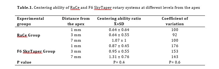
In the RaCe group, the highest centering of the canals was at the level of 7 mm from the apex (1.07), while the lowest centering of the canal was at the level of 1 mm from the apex (0.64). The difference was not significant (P=0.6).
In the F6 SkyTaper group, the highest centering of the canals was at the level of 7 mm from the apex (1.31) while the lowest centering of the canals was at the level of 1 mm from the apex (0.47). The difference was not significant (P=0.6).
The overall centering was 0.72±0.62 in the RaCe group and 0.95±1.39 in the F6 SkyTaper group. Although the centering ability of F6 SkyTaper was better than RaCe, the difference was not significant (P=0.4, Table 1)
Table.1. Centering ability of RaCe and F6 SkyTaper rotary systems at different levels from the apex
Discussion
This study showed that the centering ability of RaCe and F6 SkyTaper did not have a statistically significant difference. Deviation from the original position and shape of the canal especially in the apical third interferes with the basic rules of mechanical cleaning and shaping of the root canal system. This could end up in treatment failure and eventual tooth loss [11].
The apical third of the canals may have different degrees of curvature. Most treatment mishaps such as perforation, transportation, and zipping occur in curved parts of the root canals [12]. The curved mesiobuccal roots (25-35°) of extracted mandibular molars mimics clinical conditions. The curvature of the canals was measured by the Iqbal technique. This technique measures the curvature radius along with the curvature angle [9].
Ceyhanli et al. [13] compared centering ability of ProTaper, RaCe, and SafeSider rotary files. They found that apical transportation of RaCe and ProTaper files was significantly higher than SafeSider files. They used micro-computed tomography (CT) to compare centering of these files. On the other hand, Delgoshayi et al. [14]
compared canal transportation and
centering ability of ProTaper and SafeSider using CBCT. They concluded that ProTaper was significantly superior to SafeSider in terms of curvature preservation. Different results of the abovementioned two studies may be due to the technology of the files used, or use of micro-CT instead of CBCT. We used CBCT which is a reliable, 3D, and high-resolution modality. It is also cost-effective, non-invasive, and repeatable.
We assessed F6 SkyTaper because it has unique properties. It is designed to prepare the canals by continuous rotation and a single file system along with its S-curved design and thin central core which add more flexibility and result in faster cleaning and shaping of the canals [15]. The use of glide path before preparation by F6 SkyTaper is controversial [16,17]. The single file systems reduce the preparation time, cost, and failure related to instrumentation compared with rotary systems with multiple instruments [18].
On the other hand, RaCe seems to be the gold standard of rotary files. It consists of full-rotation instruments with a triangular cross-sectional design and altering cutting edges [19]. The present results showed that F6 SkyTaper had better centering ability than RaCe. However, the difference was not significant.
Several methods such as periapical radiographs, CBCT and micro-CT have been used to evaluate the centering ability of different endodontic instruments or canal preparation techniques [20].
In this study, CBCT was used, which is a non-invasive method and provides an accurate and repeatable 3D evaluation of the root canals before and after preparation without destroying the specimens [21].
Centering ability was analyzed by the technique described by Mesgarani et al. This analysis shows the ability of endodontic files to remain at the center of the root canal [8]. Koring et al. [22] compared the centering ability and canal transportation of five rotary systems namely HyFlex CM, ProTaper Next, F6 SkyTaper, BioRace and Mtwo using a stereomicroscope. Their results showed that F6 SkyTaper system was significantly better in maintaining the centering of the canals than BioRaCe and both systems were fast in canal preparation. Thota et al. [23] compared the centering ability and canal displacement of three rotary systems of WaveOne, ProTaper and Komet F6 SkyTaper using CBCT. They used 45 mesiobuccal canals with a 20-40° curvature. Scans were obtained at 1, 3, 5, and 7 mm from the apex. The results showed that WaveOne rotary system was significantly superior in maintaining the centering of the canal.
This study had an in vitro design; thus, the results should be interpreted with caution.
Although both systems in our study showed transportation of the canals, the difference between them was not significant.
Conclusion
Based on the results of this study, both RaCe and F6 SkyTaper had an acceptable centering ability in mesiobuccal canals of mandibular molars.
Full-Text: (191 Views)
|
Abstract
Background and Aim: Maintaining the original shape and path of the canal is among the most important criteria for optimal root canal preparation. The aim of this study was to compare the centering ability of F6 SkyTaper and RaCe rotary files in mesiobuccal canals of mandibular molars. Materials and Methods: In this experimental study, 30 mesiobuccal canals of extracted human mandibular molars with 25-30-degree curvature were randomly divided into two experimental groups (n=15) of RaCe and F6 SkyTaper. After mounting of the teeth in a putty mold, the distance between the canal walls and the outer surface of the roots in mesial and distal aspects was measured. The measurements were made at 1, 3 and 7 mm from the apex. Initial glide path in the canals was achieved using a # 15 K-file. Then, the canals in group A were prepared by RaCe rotary file #25/6% while the canals in group B were prepared by F6 Sky Taper rotary file #25/6%. Measurements were repeated and the difference between the two measurements was calculated and compared with the Mann-Whitney U test. Results: The mean centering ability was 0.72 ± 0.62 in the RaCe group and 0.95 ± 1.39 in F6 SkyTaper group. the centrality was better in F6 SkyTaper group (it was closer to 1) but the difference was not statistically significant (P=0.4). Conclusion: Both RaCe and F6 SkyTaper rotary systems partially offset the centrality of the root canal system. Key Words: Cone-Beam Computed Tomography; Root Canal Preparation; Transportation; Dental Instruments |
Introduction
The purpose of mechanical preparation of the root canal system is to remove all the infectious and necrotic debris. Mechanical preparation improves irrigation and obturation [1]. To achieve the best therapeutic results, the taper, shape, and position of the canal before and after canal preparation should be consistent. Maintaining this consistency is particularly difficult in curved canals.
Endodontic files have a tendency to straighten the curved canals, leading to problems such as ledge formation, zipping and perforation [2]. Numerous techniques and instruments have been introduced to solve this problem [3].
Nickel-titanium (NiTi) rotary files have been highly successful in this regard due to their tremendous flexibility. Biomechanical root canal preparation errors will be minimized and time will be saved with NiTi rotary files [4].
F6 SkyTaper (Komet Brasseler, Lemgo, Germany) and RaCe (FKG, La-Chaux-De-Fonds, Switzerland) are two commonly used NiTi rotary systems for root canal preparation. F6 SkyTaper rotary system is used in continuous rotation and designed for single file preparation which makes the preparation procedure easier and faster. This file can efficiently remove debris because of its unique S-curved design. It also has high flexibility due to its thin central core [5,6].
RaCe rotary files work with full rotation. These files have a triangular cross-sectional design and alternating cutting edges. The system consists of more than 30 different files in terms of size, taper and length, and root canal preparation with this system requires multiple files from path file to final file depending on the initial shape and size of the root canal [7].
In order to analyze the centering ability of endodontic instruments, two 3D images need to be taken, one before and another one after root canal preparation. Cone-beam computed tomography (CBCT) is ideal for this purpose [8].
This study aimed to compare the centering ability of F6 SkyTaper and RaCe rotary files in curved root canals using CBCT.
Materials and Methods
In this experimental study, 30 intact human mandibular molars which had been extracted for periodontal reasons were used. The present study was approved ethically by the Research Council, Dental Faculty of Islamic Azad University (IR. IAU. DENTAL. REC.1397, 46). The teeth were first disinfected in 2.5% sodium hypochlorite solution for 1 hour. After cleaning of the root surfaces from tissue residues and debris, the teeth were stored in saline. Intact mesiobuccal roots with no cracks, fracture, caries, calcification, and external/internal resorption were chosen.
The roots curvature was determined using CBCT and based on the technique described by Hartmann et al. [9]. The roots with a curvature between 20-35 degrees and 8-10 mm radius were selected for this study. For the first CBCT scan, the teeth were individually mounted in putty impression material. Then, the teeth underwent CBCT scan (Rotograph EVO 3D) with 11.2 s time, 64 kVp, and 0.9 mA and the mesiobuccal root canals were assessed on the scans at 1, 3, and 7 mm from the apex.
Measurements were made between the mesial border (A1) and distal border (B1) of the canals at 1, 3, and 7 mm from the apex before preparation. Working length was determined 0.5 mm shorter than the apex of the canals using a #10 K-file. Root canal glide path was obtained by a #15 K-file in both groups. The root canals were prepared with RaCe (#25/4% and #25/6%) or F6 SkyTaper (#25/6%) files based on the manufacturers’ instructions. Irrigation was done with 1 mL of 2.5% sodium hypochlorite solution after using each file.
Then, all specimens were placed back in their respective molds for the second CBCT.
Measurements were made between the mesial border (A2) and distal border (B2) of the canals at 1, 3, and 7 mm from the apex after preparation. The centering of the canal in each tooth was determined using the formula A1-A2 / B1-B2. A result of 1 would indicate maximum centering of the canal [10]. The Mann Whitney U test and ANOVA were used for statistical analysis.
Results
In the RaCe group, the highest centering of the canals was at the level of 7 mm from the apex (1.07), while the lowest centering of the canal was at the level of 1 mm from the apex (0.64). The difference was not significant (P=0.6).
In the F6 SkyTaper group, the highest centering of the canals was at the level of 7 mm from the apex (1.31) while the lowest centering of the canals was at the level of 1 mm from the apex (0.47). The difference was not significant (P=0.6).
The overall centering was 0.72±0.62 in the RaCe group and 0.95±1.39 in the F6 SkyTaper group. Although the centering ability of F6 SkyTaper was better than RaCe, the difference was not significant (P=0.4, Table 1)
Table.1. Centering ability of RaCe and F6 SkyTaper rotary systems at different levels from the apex
Discussion
This study showed that the centering ability of RaCe and F6 SkyTaper did not have a statistically significant difference. Deviation from the original position and shape of the canal especially in the apical third interferes with the basic rules of mechanical cleaning and shaping of the root canal system. This could end up in treatment failure and eventual tooth loss [11].
The apical third of the canals may have different degrees of curvature. Most treatment mishaps such as perforation, transportation, and zipping occur in curved parts of the root canals [12]. The curved mesiobuccal roots (25-35°) of extracted mandibular molars mimics clinical conditions. The curvature of the canals was measured by the Iqbal technique. This technique measures the curvature radius along with the curvature angle [9].
Ceyhanli et al. [13] compared centering ability of ProTaper, RaCe, and SafeSider rotary files. They found that apical transportation of RaCe and ProTaper files was significantly higher than SafeSider files. They used micro-computed tomography (CT) to compare centering of these files. On the other hand, Delgoshayi et al. [14]
compared canal transportation and
centering ability of ProTaper and SafeSider using CBCT. They concluded that ProTaper was significantly superior to SafeSider in terms of curvature preservation. Different results of the abovementioned two studies may be due to the technology of the files used, or use of micro-CT instead of CBCT. We used CBCT which is a reliable, 3D, and high-resolution modality. It is also cost-effective, non-invasive, and repeatable.
We assessed F6 SkyTaper because it has unique properties. It is designed to prepare the canals by continuous rotation and a single file system along with its S-curved design and thin central core which add more flexibility and result in faster cleaning and shaping of the canals [15]. The use of glide path before preparation by F6 SkyTaper is controversial [16,17]. The single file systems reduce the preparation time, cost, and failure related to instrumentation compared with rotary systems with multiple instruments [18].
On the other hand, RaCe seems to be the gold standard of rotary files. It consists of full-rotation instruments with a triangular cross-sectional design and altering cutting edges [19]. The present results showed that F6 SkyTaper had better centering ability than RaCe. However, the difference was not significant.
Several methods such as periapical radiographs, CBCT and micro-CT have been used to evaluate the centering ability of different endodontic instruments or canal preparation techniques [20].
In this study, CBCT was used, which is a non-invasive method and provides an accurate and repeatable 3D evaluation of the root canals before and after preparation without destroying the specimens [21].
Centering ability was analyzed by the technique described by Mesgarani et al. This analysis shows the ability of endodontic files to remain at the center of the root canal [8]. Koring et al. [22] compared the centering ability and canal transportation of five rotary systems namely HyFlex CM, ProTaper Next, F6 SkyTaper, BioRace and Mtwo using a stereomicroscope. Their results showed that F6 SkyTaper system was significantly better in maintaining the centering of the canals than BioRaCe and both systems were fast in canal preparation. Thota et al. [23] compared the centering ability and canal displacement of three rotary systems of WaveOne, ProTaper and Komet F6 SkyTaper using CBCT. They used 45 mesiobuccal canals with a 20-40° curvature. Scans were obtained at 1, 3, 5, and 7 mm from the apex. The results showed that WaveOne rotary system was significantly superior in maintaining the centering of the canal.
This study had an in vitro design; thus, the results should be interpreted with caution.
Although both systems in our study showed transportation of the canals, the difference between them was not significant.
Conclusion
Based on the results of this study, both RaCe and F6 SkyTaper had an acceptable centering ability in mesiobuccal canals of mandibular molars.
Type of Study: Original article |
Subject:
Endodontics
References
1. Sousa-Neto MD, Silva-Sousa YC, Mazzi-Chaves JF, Carvalho KKT, Barbosa AFS, Versiani MA, Jacobs R, Leoni GB. Root canal preparation using micro-computed tomography analysis: a literature review. Braz Oral Res 2018;32(suppl 1):e66. [DOI:10.1590/1807-3107bor-2018.vol32.0066]
2. Mamede-Neto I, Borges AH, Guedes OA, de Oliveira D, Pedro FL, Estrela C. Root Canal Transportation and Centering Ability of Nickel-Titanium Rotary Instruments in Mandibular Premolars Assessed Using Cone-Beam Computed Tomography. Open Dent J 2017;11:71-8.
3. Zuolo ML, Zaia AA, Belladonna FG, Silva EJNL, Souza EM, Ver-siani MA, Lopes RT, De-Deus G. Micro-CT assessment of the shaping ability of four root canal instrumentation systems in oval-shaped canals. Int Endod J 2018;51(5):564-71. [DOI:10.1111/iej.12810] [PMID]
4. Sarraf P, Kiomarsi N, Taheri FH, Moghaddamzade B, Dibaji F, Kharazifard MJ. Apical Transportation of Mesiobuccal Canals of Maxillary Molars Following Root Canal Preparation with Two Rotary Systems and Hand Files: A Cone-Beam Computed Tomo-graphic Assessment. Front Dent. 2019; 16 (4):272-8. [DOI:10.18502/fid.v14i4.2086] [PMID] [PMCID]
5. Bürklein S, Jäger PG, Schäfer E. Apical transportation and canal straightening with different continuously tapered rotary file systems in severely curved root canals: F6 SkyTaper and OneShape versus Mtwo. Int Endod J 2017; 50 (10):983-90. [DOI:10.1111/iej.12716] [PMID]
6. Khalilak Z, Sattarian I, Tour Savadkouhi S. Ex-Vivo Comparison of the Dentin Removal Ability of One Shape and F6 SkyTaper Rotary Files. J Res Dent Maxillofac Sci 2019; 4 (4):6-10. [DOI:10.29252/jrdms.4.4.6]
7. Canga M, Malagnino I, Malagnino G, Malagnino V. A Comparison of Mtwo and RaCe Rotary Instruments in the Prep-aration of Curved Canals. J Contemp Dent Pract 2020; 21(2):124-8. [DOI:10.5005/jp-journals-10024-2751] [PMID]
8. Mesgarani A, Hamidi MR, Haghanifar S, Naiemi S, Bijani A. Comparison of apical transportation and centering ability of Mtwo and Reciproc R25 in severely curved canals using cone-beam computed tomography. Dent Res J (Isfahan) 2018;15(1):57-62. [DOI:10.4103/1735-3327.223620] [PMID] [PMCID]
9. Hartmann RC, Fensterseifer M, Peters OA, de Figueiredo JAP, Gomes MS, Rossi-Fedele G. Methods for measurement of root canal curvature: a systematic and critical review. Int Endod J 2019;52(2):169-80. [DOI:10.1111/iej.12996] [PMID]
10. Rejula F, Christalin R, Ahmed W, Dinakaran S, Gopinathan AS, Babu A. Measure and compare the Degree of Root Canal Transportation and Canal-centering ability of Twist-ed, ProTaper, and Conventional Stainless Steel K Files using Spiral Computed Tomography: An in vitro Study. J Contemp Dent Pract 2017;18(6):463-9.
11. Estrela C, Pécora JD, Estrela CRA, Guedes OA, Silva BSF, Soa-res CJ, Sousa-Neto MD. Common Operative Procedural Errors and Clinical Factors Associated with Root Canal Treatment. Braz Dent J 2017;28(2):179-90. [DOI:10.1590/0103-6440201702451] [PMID]
12. Keles A, Keskin C. Deviations of Mesial Root Canals of Man-dibular First Molar Teeth at the Apical Third: A Micro-computed Tomographic Study. J Endod 2018; 44(6): 1030-2. [DOI:10.1016/j.joen.2018.02.028] [PMID]
13. Ceyhanli KT, Erdilek N, Tatar I, Cetintav B. Comparative micro-computed tomography evaluation of apical root canal transportation with the use of ProTaper, RaCe and Safesider systems in human teeth. Aust Endod J 2014;40(1):12-6. [DOI:10.1111/aej.12014] [PMID]
14. Delgoshayi N, Abbasi M, Bakhtiar H, Sakhdari S, Ghannad S, Ellini MR. Canal Transportation and Centering Ability of ProTa-per and SafeSider in Preparation of Curved Root Canals: A CBCT Evaluation. Iran Endod J 2018;13(2):240-5.
15. Kaval ME, Capar ID, Ertas H, Sen BH. Comparative evaluation of cyclic fatigue resistance of four different nickel-titanium rotary files with different cross-sectional designs and alloy properties. Clin Oral Investig 2017;21(5):1527-30. [DOI:10.1007/s00784-016-1917-x] [PMID]
16. Troiano G, Dioguardi M, Cocco A, Zhurakivska K, Ciavarella D, Muzio LL. Increase in [corrected] the glyde path diameter improves the centering ability of F6 Skytaper. Eur J Dent. 2018;12(1):89-93. [DOI:10.4103/ejd.ejd_231_17] [PMID] [PMCID]
17. Sajad M, Misgar OH, Farooq R, Purra AR, Ahangar FA. Evaluation of canal centering ability after preparation with f6 Sky Taper single continuous file systems with or without glide path files. Int J Appl Dent Sci. 2018;4(1):25-8.
18. Azim AA, Wang HH, Tarrosh M, Azim KA, Piasecki L. Com-parison between Single-file Rotary Systems: Part 1-Efficiency, Effectiveness, and Adverse Effects in Endodontic Retreatment. J Endod. 2018;44(11):1720-4. [DOI:10.1016/j.joen.2018.07.022] [PMID]
19. Aminsobhani M, Ghorbanzadeh A, Dehghan S, Niasar AN, Kharazifard MJ. A comparison of canal preparations by Mtwo and RaCe rotary files using full sequence versus one rotary file techniques; a cone-beam computed tomography analysis. Saudi Endod J. 2014;4:70-6. [DOI:10.4103/1658-5984.132722]
20. Fidler A, Plotino G, Kuralt M. A Critical Review of Methods for Quantitative Evaluation of Root Canal Transportation. J Endod. 2021 May;47(5):721-31. [DOI:10.1016/j.joen.2021.02.002] [PMID]
21. Hasheminia SM, Farhad A, Sheikhi M, Soltani P, Hendi SS, Ahmadi M. Cone-beam Computed Tomographic Analysis of Ca-nal Transportation and Centering Ability of Single-file Systems. J Endod 2018;44(12):1788-91. [DOI:10.1016/j.joen.2018.09.011] [PMID]
22. Koring S, Schwahn C, Kocher T, Steffen H. Shaping ability of five different nickel-titanium systems in simulated S-shaped canals. Endo EPT 2020;14(2):135-43.
23. Thota MM, Kakollu S, Duvvuri M, Garikapati RB. Compari-tive evaluation of canal shaping ability of three nickel titanium instrument systems using cone beam computed tomography: An in vitro study. Endodontology 2017; 29(2): 120-24. [DOI:10.4103/endo.endo_17_17]
Send email to the article author
| Rights and permissions | |
 |
This work is licensed under a Creative Commons Attribution-NonCommercial 4.0 International License. |
