


BibTeX | RIS | EndNote | Medlars | ProCite | Reference Manager | RefWorks
Send citation to:
URL: http://jrdms.dentaliau.ac.ir/article-1-339-en.html


2- Oral and Dental Disease Research Cen-ter, Oral and Maxillofacial Disease Department, School of Dentistry, Shiraz University of Medical Sciences, Shiraz, Iran , Fatemeh.lavaee@gmail.com
3- Research Committee of Shiraz Dental School, Shiraz University of Medi-cal Sciences, Shiraz, Iran
4- Research Committee of Shiraz Dental School, Shiraz University of Medical Sciences, Shiraz, Iran
|
Abstract
|
Introduction
Nowadays, diagnosis and treatment of mucosal lesions of the oral cavity is of great concern for many dentists due to the high frequency of oral mucosal lesions, the similarities between these lesions, and little information available about oral diseases.[1] Benign vascular lesions result from abnormalities of the blood vessels orproliferation of endothelial cells. A classification system of vascular lesions was introduced by Mulliken Glowacki in 1982 which was modified by the International Society for the Study of Vascular Anomalies.[2] Based on this classification system, vascular abnormalities are congenital or acquired lesions that are divided into two major groups: (I) tumors such as hemangioma, pyogenic granuloma, rapidly involuting congenital hemangioma, noninvoluting congenital hemangioma, hemangiopericytoma, tufted angioma, and Kaposiform hemangioendothelioma; and (II) vascular malformations (VMs).
Oral lesions can interfere with daily routine activities by adverse effects on biting, swallowing, and speech, and causing dry mouth, bad breath, and oral pain.[3] Since some of these lesions can be a sign of a serious systemic condition, early diagnosis of oral lesions is highly important.[4]
Vascular tumors are among the most common lesions of the oral cavity. Hemangioma is a common benign endothelial cell tumor with non-definitive neoplastic nature. Hemangioma and lymphangioma can involve both superficial and deep oral mucosa with different oral mucosal presentations. Hemangiomas are proliferative lesions characterized by increased endothelial cell proliferation. They develop after birth and disappear over the years.[2]
VMs develop due to impairments in embryonic development and are considered as a developmental abnormality.[5] Pyogenic granuloma is a benign tumor of the vascular origin in the skin or mucosal membranes. Its high growth rate and its brittle surface distinguish this type of tumor from other vascular tumors.[5,6]
Lymphangioma is known as a hamartoma of lymphatic vessels and mostly occurs in cervical and craniofacial areas.[7] Its infiltration around cervical neurovascular structures complicates its complete removal.[5,8,9]
A study conducted in 2015 on 90 elderly (over 65 years of age) residents of sanatoriums in Sari and Babol cities in Iran reported that 16.7% of them had one oral lesion and 21.1% had more than three oral lesions. The most common lesions observed in this population were reported to be fissured tongue (55.6%), varicosity (46.7%), pigmentation (26.7%), candidiasis (25.6%), and sublingual varix (21.1%).[10,11] In 2016, an epidemiological study was conducted on benign oral and facial vascular lesions in patients referred to the Oral Pathology Department of Isfahan Dental School during a 17-year period (1998-2015). They found a total number of 448 vascular lesions in 434 patients. Among all the cases studied, 265 lesions were reported in women (61.1%) and 169 lesions (38.9%) were observed in men. Of all the lesions, only 4% had a vascular origin. Most patients were women. Mandible was the most common site of involvement, and the soft palate was the least common site. They concluded that benign vascular lesions account for a small percentage of oral and maxillofacial lesions.[6] The aim of this study was to assess the prevalence of oral vascular lesions (hemangioma, VMs, pyogenic granuloma, and lymphangioma) in an Iranian population.
Materials and Methods
In this cross-sectional study, the prevalence of vascular lesions in patients referred to the Oral Medicine Department of Shiraz Dental School was investigated. A descriptive analytical study was designed to determine the frequency of oral vascular lesions. Clinical data on oral lesions were retrieved and retrospectively analyzed from the archives of the Oral Medicine Department of Shiraz School of Dentistry (Shiraz, Iran) from September 2001 to September 2017. This study was approved by Shiraz University of Medical Sciences Ethic Committee (IR.SUMS.REC.1397.803). The patients of any gender/age who were referred to the Oral Medicine department of Shiraz Dental School and diagnosed with oral vascular lesions (pyogenic granuloma, varix, hemangioma, lymphangioma, arteriovenous malformations, VMs) were included in the study. Patients with unsubstantiated or missing data were excluded from the study. A total of 176 patients diagnosed with vascular lesions in the Oral Medicine Department of Shiraz School of Dentistry (Shiraz, Iran) from September 2001 through September 2017 were retrospectively analyzed. All diagnoses had been made by specialists of the Oral Medicine Department. The records of patients referring to the Oral Medicine Department during this period were investigated and the demographic data (age, gender, etc.) were extracted from the records. Furthermore, information about the site of oral lesion was reviewed and analyzed. Data were analyzed using SPSS version 18 (SPSS Inc., Chicago, IL, USA). The mean age and gender of patients were recorded and the prevalence of vascular lesions was calculated. Associations between variables were explored using the Chi-square test and Fisher’s exact test. Moreover, to determine statistically significant differences between the variables, the non-parametric Kruskal-Wallis test was applied. P<0.05 was considered statistically significant.
Results
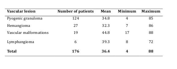
Table 1 summarizes the information of patients evaluated in this study. A total of 176 cases had oral vascular lesions among 3896 clinical patient records during the 16-year study period. Of the 176 patients, 108 (61.4%) were females and 68 (38.6%) were males. The ratio of male to female was 1:1.6. The mean age of patients was 36 ± 4.5 years (range 4-88 years).
In the present study, the prevalence of vascular lesions was as follows: pyogenic granuloma comprising 3.18% of the lesions, followed by VMs with 0.7%, hemangioma with 0.49%, and lymphangioma with 0.15% frequency. As shown in Table 1, four types of vascular lesions were observed in the study population. The most prevalent vascular lesion was pyogenic granuloma (70.45%) with a higher incidence in females. The total prevalence of oral vascular lesions was also higher in females. No significant correlation was found by the Chi-square test between gender and prevalence of vascular lesions (P=0.078, Table 1).
The frequency of different sites of involvement in patients diagnosed with oral vascular lesions is shown in Table 2. The most common site of involvement in the study population was maxillary mucosa and gingiva. There was no significant correlation between gender and the site of involvement (P=0.143) by the Chi square test as shown in Table 2.
The distribution of age was found to be almost the same among different types of oral vascular lesions. According to Table 3, no significant correlation was found between age and type of vascular lesion (P=0.174) by the Kruskal-Wallis test. The distribution of age was found to be almost the same in different types of oral vascular lesions. No significant correlation was found between age and type of vascular lesion (P=0.174) by the Chi-square test.
Table 1. Information of patients with oral vascular lesions
Table 3. Distribution of age in different vascular lesions
Table 2. Prevalence of different sites of involvement in patients with oral vascular lesions
Discussion
In this study, pyogenic granuloma was the most prevalent vascular lesion (70.45%) followed by hemangioma (15.34%), VMs (10.79%), and lymphangioma (3.4%.) Also, vascular lesions were more common in females; while no significant correlation was found between gender and prevalence of vascular lesions (P=0.078). The most common site of involvement in the evaluated lesions was maxillary mucosa and gingiva.
There was no significant correlation between gender and site of involvement (P=0.143). No significant correlation was found between age and type of vascular lesion (P=0.174). In this study, the distinction between different oral vascular lesions was based on clinical parameters from the classification by Mulliken and Glowacki.[2] Extreme variations in epidemiological data on these diseases could be attributed to the fact that most of the studies conducted on benign oral vascular lesions did not use this classification for diagnosis of oral vascular lesions.[12-14] Few studies about the frequency of oral vascular lesions are found in the literature. In our study, the most prevalent vascular lesion was pyogenic granuloma comprising 70.45% of the vascular lesions found in patients referred to the Oral Medicine Department of Shiraz Dental School, followed by VMs with 15.34%, hemangioma with 10.79%, and lymphangioma with 3.4% frequency. The total prevalence of vascular lesions amongst all the evaluated lesions of oral mucosa was reported as follows: pyogenic granuloma comprising 3.18% of the cases, followed by VMs with 0.7%, hemangioma with 0.49%, and lymphangioma with 0.15% frequency. Saravana [15] reported an incidence of 21% for pyogenic granuloma among 655 cases with tumor-like lesions of the oral cavity. In a study by Espinoza et al, [14] the prevalence of pyogenic granuloma was reported to be 0.7 % in a population of elderly Chileans. Lim et al, [16] retrospectively investigated the prevalence of oral and maxillofacial biopsies in Brazilian children and reported a prevalence of 2.56% for pyogenic granuloma and 0.96% for hemangioma. de Vasconcelos Carvalho et al. [17] conducted a similar study on the elderly Brazilian patients and reported a prevalence of 4.3% for pyogenic granuloma, and 1.87% for hemangioma. Al-Khateeb et al, (18), in a study on infants reported a relative incidence of 0.9% for hemangioma. In another study by Correa et al, [19] oral hemangioma was diagnosed in 0.9% of the cases; while, oral VMs were observed in 1.3% of the cases. In the present study, the least common oral vascular lesion in the Iranian population investigated in this study was found to be lymphangioma with a frequency of 0.15%. Similar frequencies of lymphangioma have been reported in other populations. de Vasconcelos Carvalho et al.[17] reported a prevalence of 0.19% for lymphangioma in elderly Brazilian patients.In our study, oral VMs were more frequent than oral hemangioma. This finding was in line with the results of a study by Correa et al,[19] who also reported that VMs were more common than oral hemangioma in a Brazilian population. As the results of this study indicated, pyogenic granuloma and oral hemangioma were more common in females than males. In similar studies, pyogenic granuloma and oral hemangioma were reported to be more frequent in females, as shown by Donnelly et al, [20] and Correa et al,[19] with a female to male ratio of 4:1 in patients with hemangioma. Moreover, similar to our study, Correa et al.[19] observed that VMs occurred equally in females and males. On the other hand, in our study, lymphangioma was mostly seen in males with a male to female ratio of 5:1. Sharma et al.[21] reported that hemangioma was the most common benign neoplasm in infancy, the majority arising between the 1st and the 4th week of life. The prevalence of different vascular lesions in other studies was almost the same. The referral center for oral vascular lesions in different evaluations is diverse. Hospitals, and dermatological and oral medicine centers are the most common referral centers. This can affect the reported prevalence rates. Also, the amount of patient self-care in different populations in addition to the medical insurance systems, treatment facilities, and economic condition can also affect the reported prevalence rates. The vascular lesion classification system is important for epidemiological studies as well.[21]
The results of our study revealed that there was no significant correlation between sex with either vascular lesions or site of lesion. Moreover, no significant correlation was found between age with either type of vascular lesion or site of involvement. As indicated by the findings of this study, oral vascular lesions were more common in maxillary mucosa and gingiva (29%), followed by the tongue (24.4%), mandibular mucosa and gingiva (23.3%), lips (15.4%), and buccal mucosa (8%). Saravana [15] also stated that the most commonly affected site of pyogenic granuloma was the gingiva. In contrast to the findings of the present study, Correa et al.[17] reported that oral VMs were more common in the lip and buccal mucosa. Nguyen et al.[22] reported that buccal mucosa was the most common site for oral hemangioma. The possible data biases in retrospective studies always should be taken into account. Also in this study we assessed a large number of patient records in this center, which is a referral center for oral lesions. Different populations have different epidemiological data about common oral lesions. These differences are related to the ethnicity and habits. These data are valuable for any population in order to plan long-term public health policies and also to find the most common lesions in specific populations.
Conclusion
Among oral vascular lesions, pyogenic granuloma was found to be the most prevalent lesion of the oral cavity. No significant correlation was found between age and type of vascular lesion or site of lesions.
| Rights and permissions | |
 |
This work is licensed under a Creative Commons Attribution-NonCommercial 4.0 International License. |
