

Volume 7, Issue 1 (2-2022)
J Res Dent Maxillofac Sci 2022, 7(1): 15-21 |
Back to browse issues page


Download citation:
BibTeX | RIS | EndNote | Medlars | ProCite | Reference Manager | RefWorks
Send citation to:


BibTeX | RIS | EndNote | Medlars | ProCite | Reference Manager | RefWorks
Send citation to:
Delvarani A, Arzhangi F, Mohamadi M, Toursavadkouhi S. Tubular Penetration Depth of AH26 and MTA Fillapex Root Canal Sealers in Human Single-Rooted Teeth: An Ex Vivo Study. J Res Dent Maxillofac Sci 2022; 7 (1) :15-21
URL: http://jrdms.dentaliau.ac.ir/article-1-323-en.html
URL: http://jrdms.dentaliau.ac.ir/article-1-323-en.html


1- Endodontics Department, Membership of Dental Material Research Center, Faculty of Dentistry, Islamic Azad University of Medical Sciences, Tehran, Iran
2- Dental student, Faculty of Dentistry, Islamic Azad University of Medical Sciences, Tehran, Iran.
3- Endodontics Department, Faculty of Dentistry, Islamic Azad University of Medical Sciences, Tehran, Iran
4- Endodontics Dept, Member ship of Dental Material Research Center, Faculty of Dentistry, Tehran Medical Sciences,Islamic Azad University, Tehran, Iran , s_savadkouhi@yahoo.com
Full-Text [PDF 853 kb]
(646 Downloads)
| Abstract (HTML) (1229 Views)
Full-Text: (670 Views)
Abstract
Introduction: The purpose of this study was to compare dentinal tubular penetration of two root canal sealers namely AH26 and MTA Fillapex in single-rooted teeth by scanning electron microscopy.
Materials and Methods: Thirty-five mature human single-rooted teeth were selected. Cleaning and shaping was performed. The teeth were randomly divided into 2 groups. AH26 was delivered into the canals in group 1, and MTA Fillapex was delivered into the root canals in group 2 by lateral compaction technique. The roots were sectioned at 3 mm and 5 mm from the apex. The sections were evaluated by scanning electron microscopy, and the deepest penetration depth of sealers was recorded. Statistical analysis was performed by t-test using SPSS version 19.0.
Results: The deepest tubular penetration in group 1 at 3 mm from the apex was 808 µm while it was 717 µm in group 2 at 3 mm from the apex. The difference between the two groups was not significant (P=0.4). At 5 mm from the apex, the deepest tubular infiltration in group 1 was 995 µm while it was 915 µm in group 2. The difference between the 2 groups was not significant (P=0.4).
Conclusion: Both sealers can be predictively used in different clinical situations when indicated.
Key words: Root Canal Preparation; Microscopy, Electron, Scanning; Root Canal Therapy
Introduction
One of the main goals of root canal therapy is to eradicate the microorganisms from the root canal walls and prevent reinfection. Root canal sealers are applied to fill the gaps not occupied by the gutta-percha cones .[1] Furthermore, sealers penetrate into the dentinal tubules and may entrap the residual bacteria lodged in the tubules .[2] Therefore, sealer penetration improves the treatment outcome. Presence of smear layer on the root canal walls may block the tubules and prevent sealer penetration into them .[3] Thus, it may prevent optimal adaptation of filling materials to the root canal walls .[4] Chemical bond between the root canal walls and sealers does not occur; thus, penetration of sealers into dentinal tubules may increase micromechanical bonding and resultantly improve the quality of sealing .[5] Tubular penetration might be affected by wettability, surface tension, and hydraulic properties of root canal sealers .[6] Depth of penetration of root canal sealers can be analyzed by light, confocal, or scanning electron microscopes .[7, 8] The most important advantages of the aforementioned methods include high magnification and exact determination of specific details and sealer penetration margin .[9] AH26 (DeTrey, Gmbh, Konstanz, Germany) is an epoxy resin based sealer. Good sealing property of AH26 has been previously confirmed, although evidence shows that there is no chemical bonding to root canal walls .[10] MTA Fillapex (Angelus, Londrina, Brazil) is a bio-ceramic and biocompatible root canal sealer with questionable sealing properties .[11, 12] The purpose of this study was to compare the tubular penetration of AH26 and MTA Fillapex in extracted teeth.
Materials and Methods
Thirty-five extracted sound human central incisors were collected with no root canal curvature and complete apices for this in vitro experimental study (ethical code: IR.IAU.Dental.REC1399/25). The patency of the apices was ensured by using a patency file (10#). The teeth were collected from the tooth bank of Islamic Azad University, Dental School, Tehran, Iran. Lack of root resorption and root curvature were confirmed by taking periapical radiographs. The teeth were randomly allocated to 3 groups. AH 26 was used in group 1 (n=15). MTA Fillapex was used in group 2 (n=15), and no sealer was used in the control group (n=5). Standard access cavity was prepared in all teeth using a high-speed handpiece under water spray to prevent temperature rise. Working length was determined by reduction of 1 mm of the patency file length (Dentsply Maillefer, Ballaigues, Switzerland), which passed through the apical foramen. The teeth with apical size larger than #20 K-file were excluded from this study. The root canals were shaped by the step-back technique until a master apical file size of #40; the remaining part of the canal was enlarged to #60 by reduction of working length by 0.5 mm for each file.
After removing the smear layer by 17% ethylenediaminetetraacetic acid (EDTA) and 5.25% sodium hypochlorite each for 1 minute, distilled water was used for final irrigation. Paper points were used to dry the root canals. E9 ultrasonic tip (Woodpecker, Guangxi, China) was used for 10 seconds to deliver AH26 or MTA Fillapex sealer into the root canal system in the experimental groups by a circumferential motion, and obturation was performed by the lateral compaction technique (master apical cone: #40, spreader: #25 and lateral cones: #20). The teeth were stored in an incubator for 2 weeks. To prepare the sections, the teeth were embedded in self-cure acrylic resin (AcroPars, Tehran, Iran). A CNC machine (Delta Electronics, Taoyuan, Taiwan) was used to prepare sections with 1 mm thickness perpendicular to the longitudinal axis of the tooth samples at 3 and 5 mm distance from the anatomical apex. To remove the superficial debris from the prepared sections, 17% EDTA was used (Fig. 1). After coating the prepared samples with gold, they underwent scanning electron microscopy (S-4160; Hitachi, Tokyo, Japan) to assess the maximum penetration of sealers into dentinal tubules. The highest infiltration of sealers was determined under low magnification (x20). The exact sealer penetration was ascertained under high magnification
(x500) image (Fig. 2). T-test was used to compare the deepest point of penetration among the study groups at different levels (3 and 5 mm from the apex) by SPSS version 19.0 software (Chicago, IL, USA).
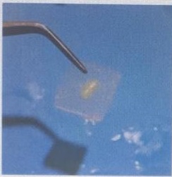
Fig 1. Prepared sections at 3&5 mm levels using CNC machine [Delta Electronic s, Taoyuan, Taiwan
Fig 2. Tubular penetration of MTA Fillapex [A, B, C] and AH26 [D, E, F] sealers in 3&5 mm levels
Results
In group 1, the maximum penetration of sealers into dentinal tubules at 3 mm from the apex was 808 µm while this value was 717 µm at 3 mm in group 2, and this difference was not significant (P= 0.4). In group 1, the maximum penetration into tubules at 5 mm from the apex was 995 µm while this value was 915 µm in group 2, and this difference was not significant (P= 0.4). In group 1, the maximum mean penetration into tubules at 3 mm from the apex (808 µm) was much lower (23%) than the penetration depth at 5 mm (995 µm), and this difference was not significant (P= 0.4). In group 2, the maximum mean penetration into tubules at 3 mm (717 µm) was much lower (27.6%) than the value at 5 mm level (915 µm), and this difference was not significant (P= 0.08) (Table 1).
Table 1. The deepest tubular penetration [Mean ± Standard deviation] of AH26 and MTA Fillapex sealers at 3&5 mm levels
Discussion
Infiltration of root canal sealers into dentinal tubules can provide a barrier against bacterial invasion into the tubules. Better infiltration provides better sealing ability .[13] Sealers do not interact chemically with the dentinal walls; thus, better infiltration of sealers can help preserve the core filling material in the root canal space .[14] Based on the results of this study, tubular infiltration of AH26 was comparable to that of MTA Fillapex at 3 and 5 mm from the apex, and infiltration of AH26 at 5 mm was higher than that at 3 mm. Higher infiltration of AH26 was due to its lower film thickness and more hydrophobic nature. The higher infiltration of sealers at 5 mm was due to density of the tubules at the middle and coronal thirds of the root .[15,16] Ultrasonic technique was used to apply the sealers into the root canals because of the optimal efficacy of this method compared with other techniques such as the use of Lentulo and master file .[17] Scanning electron microscopy can provide better images of sealer infiltration compared with confocal microscopes and stereomicroscopes .[7] Smear layer can inhibit sealer infiltration; thus, irrigating the canal with 17% EDTA and 5.25% NaOCl can increase the penetration depth of sealers .[18,19] However, the irrigation technique has no effect on infiltration of sealers .[20] Based on a study by De-Deus et al, .[19] higher penetration of sealer was observed in vertical condensation technique compared with lateral compaction or single-cone techniques. They also observed higher infiltration of sealers in the middle and coronal thirds in lateral compaction technique. In contrast, Jeong et al .[21] reported that the technique of obturation had no effect on the infiltration of calcium silicate-based sealers. Jordan et al.[22] reported that gutta-percha/AH-Plus had higher tubular infiltration compared with Resilon/Epiphany, and ethanol solution as final irrigant caused less tubular infiltration due to collagen fiber collapse and closure of tubular opening. Tuncer et al .[23] concluded that by using 17% EDTA, maleic acid, or citric acid as final irrigation, infiltration of AH26 increased due to removing the smear layer. Based on a study by Chandra et al, [24] the highest sealer infiltration was in Real Seal group and the lowest was recorded in EndoRez group. AH -Plus and RoekoSeal were between the abovementioned two, respectively. Similar to our study, the highest infiltration was in the coronal third of the root, followed by the middle and apical thirds .[21] In a study by Abdul Khader et al, [6] higher infiltration of AH-Plus and Apexit compared with Tubliseal was observed. They used lengthwise sectioning which would provide better images of sealer infiltration. Kuci et al. [4] reported that removal of the smear layer can enhance the MTA Fillapex infiltration into tubules which was not observed in AH26 sealer. In contrast to our study, MTA Fillapex group showed higher infiltration compared with AH26, probably due to different methodology, methods of evaluation, and sealer distribution techniques .[4] Based on studies by Sevimary et al, [25] and De-Deus et al, [26] the correlation between tubular infiltration of sealers and their sealing ability requires more investigations.
Conclusion
This study showed insignificantly higher infiltration of AH26 at 3 and 5 mm sections. The total penetration of sealers was greater at 5 mm compared with 3 mm from the apex. Thus, both sealers can be predictably used in different clinical situations when indicated.
Introduction: The purpose of this study was to compare dentinal tubular penetration of two root canal sealers namely AH26 and MTA Fillapex in single-rooted teeth by scanning electron microscopy.
Materials and Methods: Thirty-five mature human single-rooted teeth were selected. Cleaning and shaping was performed. The teeth were randomly divided into 2 groups. AH26 was delivered into the canals in group 1, and MTA Fillapex was delivered into the root canals in group 2 by lateral compaction technique. The roots were sectioned at 3 mm and 5 mm from the apex. The sections were evaluated by scanning electron microscopy, and the deepest penetration depth of sealers was recorded. Statistical analysis was performed by t-test using SPSS version 19.0.
Results: The deepest tubular penetration in group 1 at 3 mm from the apex was 808 µm while it was 717 µm in group 2 at 3 mm from the apex. The difference between the two groups was not significant (P=0.4). At 5 mm from the apex, the deepest tubular infiltration in group 1 was 995 µm while it was 915 µm in group 2. The difference between the 2 groups was not significant (P=0.4).
Conclusion: Both sealers can be predictively used in different clinical situations when indicated.
Key words: Root Canal Preparation; Microscopy, Electron, Scanning; Root Canal Therapy
Introduction
One of the main goals of root canal therapy is to eradicate the microorganisms from the root canal walls and prevent reinfection. Root canal sealers are applied to fill the gaps not occupied by the gutta-percha cones .[1] Furthermore, sealers penetrate into the dentinal tubules and may entrap the residual bacteria lodged in the tubules .[2] Therefore, sealer penetration improves the treatment outcome. Presence of smear layer on the root canal walls may block the tubules and prevent sealer penetration into them .[3] Thus, it may prevent optimal adaptation of filling materials to the root canal walls .[4] Chemical bond between the root canal walls and sealers does not occur; thus, penetration of sealers into dentinal tubules may increase micromechanical bonding and resultantly improve the quality of sealing .[5] Tubular penetration might be affected by wettability, surface tension, and hydraulic properties of root canal sealers .[6] Depth of penetration of root canal sealers can be analyzed by light, confocal, or scanning electron microscopes .[7, 8] The most important advantages of the aforementioned methods include high magnification and exact determination of specific details and sealer penetration margin .[9] AH26 (DeTrey, Gmbh, Konstanz, Germany) is an epoxy resin based sealer. Good sealing property of AH26 has been previously confirmed, although evidence shows that there is no chemical bonding to root canal walls .[10] MTA Fillapex (Angelus, Londrina, Brazil) is a bio-ceramic and biocompatible root canal sealer with questionable sealing properties .[11, 12] The purpose of this study was to compare the tubular penetration of AH26 and MTA Fillapex in extracted teeth.
Materials and Methods
Thirty-five extracted sound human central incisors were collected with no root canal curvature and complete apices for this in vitro experimental study (ethical code: IR.IAU.Dental.REC1399/25). The patency of the apices was ensured by using a patency file (10#). The teeth were collected from the tooth bank of Islamic Azad University, Dental School, Tehran, Iran. Lack of root resorption and root curvature were confirmed by taking periapical radiographs. The teeth were randomly allocated to 3 groups. AH 26 was used in group 1 (n=15). MTA Fillapex was used in group 2 (n=15), and no sealer was used in the control group (n=5). Standard access cavity was prepared in all teeth using a high-speed handpiece under water spray to prevent temperature rise. Working length was determined by reduction of 1 mm of the patency file length (Dentsply Maillefer, Ballaigues, Switzerland), which passed through the apical foramen. The teeth with apical size larger than #20 K-file were excluded from this study. The root canals were shaped by the step-back technique until a master apical file size of #40; the remaining part of the canal was enlarged to #60 by reduction of working length by 0.5 mm for each file.
After removing the smear layer by 17% ethylenediaminetetraacetic acid (EDTA) and 5.25% sodium hypochlorite each for 1 minute, distilled water was used for final irrigation. Paper points were used to dry the root canals. E9 ultrasonic tip (Woodpecker, Guangxi, China) was used for 10 seconds to deliver AH26 or MTA Fillapex sealer into the root canal system in the experimental groups by a circumferential motion, and obturation was performed by the lateral compaction technique (master apical cone: #40, spreader: #25 and lateral cones: #20). The teeth were stored in an incubator for 2 weeks. To prepare the sections, the teeth were embedded in self-cure acrylic resin (AcroPars, Tehran, Iran). A CNC machine (Delta Electronics, Taoyuan, Taiwan) was used to prepare sections with 1 mm thickness perpendicular to the longitudinal axis of the tooth samples at 3 and 5 mm distance from the anatomical apex. To remove the superficial debris from the prepared sections, 17% EDTA was used (Fig. 1). After coating the prepared samples with gold, they underwent scanning electron microscopy (S-4160; Hitachi, Tokyo, Japan) to assess the maximum penetration of sealers into dentinal tubules. The highest infiltration of sealers was determined under low magnification (x20). The exact sealer penetration was ascertained under high magnification
(x500) image (Fig. 2). T-test was used to compare the deepest point of penetration among the study groups at different levels (3 and 5 mm from the apex) by SPSS version 19.0 software (Chicago, IL, USA).
Fig 1. Prepared sections at 3&5 mm levels using CNC machine [Delta Electronic s, Taoyuan, Taiwan
Fig 2. Tubular penetration of MTA Fillapex [A, B, C] and AH26 [D, E, F] sealers in 3&5 mm levels
![Fig 2. Tubular penetration of MTA Fillapex [A, B, C] and AH26 [D, E, F] sealers in 3&5 mm levels](./files/site1/images/20221/savadkouhi-A-10-352-2.jpg)
Results
In group 1, the maximum penetration of sealers into dentinal tubules at 3 mm from the apex was 808 µm while this value was 717 µm at 3 mm in group 2, and this difference was not significant (P= 0.4). In group 1, the maximum penetration into tubules at 5 mm from the apex was 995 µm while this value was 915 µm in group 2, and this difference was not significant (P= 0.4). In group 1, the maximum mean penetration into tubules at 3 mm from the apex (808 µm) was much lower (23%) than the penetration depth at 5 mm (995 µm), and this difference was not significant (P= 0.4). In group 2, the maximum mean penetration into tubules at 3 mm (717 µm) was much lower (27.6%) than the value at 5 mm level (915 µm), and this difference was not significant (P= 0.08) (Table 1).
Table 1. The deepest tubular penetration [Mean ± Standard deviation] of AH26 and MTA Fillapex sealers at 3&5 mm levels
![Table 1. The deepest tubular penetration [Mean ± Standard deviation] of AH26 and MTA Fillapex sealers at 3&5 mm levels](./files/site1/images/20221/savad1.jpg)
Discussion
Infiltration of root canal sealers into dentinal tubules can provide a barrier against bacterial invasion into the tubules. Better infiltration provides better sealing ability .[13] Sealers do not interact chemically with the dentinal walls; thus, better infiltration of sealers can help preserve the core filling material in the root canal space .[14] Based on the results of this study, tubular infiltration of AH26 was comparable to that of MTA Fillapex at 3 and 5 mm from the apex, and infiltration of AH26 at 5 mm was higher than that at 3 mm. Higher infiltration of AH26 was due to its lower film thickness and more hydrophobic nature. The higher infiltration of sealers at 5 mm was due to density of the tubules at the middle and coronal thirds of the root .[15,16] Ultrasonic technique was used to apply the sealers into the root canals because of the optimal efficacy of this method compared with other techniques such as the use of Lentulo and master file .[17] Scanning electron microscopy can provide better images of sealer infiltration compared with confocal microscopes and stereomicroscopes .[7] Smear layer can inhibit sealer infiltration; thus, irrigating the canal with 17% EDTA and 5.25% NaOCl can increase the penetration depth of sealers .[18,19] However, the irrigation technique has no effect on infiltration of sealers .[20] Based on a study by De-Deus et al, .[19] higher penetration of sealer was observed in vertical condensation technique compared with lateral compaction or single-cone techniques. They also observed higher infiltration of sealers in the middle and coronal thirds in lateral compaction technique. In contrast, Jeong et al .[21] reported that the technique of obturation had no effect on the infiltration of calcium silicate-based sealers. Jordan et al.[22] reported that gutta-percha/AH-Plus had higher tubular infiltration compared with Resilon/Epiphany, and ethanol solution as final irrigant caused less tubular infiltration due to collagen fiber collapse and closure of tubular opening. Tuncer et al .[23] concluded that by using 17% EDTA, maleic acid, or citric acid as final irrigation, infiltration of AH26 increased due to removing the smear layer. Based on a study by Chandra et al, [24] the highest sealer infiltration was in Real Seal group and the lowest was recorded in EndoRez group. AH -Plus and RoekoSeal were between the abovementioned two, respectively. Similar to our study, the highest infiltration was in the coronal third of the root, followed by the middle and apical thirds .[21] In a study by Abdul Khader et al, [6] higher infiltration of AH-Plus and Apexit compared with Tubliseal was observed. They used lengthwise sectioning which would provide better images of sealer infiltration. Kuci et al. [4] reported that removal of the smear layer can enhance the MTA Fillapex infiltration into tubules which was not observed in AH26 sealer. In contrast to our study, MTA Fillapex group showed higher infiltration compared with AH26, probably due to different methodology, methods of evaluation, and sealer distribution techniques .[4] Based on studies by Sevimary et al, [25] and De-Deus et al, [26] the correlation between tubular infiltration of sealers and their sealing ability requires more investigations.
Conclusion
This study showed insignificantly higher infiltration of AH26 at 3 and 5 mm sections. The total penetration of sealers was greater at 5 mm compared with 3 mm from the apex. Thus, both sealers can be predictably used in different clinical situations when indicated.
Type of Study: Original article |
Subject:
Oral medicine
Send email to the article author
| Rights and permissions | |
 |
This work is licensed under a Creative Commons Attribution-NonCommercial 4.0 International License. |
