


BibTeX | RIS | EndNote | Medlars | ProCite | Reference Manager | RefWorks
Send citation to:
URL: http://jrdms.dentaliau.ac.ir/article-1-310-en.html


2- Private Practice, Tehran, Iran. ,
3- Faculty of Dentistry, AJA University of Medical Sciences, Tehran, Iran.
Abstract
Background: Compressive strength of acrylic resin base is an effective factor on durability of a prosthesis. The purpose of this in vitro study was to compare the compressive strength of three different types of heat-cure acrylic resins.
Materials and Methods: In this in vitro experimental study, 60 acrylic samples were fabricated from Acropars, Acrosun, and Meliodent acrylic resins (n=20 from each). The specimens were placed in a universal testing machine, and force was applied until their fracture. The load at fracture was recorded as the compressive strength. Data were analyzed using one-way ANOVA followed by the Tukey’s test.
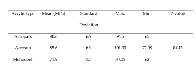
Results: The mean compressive strength was 80.6 ± 6.9, 85.6 ± 6.9, and 71.9 ± 5.3 MPa for Acropars, Acrosun, and Meliodent, respectively. The compressive strength of Acrosun was significantly higher than that of other groups (P=0.047).
Conclusion: In general, the results showed that the highest and the lowest compressive strength values were related to Acrosun and Meliodent, respectively. These results indicated the optimal compressive strength of Iranian acrylic resin.
Keywords: Acrylic Resins; Compressive Strength; Polymethyl Methacrylate
Introduction
Although dental implants are a more favorable option for replacement of the lost teeth, removable dentures are still considered as an alternative treatment for patients suffering from systemic diseases or severe jawbone atrophy (1). Various substances have been introduced as denture-base materials such as metal and heat-cure resins. Metal is not a desirable choice compared with other available materials since it cannot meet the esthetic requirements, and cannot bond to other denture structures (2). Acrylic denture bases are more popular due to their physical and chemical properties which enable relining and have optimal handling and esthetic properties as well (3). Acrylic resin materials are low weight and cheap, and have acceptable biocompatibility, esthetics, and color match with the natural tissue (4). Some concerns exist regarding the fracture of acrylic resin under heavy occlusal forces or trauma (5).
Denture base acrylic resin is mainly composed of polymethyl methacrylate (PMMA). PMMA is the preferred material for removable denture base and implant-supported restorations. Pure PMMA is a clear thermoplastic material that provides the opportunity of color matching with the tissue by adding plasticizers and cadmium. Its polymerization can be activated chemically or by light irradiation, microwave energy, or hot water. Activation of heat-cure acrylic resins is done by releasing free radicals (6). These materials are supplied in the form of powder and liquid. Many attempts have been made for the enhancement of PMMA characteristics. Addition of fillers is a practical method for this purpose. Many factors may contribute to fracture of denture base such as high frenal attachment, heavy occlusal loads, trauma, insufficient polymerization, residual monomers, or poor denture adaptation. Therefore, many studies have been conducted on the properties of denture base materials, location and time of crack propagation, and reasons for fracture (7-9).
Compressive strength refers to the strength and stiffness of a cylindrical sample that is vertically supported and is compassed in the same direction. To reduce the risk of fracture of the denture base, multiple factors such as preparation techniques, and physical and chemical properties of materials should be considered (10, 11). Also, a compressive resistance test should be performed (12). Because of the importance of the subject, the aim of this in vitro study was to evaluate and compare the compressive strength of three different types of heat-cure acrylic resins namely Acropars, Acrosun, and Meliodent.
Materials and Methods
In this in vitro experimental study, 60 acrylic samples were fabricated from Acropars, Acrosun, and Meliodent (n=20 from each) using molds. Cylindrical specimens were fabricated in 6 aluminum molds measuring 4 mm x 10 mm. After preparation of the molds, petroleum jelly was applied on the walls of the molds to prevent the attachment of wax to the molds in the waxing stage; then, melted modeling wax was poured into the cylindrical molds while using a dental catheter to prevent void and defect formation. Next, with the help of an expert technician, the process of flasking was done with type 2 plaster (13). Flasks were placed under the pressure of a hydraulic press machine for 10 minutes; then, they were placed in cold water and after that, they were sintered. The flasks were placed in water at 70°C temperature for 8 hours. Then, they were placed in water at 100°C temperature for 1 hour. The water temperature was checked by a thermometer during the entire process (14). Finally, all specimens remained at room temperature for 24 hours to cool down (15). The flasks were opened carefully to remove the samples. A Sandblaster was used to remove the plaster attached to the samples. All samples were measured to be of the same size.
The study groups were as follows:
- Group A: Acropars acrylic resin (Marlic Co., Tehran, Iran) (n=20)
- Group B: Acrosun acrylic resin (Betadent Co., Iran) (n=20)
- Group C: Meliodent acrylic resin (Bayer Co., New Burg, Germany) (n=20)
All specimens were fabricated under the same conditions with the powder-liquid ratio of 3:1 according to the manufacturer’s instructions. To measure the compressive strength, the samples were placed in a universal testing machine and load was applied at a crosshead speed of 1 mm/minute until the fracture of acrylic resin sample; the load at failure was recorded as compressive strength (16).
Statistical analysis:
Data analysis was performed using SPSS version 22. ANOVA was used tocompressive strength of the three groups . The Tukey’s test was applied for pairwise comparisons. The significance level was set at 0.05.
Results
The compressive strength of the three experimental groups is shown in Table 1.
The results indicated that Acrosun had the maximum and Meliodent had the minimum mean compressive strength. One-way ANOVA showed a significant difference in compressive strength of the three groups (P=0.047). Thus, pairwise comparisons were performed by the Tukey’s test, which showed a significant difference in the compressive strength of Acropars and Acrosun (P=0.047), Acropars and Meliodent (P=0.00), and Acrosun and Meliodent (P=0.00) groups (Table 2).
Table 1. Compressive strength of the three groups
Table 2. Pairwise comparisons of the groups by the Tukey’s test
Discussion
Fracture of denture base is a common problem. Therefore, various methods have been used to reduce the risk of fracture and provide proper masticatory function, optimal esthetics, and speech. Auto-polymerized acrylic resins have shown lower compressive strength in comparison with heat-cure acrylic resins, which is due to the higher amount of residual monomers in auto-polymerized acrylic resins (17). Assessment of the compressive and flexural strength of denture base materials is beneficial since the forces applied to dentures during mastication can be simulated as such. Previous studies did not agree on the mechanical and chemical properties of acrylic resins. Gharehchahi et al. (18) evaluated the bond strength of two heat-cure acrylic resins namely glass-reinforced Meliodent and Acropars. In contrast to the results of the present study, they showed no significant difference between the two acrylic resins. However, Gungor et al. (19) found different flexural and compressive strength values of denture base materials depending on their polymerization method. They reported that the flexural and compressive strengths of polymerized acrylic resins were higher when using short cycle polymerization, which included 90 minutes at 73°C and then 30 minutes at 100°C compared with long polymerization cycle (9 hours of polymerization). Their results are consistent with the results of the present study despite different methodologies.
In another study, Hashem et al. (17) found that Eclipse acrylic resin had higher compressive and flexural strength values compared with Meliodent acrylic resin. This might be due to higher polymerization temperature or crystalline formulation of the former acrylic resin, resulting in fewer voids and gaps in it. Durkan and Oyar (13) stated that the highest flexural and compressive strength values were related to Paladin acrylic resin, and the lowest values was recorded for Meliodent, which is consistent with our results. According to a study by Ellakwa et al, (3) Al2O3 fillers, as components added to denture base materials, have the potential to increase the bond strength and heat dissipation, and also, improve the flexural strength and heat transfer properties of acrylic resin base. These properties increase patient satisfaction. Begum et al. (20) evaluated the impact strength and dimensional accuracy of heat-cure PMMA reinforced with ZrO2 nanoparticles, and reported that reinforcement of heat-cure PMMA with ZrO2 nanoparticles significantly increased the dimensional accuracy and decreased the impact strength. Recently, the advent of computer-aided design and computer-aided manufacturing (CAD/CAM) technology enabled the fabrication of complete dentures from PMMA blocks. Accordingly, Al‐Dwairi et al. (21) compared the flexural strength, impact strength, and flexural modulus of CAD/CAM and heat-cure PMMA. Their results showed that since CAD/CAM PMMA specimens exhibited improved flexural strength, flexural modulus, and impact strength in comparison with the conventional heat‐cure groups, CAD/CAM dentures are expected to be more durable. Khan et al. (22) reported that the flexural strength of heat-cure PMMA denture base did not statistically improve after reinforcement with zinc powder in different concentrations. Moreover, Maheshwari and Parihar (23) reported that heat-cure acrylic resin had higher compressive and flexural strength, and hardness in comparison with other types of acrylic resins, which was in line with our study.
In the present study, samples were fabricated in a size larger than the actual size of restorations in the oral cavity to enhance measurement of their compressive strength. In clinical conditions, the maximum thickness of acrylic resin in the palate area is 2.5 mm, and increasing the thickness of specimens increases the likelihood of void formation or incomplete polymerization and can compromise the accuracy of the results. To minimize this error, sintering of the samples was performed under similar conditions. Not measuring the flexural strength was a limitation of this study. Also, considering the in vitro design of this study, it could not completely simulate the intraoral conditions. Therefore, clinical studies are required to confirm the present results.
Conclusion
Considering the limitations of this study, the following results were drawn: Acrosun had the highest compressive strength compared with Meliodent and Acropars. The lowest compressive strength was related to Meliodent. These results confirmed the optimal compressive strength of Iranian acrylic resin. Dental clinicians can use it considering its cost-effectiveness and easy availability.
| Rights and permissions | |
 |
This work is licensed under a Creative Commons Attribution-NonCommercial 4.0 International License. |
