


BibTeX | RIS | EndNote | Medlars | ProCite | Reference Manager | RefWorks
Send citation to:
URL: http://jrdms.dentaliau.ac.ir/article-1-299-en.html


2- Private Practice, Tehran, Iran
3- Dental Implant Research Center, Dental Faculty, Tehran Medical Sciences, Islamic Azad University, Tehran, Iran ,
Abstract
Background and Aim: The aim of this study was to compare the cytotoxicity of ProRoot MTA and Endocem at different times and concentrations on human gingival fibroblasts.
Materials and Methods: In this in-vitro study, ProRoot MTA and Endocem extracts with concentrations of 3, 6, 12, 25, and 50 mg/ml in the unhardened state (solution) at 24, 48, and 72 hours on human gingival fibroblasts were transferred to 96-well plates in Dulbecco's Modified Eagle's medium (DMEM). For each material at any time and any concentration, three wells were used, and 90 samples were examined. Also, to calibrate the cytotoxicity measuring device at 24, 48, and 72 hours, 3 wells for positive control and 3 wells for negative control have been considered, which include a total of 9 positive control wells and 9 negative control wells. Samples were analyzed by MTT assay. Data were analyzed by analysis of variance (ANOVA).
Results: In the present study, the samples in the two groups at different times and concentrations did not show a significant difference in terms of cytotoxicity (P<0.05). The highest cytotoxicity was related to Endocem at 24 hours and a concentration of 50 mg/ml, and the lowest was related to ProRoot MTA at 72 hours and a concentration of 6 mg/ml.
Conclusion: According to the research, in general, the degree of cytotoxicity of Endocem is comparable to that of ProRoot MTA.
Keywords: Cytotoxicity, Endocem, ProRoot MTA, MTT Assay
Introduction
One of the concerns of dentists has always been the toxicity of dental materials because these substances are in contact with the periodontium and living tissues around the teeth; lack of biocompatibility of these substances can lead to inflammatory reactions. (1) An ideal root-end filling material must have physical, chemical, and biological properties such as antimicrobial effect, biocompatibility, effective sealing capability, moisture and solubility resistance, short setting time, sufficient radiopacity, and ease of use, among which, the biocompatibility is a factor that affects the prognosis of treatment. (2-4)
Mineral trioxide aggregate (MTA) is a powder composed of hydrophilic fine particles that harden in the presence of moisture. This material was first used as a root-end filling material, but today, it is also used for perforation repair, pulp capping, pulpotomy, and apexification. (5,6) The first MTA produced was ProRoot MTA, and so far, many studies have confirmed the superior biological properties of this cement compared to other endodontic cements. (2-4) However, problems such as difficulty in use, long setting time, and discoloration of teeth or soft tissue have been reported for this cement. (5,6) These problems led to the introduction of new materials to the market. One of these materials is Endocem. Its chemical composition is similar to that of MTA, it has no chemical accelerator, and its small particle size causes a rapid setting. (7,8) To date, little research has been done on the biocompatibility and physical properties of Endocem in the laboratory. Endocem cytotoxicity has been investigated in quantitative studies. (9)
There are several methods to evaluate the cytotoxicity of substances, among which, MTT assay has high accuracy and validity. (10) In the study of living tissues, the use of periodontal ligament (PDL) cells has high validity as a sample in measuring the degree of cytotoxicity, and these cells are close to the site where the substance is used. (11)
The aim of this study was to evaluate the cytotoxicity of Endocem and compare it to that of ProRoot MTA, which has been accepted as a biocompatible substance. (12)
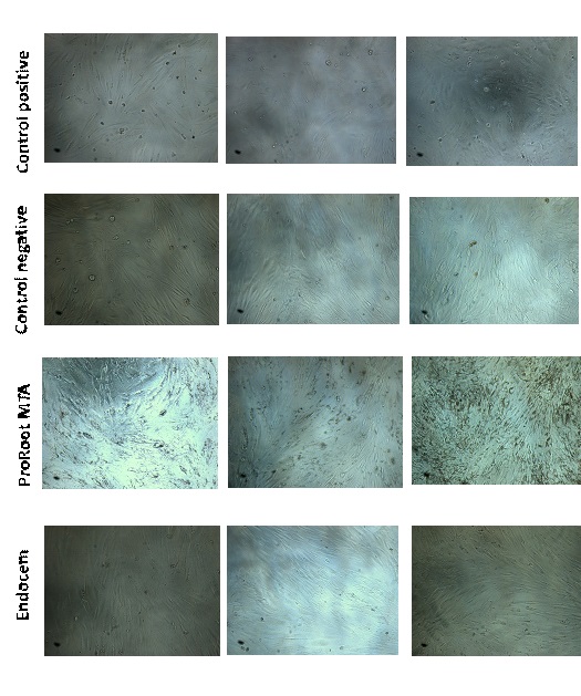
Figure 1. Morphological changes of gingival fibroblast cells in contact with experimental materials: Positive control (1A: 24h, 1B: 48h, 1C: 72h), Negative control (1A: 24h, 1B: 48h, 1C: 72h), ProRoot MTA (1A: 24h, 1B: 48h, 1C: 72h), and Endocem (1A: 24h, 1B: 48h, 1C: 72h)
Materials and Methods
This study was performed experimentally in vitro. All prepared materials were kept in the same culture medium and laboratory environment. A person with the necessary knowledge and skills, who was blind to the grouping of the tested samples, assessed cytotoxicity. All the work steps, including mixing the materials, preparing the plate, and placing them in the culture medium, were performed under germ-free conditions.
Preparation of gingival fibroblasts:
In this study, gingival fibroblasts isolated by the explants technique from extracted third molars were used. Extracted wisdom teeth were transferred to the laboratory in conical tubes containing 1% penicillin, 1% streptomycin, 1% antifungal agent, and 10% fetal serum. The gingiva of the extracted wisdom tooth was carefully removed and transferred to the phosphate buffer saline (PBS) medium. After several washings in culture medium to ensure no contamination, the sent tissue was placed for 24 hours in Dulbecco's Modified Eagle Medium (DMEM) to which 1% penicillin, 1% streptomycin, 1% antifungal agent, and 10% glutamine serum were added, and was stored at 5% CO2 and saturated humidity at 37°C. (13,14) To isolate fibroblast cells, collagenase (2 mg per cc of Hanks containing 5% of serum) and pronase (10 mg per cc of Hanks containing 5% of serum) were used, and the cells prepared for culture were transferred to DMEM. (15,16)
Preparation of cell culture medium:
To prepare the cell culture medium, in this study, 133.8g DMEM, 6.1mg penicillin, 3.7g sodium bicarbonate, 1.1g sodium pyruvate, 10mg streptomycin, and 0.02 to 0.06g L-glutamine were poured into distilled water. (17)
The cells were passaged 5 times to reach the required number for the experiment. To ensure cell viability at each stage of proliferation, 1ml of the cell-containing solution was placed on a slide, and trypan blue staining confirmed cell viability. (17)
Preparation of samples:
In this study, ProRoot MTA and Endocem materials were mixed in proportion of three parts powder and one part liquid according to the factory instructions, and then, 5ml of pure DMEM was added to 1g of the tested material, and the resulting medium was incubated at 37°C and 100% humidity for 24 hours. Next, the solutions were diluted by DMEM with concentrations of 3, 6, 12, 25, and 50 mg/ml. (3) In addition, 200μl of pure culture medium was used as a negative control, and 200μl of culture medium containing 2% dimethyl sulfoxide (DMSO) dissolved in DMEM was used as a positive control.
For each material studied at each time, three separate wells were considered. Considering that the experiment included three time intervals, nine wells for each material, three wells for positive control, and three wells for negative control were considered.
It should be noted that the whole experiment was repeated three times. The cells were placed in 96-well plates at a density of 5000 and incubated for 24 hours at 37°C, and then, the culture medium was replaced with 200μl of the prepared samples. After 24, 48, and 72 hours, cell viability was assessed by MTT assay. First, the culture medium was removed, and then, 100μl of MTT solution was added to each well of the plate, and the cells were incubated for another 1 hour. Mitochondrial enzymes reduced MTT and converted it to purple formazan crystals. Then, 100μl of DMSO solution was added to each well. The plates were stirred for 10 minutes to dissolve the formazan crystals and then prepared for spectrophotometric analysis at 570 nm.
The number of surviving cells has been calculated according to the following formula: Percentage of living cells=  ×100.
×100.
If the uptake of the sample (cells to which the test substance was added) is greater than the uptake of the control group (cells that have not been examined in the presence of the drug or substance), it indicates a higher proliferation process than the control group. If the absorbance of the sample is less than the absorption of the control group, it indicates the process of cell death in the sample and the cytotoxicity of the substance used in this concentration. If the absorption of the sample is equal to the absorption of the control group, it indicates the ineffectiveness of the substance used on cell proliferation and cell death. Therefore, a larger number indicates less toxicity. Data were analyzed by analysis of variance (ANOVA).
Results
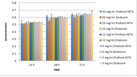
The degree of cytotoxicity of the two substances by concentration and time is presented in Table 1. The lowest levels of cytotoxicity at 72 hours were related to ProRoot MTA (6mg/ml = 0.645±0.008 OD), Endocem (3mg/ml = 0.639±0.0050 OD), and ProRoot MTA (50mg/ml = 0.635±0.016), respectively. The highest rate of cytotoxicity at 24 hours was related to Endocem (50mg/ml = 0.508±0.003 OD), Endocem (25 mg/ml = 0.519±0.025 OD), and ProRoot MTA (50 mg/ml = 0.519±0.037 OD), respectively. There was no significant difference between the two groups of samples regarding cytotoxicity at different times and concentrations (P<0.05).
Cell morphology and cell density of each substance at each concentration and time were assessed using an inverted light microscope at ×100 magnification (CKX41, Olympus, Tokyo, Japan; Figure 1).
Table 1. Distribution of the studied samples according to the degree of cytotoxicity (optical density) at different concentrations and times
In most groups, the cells attached to the plate in the first 24 hours after placement, except in the Endocem and positive control groups. The ProRoot MTA and negative control groups showed high cell density from the initial stage even at the material boundaries. The Endocem group did not show cell adhesion immediately after placement. However, for up to 48 hours, the cell density of the Endocem group increased. 72 hours later, no significant difference was observed between the groups in terms of cell number. As with the MTT test results, in the early hours of exposure of solutions to the cells in the culture medium, the number of cells decreases due to toxicity, the cells lose their morphology, and are removed from the bottom of the culture medium. These changes are more pronounced in the fresh Endocem solution group and indicate the transient toxicity of this substance in the first 24 hours.
Discussion
The present study showed that the cytotoxicity of Endocem is comparable to that of ProRoot MTA. Endocem has low transient cytotoxicity in the early hours after mixing, which improves over time. Considering the difficult use and long setting time of ProRoot MTA, Endocem can be suggested as an alternative to this material. Koseoglu et al (3) have shown that there is no significant difference in cytotoxicity and cell viability between Endocem and ProRoot MTA, which confirms the present findings. (3) Studies by Song et al and Chung et al (4), like the present study, showed that Endocem was more toxic, but the difference was significant. (2,4)
The results of the present study display that the cells exposed to Endocem differed in shape and number from cells exposed to ProRoot MTA in the first 24 hours after culture, but within 48 hours after replacement, the morphology and cell distribution are similar. The cells in contact with the fresh Endocem mixture have lost their spindle shape, become rounded, and their density has decreased, which means that the initial Endocem mixture is toxic. This can be explained by the high pH and heat of the cement surface produced in the initial mixture. High pH and heat can directly damage cells through apoptosis or necrosis and indirectly cause culture proteins to denature. (2) Primary cytotoxicity tends to decrease over time so that the Endocem group showed adhesion and cell proliferation 48 hours after mixing time.
In the ProRoot MTA group, samples at different times and concentrations did not show significant differences in cytotoxicity and cell viability. Cell adhesion occurred immediately after exposure to the culture medium and only a small number of cells were deformed. The highest OD level belonged to this group, and therefore, ProRoot MTA showed the lowest cytotoxicity and the highest biocompatibility. Previous studies confirm the findings regarding ProRoot MTA and refer to it as a substance with the highest biological properties. (2-4,8,12,18-21)
ProRoot MTA includes 55% (C3S), 19% (C2S), 10% (C3A), 7% (C4AF), 2.8% (MgO), 2.9% (SO3), and 1% (CaO). (18) The combination of MTA with water results in the formation of a gel that hardens between 1 and 9 hours. (16) The small particle size of the Endocem increases the contact surface of the particles during mixing, resulting in a rapid setting. (7) According to a recent study by Choi et al, The initial setting time of this material is 4 minutes. (7) The rapid setting of this material increases washout resistance, which may reduce the release of ions. Endocem contains 40% calcium carbonate (the main component); this extra amount of calcium carbonate indicates that this material is pozzolan. (3)
In the present study, gingival fibroblasts were used because, according to previous studies, an ideal repair after an endodontic surgery includes alveolar bone regeneration and PDL regrowth along the root surface. The behavior of PDL cells and how they react when in direct contact with root-end filling materials is of utmost importance. (2) Therefore, Song et al and Chung et al also used fibroblasts attached to extracted teeth to simulate the clinical condition. (2,4)
In the present study, an indirect extraction method was used to evaluate the material on gingival fibroblast cells, which is a valuable method according to previous studies, because, in the clinic, a fresh mixture of cement is used, which is possibly more toxic to living tissues compared to hardened cement; this is closer to the oral conditions. (2,4) Chung et al have stated that one of the limitations of their study is the use of hard cement instead of a fresh solution of materials. (4)
In the present study, different concentrations of substances (3, 6, 12, 25, and 50 mg/ml) were used to more precisely evaluate the degree of cytotoxicity, while in most previous studies, only one concentration of substances was examined. (2-4) Also, the toxicity of the substances was evaluated at 3 different times (24, 48, and 72 hours after mixing), which is a feature of studies by Song et al, Chung et al, and Koseoglu et al. (2-4)
Today, various methods, such as antigen analysis, labeled thymidine, bromodeoxyuridine, propidium iodide, tetrazolium salts (MTT, XTT, MTS, and WST-1), Alamar Blue, ViaLight, and the oxygen biosensor system methods are used to study the rate of cell proliferation. These methods can be used according to the needs of the application and laboratory equipment, but in the meantime, yellow tetrazolium (MTT) is known as a cost-effective, valid, and well-known method among researchers. In the present study, we used MTT assay to evaluate the toxicity of the materials, which has high accuracy and validity among other methods. (7) Chung et al and Koseoglu et al used XTT assay and Song et al used WST-1 assay. (2-4)
In the present study, as a scale for assessing material biocompatibility, we observed the adhesion and morphology of gingival fibroblast cells when in direct contact with the test material, because cell adhesion to the material is important for survival and proliferation. (2) Therefore, many studies evaluate the toxicity of substances along with cell morphology analysis. (2-4) For this purpose, we used an inverted light microscope at 100× magnification. These types of microscopes are suitable for examining thick specimens, such as cultured cells, because the lenses can be closer to where the cell grows, so they are more accurate. Most previous studies have used conventional light microscopes or electron microscopes. (2-4,7)
Comparisons regarding the mechanical and chemical properties, dimensional stability, operating time, and fluidity of MTAs are recommended to help select the appropriate material for dental treatment.
Conclusion
According to the present research, the cytotoxicity of Endocem is comparable to that of ProRoot MTA. Considering the difficult use and long setting time of ProRoot MTA, Endocem can be suggested as an alternative.
| Rights and permissions | |
 |
This work is licensed under a Creative Commons Attribution-NonCommercial 4.0 International License. |
