

Volume 6, Issue 1 (3-2021)
J Res Dent Maxillofac Sci 2021, 6(1): 19-23 |
Back to browse issues page

Download citation:
BibTeX | RIS | EndNote | Medlars | ProCite | Reference Manager | RefWorks
Send citation to:


BibTeX | RIS | EndNote | Medlars | ProCite | Reference Manager | RefWorks
Send citation to:
Jalili Sadrabad M, Sahraeeyan Y, Sohrabpoor F, Mirmohammadkhani M, Sohanian S. Evaluation of Dysplasia in Oral Lichen Planus Patients of Oral Medicine Department of Semnan University of Medical Sciences from 2013 to 2018. J Res Dent Maxillofac Sci 2021; 6 (1) :19-23
URL: http://jrdms.dentaliau.ac.ir/article-1-296-en.html
URL: http://jrdms.dentaliau.ac.ir/article-1-296-en.html


1- Assistant Professor, Oral Medicine Dept, Dental School
2- Dental Student, Student Research Committee, Dental School
3- Social Determinants of Health Research Center, Department of Epidemiology & Statistics, Faculty of Medicine
4- Assistant Professor, Oral and Maxillofacial Pathology Dept, Dental School, Semnan University of Medical Sciences, Semnan, Iran. , dr.sh.sohanian@gmail.com
Full-Text [PDF 241 kb]
(895 Downloads)
| Abstract (HTML) (1721 Views)
Full-Text: (544 Views)
Abstract
Background and Aim: Oral lichen planus (OLP) is a chronic mucocutaneous disease, which is considered a precancerous condition. OLP is a multifactorial disease with no conclusive treatment. Timely diagnosis and treatment of oral lesions are necessary steps to prevent malignant transformations and severe complications of oral cancer. This study aimed to evaluate the frequency and severity of malignant transformations in OLP.
Materials and Methods: In this study, 2400 files of patients who presented to the Oral Medicine Department of the Dental School of Semnan University of Medical Sciences from 2013 to 2018 were evaluated. Data were statistically analyzed using SPSS.
Results: Forty patients (9 males and 31 females with the mean age of 45.2 years) were diagnosed with OLP (1.6%). The most common type of OLP was reticular (75%), and the least common was the ulcerative type. Since toluidine blue staining was positive for 4 patients (10%), a biopsy was done. The histopathological examinations showed one case with dysplasia (2.5%) and one case of carcinoma (2.5%).
Conclusion: According to the results, carcinoma and dysplasia can develop in OLP patients. Regular follow-ups are strongly recommended for timely diagnosis and treatment of malignant transformations.
Keywords: Lichen Planus, Oral, Squamous Cell Carcinoma, Mouth Neoplasms
Introduction
Oral lichen planus (OLP) is a chronic mucocutaneous disease, which frequently affects the oral mucosa with a wide spectrum of clinical manifestations. It is more common in middle-aged women. OLP has been reported in other parts of the body, such as the flexor areas of the skin (elbows and groin), nails, and esophageal and genital mucosa. There are different concepts regarding the main etiology of OLP, such as immune system involvement, T cell activation, and stress. Nevertheless, studies show that OLP is a multifactorial process. (1-5)
Clinically, OLP presents as white and red components with different subtypes, such as reticular, papular, plaque-like, bullous, erythematous, and ulcerative types. (6) The presence of reticular and papular components is essential for the clinical diagnosis of OLP. Due to its unknown etiology, OLP has no definitive treatment. Palliative treatments are used to subside the symptoms. Also, physicians may prescribe drugs, such as steroids, for patient comfort. (7) In addition to side effects such as pain, burning, mucosal irritation, and the possibility of involvement of other mucous membranes and skin, many researchers consider OLP as a precancerous condition. The percentage of OLP malignant transformations has been reported to be between 0% and 10% in previous studies. (8,9) A study performed in Iran showed a significant rate of dysplastic changes in OLP (10.7%); therefore, it is rational to periodically follow-up the patients, not only to monitor the development of the disease but also to avoid possible initial misdiagnoses. (10)
Timely diagnosis and treatment of oral lesions are necessary steps to prevent malignant transformations and severe complications of oral cancer. This study aimed to evaluate the frequency and severity of malignant transformations in OLP patients who presented to the Oral Medicine Department of the Dental School of Semnan University of Medical Sciences.
Materials and Methods
In this cross-sectional study (ethical approval code: IR.SEMUMS.REC.1396.251), the archived files of patients who presented to the Oral Medicine Department of the Dental School of Semnan University of Medical Sciences were reviewed, and the patients were clinically examined. The demographic data, medical history, medications, and the intraoral soft tissue characteristics were recorded in datasheets. Incomplete files and patients with severe oral mucosal diseases other than OLP were excluded. Written consent forms were received from all patients. Data were statistically analyzed using SPSS (SPSS Inc., Chicago, IL, USA).
Results
The examination of 2400 cases showed a prevalence rate of 1.6% (40 cases) for OLP (31 females (77.5%) and 9 males (22.5%) with the mean age of 45.2 years). In terms of the educational level, 26 patients had a diploma (65%) while 11 patients had not (27.5%); three patients had a bachelor's degree (7.5%). Reticular subtype affected most patients. The least common form of OLP was the ulcerative type (Table 1).
Table 1. Frequency of different types of oral lichen planus (OLP) among patients
OLP was mostly reported in housekeepers and least reported in workers. Twenty-three patients were housekeepers (57.5%), 6 were employees (15%), 6 were self-employed (15%), 3 were students (7.5%), and 2 were workers (5%).
In terms of the underlying disease, five patients had hypertension (12.5%), and one patient was diabetic (2.5%). Two patients were (5%) smokers, and cutaneous involvement was reported in only one patient (2.5%). Three patients were taking levothyroxine (7.5%), and 3 patients were on propranolol (7.5%).
Toluidine blue staining was positive for four patients (10%). The histopathological examinations revealed dysplasia in one patient (2.5%) and carcinoma in another patient (2.5%).
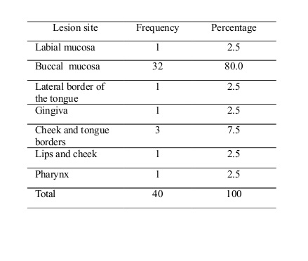
The frequency of OLP based on the location of the lesion is presented in Table 2.
Table 2. Frequency of oral lichen planus (OLP) according to the location of the lesion
Discussion
Despite its significance, few studies have been conducted concerning OLP in different parts of Iran, and therefore, there is a lack of accurate statistics of its risk of malignant transformations.
In this study, the prevalence of OLP among patients who presented to the Oral Medicine Department of the Dental School of Semnan University of Medical Sciences from 2013 to 2018 was reported to be 1.6%. Other studies have reported the prevalence of OLP to be between 0.22% and 0.7%. (11-13) This difference is probably due to different sample sizes and statistical populations, as well as various risk factors in different societies.
Similar to other studies that have reported a higher prevalence of OLP in women, in the current study, women were 3.44 times more likely to develop the disease than men were. (12-14) In the present study, the mean age of OLP patients was 45.2 years. Pakfetrat et al (16.41 years), Oliveira Alves et al (08.54±13.14 years), Fitzpatrick et al (52 years), and Mardani et al (54.3 years) also reported most patients to be middle-aged. (11-13,15-17) The rate of cutaneous involvement was 2.5% in our study but Fitzpatrick et al reported a rate of 25%. (15)
The most common OLP site was the buccal mucosa, which was similar to the reports of previous studies. (12-14) The most common OLP subtype in the present study was the reticular form, which was similar to the findings of previous studies. (12,13) Mardani et al reported erosive OLP as the most common form since they biopsied and further examined red lesions. (11)
From the samples, 10% required toluidine blue staining; the results indicated a 5% rate of malignant transformation as dysplasia was detected in one patient (2.5%) and carcinoma was observed in another (2.5%). Fitzpatrick et al (1.09%), Mardani et al (1.03%), Tovaru et al (0.95%), and Guan et al (0.6%) have reported lower rates of malignant transformations compared to the present study. (11,14-17) Aghbari et al and Idrees et al found a higher incidence of malignant transformations among smokers, alcoholics, and hepatitis C virus (HCV)-infected patients; (18,19) nevertheless, such associations need further research. This discrepancy shows the need for supplementary studies as to why the risk of OLP malignant transformations in the current study is higher than that reported by previous studies. Since stress plays a significant role in malignant transformations, future studies should focus on examining stress as an etiologic factor in the development of squamous cell carcinoma (SCC) in OLP.
In a study conducted on an Iranian population, the prevalence of dysplasia was reported to be 10.7% among 112 OLP cases studied. (10) The high prevalence of dysplasia in the cited study (in comparison with the present research) emphasizes the importance of monitoring OLP patients.
Conclusion
According to the results, the incidence rate of carcinoma and dysplasia in OLP patients is 2.5%. It is essential to monitor the patients regularly for malignant transformations. Timely and non-invasive treatments are necessary to improve the quality of life of these patients.
Background and Aim: Oral lichen planus (OLP) is a chronic mucocutaneous disease, which is considered a precancerous condition. OLP is a multifactorial disease with no conclusive treatment. Timely diagnosis and treatment of oral lesions are necessary steps to prevent malignant transformations and severe complications of oral cancer. This study aimed to evaluate the frequency and severity of malignant transformations in OLP.
Materials and Methods: In this study, 2400 files of patients who presented to the Oral Medicine Department of the Dental School of Semnan University of Medical Sciences from 2013 to 2018 were evaluated. Data were statistically analyzed using SPSS.
Results: Forty patients (9 males and 31 females with the mean age of 45.2 years) were diagnosed with OLP (1.6%). The most common type of OLP was reticular (75%), and the least common was the ulcerative type. Since toluidine blue staining was positive for 4 patients (10%), a biopsy was done. The histopathological examinations showed one case with dysplasia (2.5%) and one case of carcinoma (2.5%).
Conclusion: According to the results, carcinoma and dysplasia can develop in OLP patients. Regular follow-ups are strongly recommended for timely diagnosis and treatment of malignant transformations.
Keywords: Lichen Planus, Oral, Squamous Cell Carcinoma, Mouth Neoplasms
Introduction
Oral lichen planus (OLP) is a chronic mucocutaneous disease, which frequently affects the oral mucosa with a wide spectrum of clinical manifestations. It is more common in middle-aged women. OLP has been reported in other parts of the body, such as the flexor areas of the skin (elbows and groin), nails, and esophageal and genital mucosa. There are different concepts regarding the main etiology of OLP, such as immune system involvement, T cell activation, and stress. Nevertheless, studies show that OLP is a multifactorial process. (1-5)
Clinically, OLP presents as white and red components with different subtypes, such as reticular, papular, plaque-like, bullous, erythematous, and ulcerative types. (6) The presence of reticular and papular components is essential for the clinical diagnosis of OLP. Due to its unknown etiology, OLP has no definitive treatment. Palliative treatments are used to subside the symptoms. Also, physicians may prescribe drugs, such as steroids, for patient comfort. (7) In addition to side effects such as pain, burning, mucosal irritation, and the possibility of involvement of other mucous membranes and skin, many researchers consider OLP as a precancerous condition. The percentage of OLP malignant transformations has been reported to be between 0% and 10% in previous studies. (8,9) A study performed in Iran showed a significant rate of dysplastic changes in OLP (10.7%); therefore, it is rational to periodically follow-up the patients, not only to monitor the development of the disease but also to avoid possible initial misdiagnoses. (10)
Timely diagnosis and treatment of oral lesions are necessary steps to prevent malignant transformations and severe complications of oral cancer. This study aimed to evaluate the frequency and severity of malignant transformations in OLP patients who presented to the Oral Medicine Department of the Dental School of Semnan University of Medical Sciences.
Materials and Methods
In this cross-sectional study (ethical approval code: IR.SEMUMS.REC.1396.251), the archived files of patients who presented to the Oral Medicine Department of the Dental School of Semnan University of Medical Sciences were reviewed, and the patients were clinically examined. The demographic data, medical history, medications, and the intraoral soft tissue characteristics were recorded in datasheets. Incomplete files and patients with severe oral mucosal diseases other than OLP were excluded. Written consent forms were received from all patients. Data were statistically analyzed using SPSS (SPSS Inc., Chicago, IL, USA).
Results
The examination of 2400 cases showed a prevalence rate of 1.6% (40 cases) for OLP (31 females (77.5%) and 9 males (22.5%) with the mean age of 45.2 years). In terms of the educational level, 26 patients had a diploma (65%) while 11 patients had not (27.5%); three patients had a bachelor's degree (7.5%). Reticular subtype affected most patients. The least common form of OLP was the ulcerative type (Table 1).
Table 1. Frequency of different types of oral lichen planus (OLP) among patients
OLP was mostly reported in housekeepers and least reported in workers. Twenty-three patients were housekeepers (57.5%), 6 were employees (15%), 6 were self-employed (15%), 3 were students (7.5%), and 2 were workers (5%).
In terms of the underlying disease, five patients had hypertension (12.5%), and one patient was diabetic (2.5%). Two patients were (5%) smokers, and cutaneous involvement was reported in only one patient (2.5%). Three patients were taking levothyroxine (7.5%), and 3 patients were on propranolol (7.5%).
Toluidine blue staining was positive for four patients (10%). The histopathological examinations revealed dysplasia in one patient (2.5%) and carcinoma in another patient (2.5%).
The frequency of OLP based on the location of the lesion is presented in Table 2.
Table 2. Frequency of oral lichen planus (OLP) according to the location of the lesion
Discussion
Despite its significance, few studies have been conducted concerning OLP in different parts of Iran, and therefore, there is a lack of accurate statistics of its risk of malignant transformations.
In this study, the prevalence of OLP among patients who presented to the Oral Medicine Department of the Dental School of Semnan University of Medical Sciences from 2013 to 2018 was reported to be 1.6%. Other studies have reported the prevalence of OLP to be between 0.22% and 0.7%. (11-13) This difference is probably due to different sample sizes and statistical populations, as well as various risk factors in different societies.
Similar to other studies that have reported a higher prevalence of OLP in women, in the current study, women were 3.44 times more likely to develop the disease than men were. (12-14) In the present study, the mean age of OLP patients was 45.2 years. Pakfetrat et al (16.41 years), Oliveira Alves et al (08.54±13.14 years), Fitzpatrick et al (52 years), and Mardani et al (54.3 years) also reported most patients to be middle-aged. (11-13,15-17) The rate of cutaneous involvement was 2.5% in our study but Fitzpatrick et al reported a rate of 25%. (15)
The most common OLP site was the buccal mucosa, which was similar to the reports of previous studies. (12-14) The most common OLP subtype in the present study was the reticular form, which was similar to the findings of previous studies. (12,13) Mardani et al reported erosive OLP as the most common form since they biopsied and further examined red lesions. (11)
From the samples, 10% required toluidine blue staining; the results indicated a 5% rate of malignant transformation as dysplasia was detected in one patient (2.5%) and carcinoma was observed in another (2.5%). Fitzpatrick et al (1.09%), Mardani et al (1.03%), Tovaru et al (0.95%), and Guan et al (0.6%) have reported lower rates of malignant transformations compared to the present study. (11,14-17) Aghbari et al and Idrees et al found a higher incidence of malignant transformations among smokers, alcoholics, and hepatitis C virus (HCV)-infected patients; (18,19) nevertheless, such associations need further research. This discrepancy shows the need for supplementary studies as to why the risk of OLP malignant transformations in the current study is higher than that reported by previous studies. Since stress plays a significant role in malignant transformations, future studies should focus on examining stress as an etiologic factor in the development of squamous cell carcinoma (SCC) in OLP.
In a study conducted on an Iranian population, the prevalence of dysplasia was reported to be 10.7% among 112 OLP cases studied. (10) The high prevalence of dysplasia in the cited study (in comparison with the present research) emphasizes the importance of monitoring OLP patients.
Conclusion
According to the results, the incidence rate of carcinoma and dysplasia in OLP patients is 2.5%. It is essential to monitor the patients regularly for malignant transformations. Timely and non-invasive treatments are necessary to improve the quality of life of these patients.
Type of Study: Original article |
Subject:
Oral medicine
References
1. [1] Al-Hashimi I, Schifter M, Lockhart PB, Wray D, Brennan M, Migliorati CA, et al. Oral lichen planus and oral lichenoid lesions: diagnostic and therapeutic considerations. Oral Surg Oral Med Oral Pathol Oral Radiol Endod. 2007 Mar;103 Suppl:S25.e1-12. [DOI:10.1016/j.tripleo.2006.11.001] [PMID]
2. [2] Farhi D, Dupin N. Pathophysiology, etiologic factors, and clinical management of oral lichen planus, part I: facts and controversies. Clin Dermatol. 2010 Jan-Feb;28(1):100-8. [DOI:10.1016/j.clindermatol.2009.03.004] [PMID]
3. [3] Gorouhi F, Solhpour A, Beitollahi JM, Afshar S, Davari P, Hashemi P, Nassiri Kashani M, Firooz A. Randomized trial of pimecrolimus cream versus triamcinolone acetonide paste in the treatment of oral lichen planus. J Am Acad Dermatol. 2007 Nov;57(5):806-13. [DOI:10.1016/j.jaad.2007.06.022] [PMID]
4. [4] Irani S, Monsef Esfahani A, Bidari Zerehpoush F. Detection of Helicobacter pylori in Oral Lesions. J Dent Res Dent Clin Dent Prospects. 2013 Fall;7(4):230-7.
5. [5] Ismail SB, Kumar SK, Zain RB. Oral lichen planus and lichenoid reactions: etiopathogenesis, diagnosis, management and malignant transformation. J Oral Sci. 2007 Jun;49(2):89-106. [DOI:10.2334/josnusd.49.89] [PMID]
6. [6] Jones KB, Jordan R. White lesions in the oral cavity: clinical presentation, diagnosis, and treatment. Semin Cutan Med Surg. 2015 Dec;34(4):161-70. [DOI:10.12788/j.sder.2015.0180] [PMID]
7. [7] Glick M, William M. Burket's Oral Medicine. 12th ed. People's Medical Publishing House, USA, Ltd., 2015:105-9.
8. [8] van der Meij EH, Mast H, van der Waal I. The possible premalignant character of oral lichen planus and oral lichenoid lesions: a prospective five-year follow-up study of 192 patients. Oral Oncol. 2007 Sep;43(8):742-8. [DOI:10.1016/j.oraloncology.2006.09.006] [PMID]
9. [9] González-Moles MÁ, Ruiz-Ávila I, González-Ruiz L, Ayén Á, Gil-Montoya JA, Ramos-García P. Malignant transformation risk of oral lichen planus: A systematic review and comprehensive meta-analysis. Oral Oncol. 2019 Sep;96:121-30. [DOI:10.1016/j.oraloncology.2019.07.012] [PMID]
10. [10] Irani S, Esfahani AM, Ghorbani A. Dysplastic change rate in cases of oral lichen planus: A retrospective study of 112 cases in an Iranian population. J Oral Maxillofac Pathol. 2016 Sep-Dec;20(3):395-9. [DOI:10.4103/0973-029X.190911] [PMID] [PMCID]
11. [11] Mardani M, Andisheh Tadbir A, Ahmadi R. The Prevalence of Malignant Transformation in Oral Lichen Planus in Two Main Centers in Shiraz (2006-2009). J Mazandaran Univ Med Sci. 2011;21(84):145-8.
12. [12] Pakfetrat A, Basir Shabestari S, Falaki F. Five year clinical and epidemiologic findings of oral lichen planus patients referred to oral medicine department of Mashhad dental school-Iran. J Mash Dent Sch 2008;32(3):195-8.
13. [13] Oliveira Alves MG, Almeida JD, Balducci I, Guimarães Cabral LA. Oral lichen planus: A retrospective study of 110 Brazilian patients. BMC Res Notes. 2010 Jun 3;3:157. [DOI:10.1186/1756-0500-3-157] [PMID] [PMCID]
14. [14] Tovaru S, Parlatescu I, Gheorghe C, Tovaru M, Costache M, Sardella A. Oral lichen planus: a retrospective study of 633 patients from Bucharest, Romania. Med Oral Patol Oral Cir Bucal. 2013 Mar 1;18(2):e201-6. [DOI:10.4317/medoral.18035] [PMID] [PMCID]
15. [15] Fitzpatrick SG, Hirsch SA, Gordon SC. The malignant transformation of oral lichen planus and oral lichenoid lesions: a systematic review. J Am Dent Assoc. 2014 Jan;145(1):45-56. [DOI:10.14219/jada.2013.10] [PMID]
16. [16] Guan G, Mei L, Polonowita A, Hussaini H, Seo B, Rich AM. Malignant transformation in oral lichen planus and lichenoid lesions: a 14-year longitudinal retrospective cohort study of 829 patients in New Zealand. Oral Surg Oral Med Oral Pathol Oral Radiol. 2020 Oct;130(4):411-8. [DOI:10.1016/j.oooo.2020.07.002] [PMID]
17. [17] Aghbari SMH, Abushouk AI, Attia A, Elmaraezy A, Menshawy A, Ahmed MS, Elsaadany BA, Ahmed EM. Malignant transformation of oral lichen planus and oral lichenoid lesions: A meta-analysis of 20095 patient data. Oral Oncol. 2017 May;68:92-102. [DOI:10.1016/j.oraloncology.2017.03.012] [PMID]
18. [18] Idrees M, Kujan O, Shearston K, Farah CS. Oral lichen planus has a very low malignant transformation rate: A systematic review and meta-analysis using strict diagnostic and inclusion criteria. J Oral Pathol Med. 2020 Jan 25. [DOI:10.1111/jop.12996] [PMID]
Send email to the article author
| Rights and permissions | |
 |
This work is licensed under a Creative Commons Attribution-NonCommercial 4.0 International License. |
