

Volume 2, Issue 3 (7-2017)
J Res Dent Maxillofac Sci 2017, 2(3): 10-15 |
Back to browse issues page


Download citation:
BibTeX | RIS | EndNote | Medlars | ProCite | Reference Manager | RefWorks
Send citation to:


BibTeX | RIS | EndNote | Medlars | ProCite | Reference Manager | RefWorks
Send citation to:
Farhadi S, Jolehar M, Babaeiha K. Evaluation of Micronucleus Frequency and Related Factors in the Buccal Mucosa of Cone Beam Computed Tomography-Exposed
Subjects. J Res Dent Maxillofac Sci 2017; 2 (3) :10-15
URL: http://jrdms.dentaliau.ac.ir/article-1-168-en.html
URL: http://jrdms.dentaliau.ac.ir/article-1-168-en.html


1- Assistant professor, Oral & Maxillofacial pathology dept, Dental branch of Tehran, Islamic Azad University, Tehran, Iran
2- Assistant professor, Oral & Maxillofacial pathology dept, Dental branch of Tehran, Islamic Azad University, Tehran, Iran.
3- Dentist , kimibabaiha@yahoo.com
2- Assistant professor, Oral & Maxillofacial pathology dept, Dental branch of Tehran, Islamic Azad University, Tehran, Iran.
3- Dentist , kimibabaiha@yahoo.com
Full-Text [PDF 288 kb]
(1498 Downloads)
| Abstract (HTML) (4378 Views)
Abstract
Background and aim: The ionizing radiation has been recognized to have deleterious effects on the DNA and induce cell death. Due to the importance of evaluating the DNA alterations in buccal mucosa cells induced by the ionizing radiation and by considering the limited volume of the samples used in previous studies, the present study aimed to assess the micronucleus (MN) incidence in the buccal mucosa of cone beam computed tomography (CBCT)-exposed patients of an oral and maxillofacial radiology center during 2016.
Materials and Methods: This descriptive study was conducted by using the target-based sampling. All the subjects, corresponding to the inclusion criteria, had been exposed to CBCT radiation according to their treatment plans. The specimens were scraped from the buccal mucosa of the participants by a damp spatula and were subsequently stained by Papanicolaou staining process. The percentage of the micronucleated cells was reported after evaluation under a light microscope. Also, the multiple linear regression (MLR) method was approached to statistically analyze the correlation between the MN frequency and the age and gender of the participants.
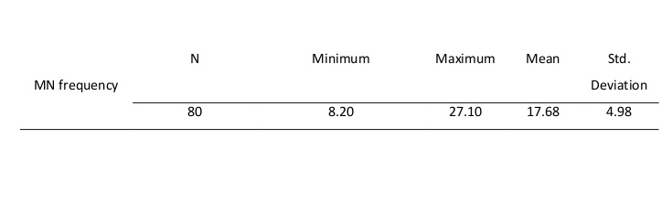
Results: The lowest and highest percentages of the MN frequency were respectively equal to 8.20% and 27.10% with the mean MN frequency of 17.68±4.98. Also, there was no significant correlation between the age and gender of the subjects and the MN frequency (P>0.05).
Conclusion: Based on the results of the present study, there was no significant correlation between the age and gender of the participants and the MN frequency.
Keywords: Micronucleus, Cytotoxicity, Cone Beam Computed Tomography
Introduction
Cancer is among the principal life-threatening diseases throughout the world. (1) It is normally diagnosed in advanced stages since there are no early diagnostic signs and therefore, the chance of survival decreases despite the advanced levels of medical care. (2) It seems essential to use noninvasive diagnostic methods in the early stages of cancer formation.
During the last decade, X-rays have been vastly employed in dental imaging. However, the effects of the ionizing radiation on the structure of the DNA and protein-DNA crosslinking, which cause cell death, cannot be overlooked. (3, 4)
Periapical and panoramic radiography are extensively used in diagnostic and therapeutic dentistry. However, more sophisticated and precise techniques such as computed tomography (CT), magnetic resonance imaging (MRI), and soft tissue scans are also being used nowadays. Cone beam computed tomography (CBCT) is a new technique, which provides CT scans by the use of cone-shaped radiation (4).
CBCT images are similar to the images obtained by medical CT, except that the radiation dose emitted from CBCT units is generally lower than that of medical CT. CBCT provides images of the inner bone structures of the head and neck. CBCT units require less space and equipment than medical CT. (5)
The alterations in the cell nuclei occur in the early stages of cancer. These alterations in the buccal mucosa cells were first identified by Stich and Rosin in 1983. (6) Today, the alterations in the cell nuclei are widely used as biomarkers to highlight the genetic damages in the cells (7), which allow the identification of different pre-neoplastic conditions in the cell nuclei prior to the clinical manifestations. (8) The nuclear alterations in the cytological specimens exfoliated from the buccal mucosa of patients have also been examined. This method is simple, noninvasive, and relatively painless. (9) Therefore, the micronucleus (MN) scoring is one of the most sensitive markers of DNA damage and is considered one of the most trusted biomarkers of the pathologies induced by occupational and environmental exposures in numerous human and animal epidemiological studies. (10,11) The MN scoring can be used for the evaluation of the chromosomal damage in routine cytopathological activities. (8) The advantage of this method is its simplicity since the MN assessment is fast and does not require special expertise. (1)
Kamboj and Mahajan showed that analyzing the epithelial cells of the buccal mucosa is a reliable indicator for the early evaluation of malignant and pre-malignant lesions. (1) The MN scoring of the epithelial cells in the buccal mucosa is considered a method for verifying the risk of carcinogenesis. (12-14)
Some studies have been conducted in the field of maxillofacial surgery on the effects of CBCT, but the number of studies on the effects of CBCT on the frequency of MN is very limited. (4,5) Therefore, by considering the importance of analyzing the alterations of the buccal mucosa cells and the limited number and volume of the samples used in previous related analyses, The present study was performed to assess the MN incidence in the buccal mucosa of CBCT-exposed subjects in an oral and maxillofacial radiology center during 2016.
Materials and Methods
This descriptive study was conducted by the use of target-based sampling. All the subjects had been exposed to the ionizing radiation by ProMax® 3D SCARA3 CBCT unit (Planmeca Co., Helsinki, Finland) with a dose area product (DAP)=445-994 mGy/cm2 and a CT dose index (CT DI)=4.1-8 mGy at the same radiology center. In addition, all the subjects met the inclusion criteria, which consisted of no alcohol consumption, no systemic diseases, no smoking habits, no medication consumption, no recent viral infection, no history of radiotherapy and/or chemotherapy and no jobs that involve contact with chemicals. (4) The sample size for each of the independent variables was calculated to be 10 samples and at least 40 samples of each gender were evaluated. To collect the data, after obtaining informed consent, the patients underwent a biopsy and the buccal mucosa cells were scraped by using a damp spatula and were observed under a light microscope. Before the sampling, the subjects were asked to wash their mouths thoroughly with water. The buccal mucosa cells were exfoliated by the use of the spatula and were spread over clean glass slides. The smears on the slides were fixed by the use of the Patofix cytology smear spray. The slides were then air-dried at room temperature. Papanicolaou staining was then performed to measure the MN frequency. 500 cells per sample were counted and the presence of the micronucleated cells was determined and was reported in percentage. (15)
Results
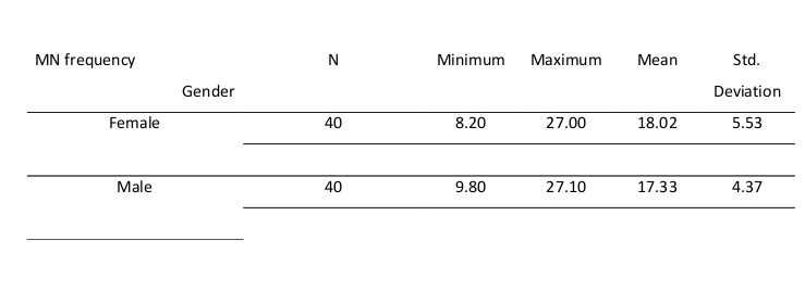
In this study, 80 individuals (40 women and 40 men), who met the inclusion criteria, were exposed to a single predetermined dose of CBCT radiation. The lowest and highest percentages of the MN incidence were respectively equal to 8.20% and 27.10%, and the mean MN frequency was 17.68±4.98. The mean and standard deviation (SD) of the MN frequency equaled to 18.02±5.53 in women and 17.33±4.37 in men. The youngest participant was 19 years old and the oldest was 86 years old. The average age of the subjects was 45.02±14.52 years. (Tables 1 and 2)
The regression analysis showed no significant correlation between the age (P=0.372) and gender (P=0.491) of the subjects and the MN frequency (P>0.05). Figure 1 shows the micronucleated cells in the studied samples.
Figure 1: Micronucleated cells in the buccal mucosa smears of CBCT-exposed subjects (Papanicolaou staining, ×400 magnification).
Table 1: Micronucleus (MN) frequency
Table 2: Distribution of micronucleus (MN) frequency categorized by gender
Discussion
In this study, the MN frequency was equal to 17.68±4.98. The results showed that there was no significant correlation between the age and gender of the participants and the MN frequency.
In 2010, Carlin et al conducted a study on the relationship between the MN frequency and CBCT radiation and measured the MN rate in 10 men and 9 women before and after exposure to CBCT radiation. The average MN frequency after the irradiation was 0.04% and there were no significant differences in the MN scores before and after the irradiation. (5)
In 2012, Ribeiro examined the DNA damage and cell death in adult buccal mucosa cells after exposure to CBCT radiation. (16) Although the mentioned study did not show any significant statistical differences in the MN prevalence before and after exposure to CBCT radiation, it indicated an increase in the incidence of other nuclear changes closely correlated with cytotoxicity such as karyorrhexis, pyknosis, and karyolysis. (16) On the other hand, Lorenzoni et al investigated the mutagenicity (MN) and cytotoxicity-related alterations (karyorrhexis, pyknosis, and karyolysis) in the mucous membrane lining of the cheek of 49 children exposed to CBCT or conventional radiography for orthodontic treatment. (4) The average MN frequency after irradiation was 0.025%. The cited study also showed that the MN frequency was similar in the pre- and post-intervention groups. However, the mentioned assay indicated that the irradiation caused other nuclear changes related to cytotoxicity such as karyorrhexis, pyknosis and karyolysis in both groups (P<0.05). They concluded that the CBCT-exposed group showed a greater rate of cell death than the conventionally-radiographed group (P<0.44). (4) In most studies, the MN abnormalities have been compared before and after the exposure to CBCT radiation. (2,3,10)
A search of the literature revealed studies on the nuclear abnormalities that were merely based on the incidence of the abnormalities without any reference to the demographic characteristics of the subjects such as age and gender; thus, our study is probably the first to consider these factors. The results of the present research can be used as a basis for future studies in this field.
The epithelial cells of the buccal mucosa are a good resource for early detection of the genotoxic effects of the carcinogenic agents that enter the body through inhalation, injection, and direct contact. (17-19) Malignancies are the sixth most common cancer worldwide, and 90% of human cancers originate from the epithelial cells. These scientific facts emphasize the importance of the MN scoring in clinical pathology. This method is inexpensive, noninvasive and simple, and evaluates the elevated MN frequency, due to the carcinogenic stimuli, long before the manifestation of the clinical symptoms.
The rapid turnover of the epithelial cell cycle (7 to 16 days) gives researchers the opportunity to verify the damages in the basal layer of the epithelial tissue within a maximum of 16 days of cell renewal (exfoliation). (20) Therefore, in the current study, similar to most of the previous studies, the sampling was performed 10 days after exposure to radiation.
The Biomonitoring of the buccal cells is often a complex study due to the diversity of the influential variables including the age, lifestyle, oral hygiene, smoking habits, alcohol consumption, exposure to various chemicals, and the impact of some viruses that can affect the epithelial cells in the oral mucosa. For a more precise evaluation of the genotoxic effects of radiation, we tried to obtain our samples from individuals that had not been exposed to the other known influencing factors.
Nonetheless, the environmental variables such as airborne particles, background ionizing radiation, and other environmental exposures related to the geographic habitat of the individuals could also influence the results. The percentage of the micronucleated cells in the present study was higher than that in similar studies.
It should be noted that the differences in the radiation dose, duration of irradiation, variables related to the CBCT equipment, and also the biopsy site can interfere with the results of the MN scoring in the individuals exposed to CBCT radiation. The present study showed no significant correlation between the MN frequency and the age and gender of the subjects, which was in line with the results of previous reports; although, few studies have considered the age-related effects (21-24).
Conclusion
It seems that there is no significant correlation between the age and gender of the individuals and the MN frequency in the buccal mucosa cells.
Full-Text: (792 Views)
Abstract
Background and aim: The ionizing radiation has been recognized to have deleterious effects on the DNA and induce cell death. Due to the importance of evaluating the DNA alterations in buccal mucosa cells induced by the ionizing radiation and by considering the limited volume of the samples used in previous studies, the present study aimed to assess the micronucleus (MN) incidence in the buccal mucosa of cone beam computed tomography (CBCT)-exposed patients of an oral and maxillofacial radiology center during 2016.
Materials and Methods: This descriptive study was conducted by using the target-based sampling. All the subjects, corresponding to the inclusion criteria, had been exposed to CBCT radiation according to their treatment plans. The specimens were scraped from the buccal mucosa of the participants by a damp spatula and were subsequently stained by Papanicolaou staining process. The percentage of the micronucleated cells was reported after evaluation under a light microscope. Also, the multiple linear regression (MLR) method was approached to statistically analyze the correlation between the MN frequency and the age and gender of the participants.
Results: The lowest and highest percentages of the MN frequency were respectively equal to 8.20% and 27.10% with the mean MN frequency of 17.68±4.98. Also, there was no significant correlation between the age and gender of the subjects and the MN frequency (P>0.05).
Conclusion: Based on the results of the present study, there was no significant correlation between the age and gender of the participants and the MN frequency.
Keywords: Micronucleus, Cytotoxicity, Cone Beam Computed Tomography
Introduction
Cancer is among the principal life-threatening diseases throughout the world. (1) It is normally diagnosed in advanced stages since there are no early diagnostic signs and therefore, the chance of survival decreases despite the advanced levels of medical care. (2) It seems essential to use noninvasive diagnostic methods in the early stages of cancer formation.
During the last decade, X-rays have been vastly employed in dental imaging. However, the effects of the ionizing radiation on the structure of the DNA and protein-DNA crosslinking, which cause cell death, cannot be overlooked. (3, 4)
Periapical and panoramic radiography are extensively used in diagnostic and therapeutic dentistry. However, more sophisticated and precise techniques such as computed tomography (CT), magnetic resonance imaging (MRI), and soft tissue scans are also being used nowadays. Cone beam computed tomography (CBCT) is a new technique, which provides CT scans by the use of cone-shaped radiation (4).
CBCT images are similar to the images obtained by medical CT, except that the radiation dose emitted from CBCT units is generally lower than that of medical CT. CBCT provides images of the inner bone structures of the head and neck. CBCT units require less space and equipment than medical CT. (5)
The alterations in the cell nuclei occur in the early stages of cancer. These alterations in the buccal mucosa cells were first identified by Stich and Rosin in 1983. (6) Today, the alterations in the cell nuclei are widely used as biomarkers to highlight the genetic damages in the cells (7), which allow the identification of different pre-neoplastic conditions in the cell nuclei prior to the clinical manifestations. (8) The nuclear alterations in the cytological specimens exfoliated from the buccal mucosa of patients have also been examined. This method is simple, noninvasive, and relatively painless. (9) Therefore, the micronucleus (MN) scoring is one of the most sensitive markers of DNA damage and is considered one of the most trusted biomarkers of the pathologies induced by occupational and environmental exposures in numerous human and animal epidemiological studies. (10,11) The MN scoring can be used for the evaluation of the chromosomal damage in routine cytopathological activities. (8) The advantage of this method is its simplicity since the MN assessment is fast and does not require special expertise. (1)
Kamboj and Mahajan showed that analyzing the epithelial cells of the buccal mucosa is a reliable indicator for the early evaluation of malignant and pre-malignant lesions. (1) The MN scoring of the epithelial cells in the buccal mucosa is considered a method for verifying the risk of carcinogenesis. (12-14)
Some studies have been conducted in the field of maxillofacial surgery on the effects of CBCT, but the number of studies on the effects of CBCT on the frequency of MN is very limited. (4,5) Therefore, by considering the importance of analyzing the alterations of the buccal mucosa cells and the limited number and volume of the samples used in previous related analyses, The present study was performed to assess the MN incidence in the buccal mucosa of CBCT-exposed subjects in an oral and maxillofacial radiology center during 2016.
Materials and Methods
This descriptive study was conducted by the use of target-based sampling. All the subjects had been exposed to the ionizing radiation by ProMax® 3D SCARA3 CBCT unit (Planmeca Co., Helsinki, Finland) with a dose area product (DAP)=445-994 mGy/cm2 and a CT dose index (CT DI)=4.1-8 mGy at the same radiology center. In addition, all the subjects met the inclusion criteria, which consisted of no alcohol consumption, no systemic diseases, no smoking habits, no medication consumption, no recent viral infection, no history of radiotherapy and/or chemotherapy and no jobs that involve contact with chemicals. (4) The sample size for each of the independent variables was calculated to be 10 samples and at least 40 samples of each gender were evaluated. To collect the data, after obtaining informed consent, the patients underwent a biopsy and the buccal mucosa cells were scraped by using a damp spatula and were observed under a light microscope. Before the sampling, the subjects were asked to wash their mouths thoroughly with water. The buccal mucosa cells were exfoliated by the use of the spatula and were spread over clean glass slides. The smears on the slides were fixed by the use of the Patofix cytology smear spray. The slides were then air-dried at room temperature. Papanicolaou staining was then performed to measure the MN frequency. 500 cells per sample were counted and the presence of the micronucleated cells was determined and was reported in percentage. (15)
Results
In this study, 80 individuals (40 women and 40 men), who met the inclusion criteria, were exposed to a single predetermined dose of CBCT radiation. The lowest and highest percentages of the MN incidence were respectively equal to 8.20% and 27.10%, and the mean MN frequency was 17.68±4.98. The mean and standard deviation (SD) of the MN frequency equaled to 18.02±5.53 in women and 17.33±4.37 in men. The youngest participant was 19 years old and the oldest was 86 years old. The average age of the subjects was 45.02±14.52 years. (Tables 1 and 2)
The regression analysis showed no significant correlation between the age (P=0.372) and gender (P=0.491) of the subjects and the MN frequency (P>0.05). Figure 1 shows the micronucleated cells in the studied samples.
Figure 1: Micronucleated cells in the buccal mucosa smears of CBCT-exposed subjects (Papanicolaou staining, ×400 magnification).
.jpg)
Table 1: Micronucleus (MN) frequency
Table 2: Distribution of micronucleus (MN) frequency categorized by gender
Discussion
In this study, the MN frequency was equal to 17.68±4.98. The results showed that there was no significant correlation between the age and gender of the participants and the MN frequency.
In 2010, Carlin et al conducted a study on the relationship between the MN frequency and CBCT radiation and measured the MN rate in 10 men and 9 women before and after exposure to CBCT radiation. The average MN frequency after the irradiation was 0.04% and there were no significant differences in the MN scores before and after the irradiation. (5)
In 2012, Ribeiro examined the DNA damage and cell death in adult buccal mucosa cells after exposure to CBCT radiation. (16) Although the mentioned study did not show any significant statistical differences in the MN prevalence before and after exposure to CBCT radiation, it indicated an increase in the incidence of other nuclear changes closely correlated with cytotoxicity such as karyorrhexis, pyknosis, and karyolysis. (16) On the other hand, Lorenzoni et al investigated the mutagenicity (MN) and cytotoxicity-related alterations (karyorrhexis, pyknosis, and karyolysis) in the mucous membrane lining of the cheek of 49 children exposed to CBCT or conventional radiography for orthodontic treatment. (4) The average MN frequency after irradiation was 0.025%. The cited study also showed that the MN frequency was similar in the pre- and post-intervention groups. However, the mentioned assay indicated that the irradiation caused other nuclear changes related to cytotoxicity such as karyorrhexis, pyknosis and karyolysis in both groups (P<0.05). They concluded that the CBCT-exposed group showed a greater rate of cell death than the conventionally-radiographed group (P<0.44). (4) In most studies, the MN abnormalities have been compared before and after the exposure to CBCT radiation. (2,3,10)
A search of the literature revealed studies on the nuclear abnormalities that were merely based on the incidence of the abnormalities without any reference to the demographic characteristics of the subjects such as age and gender; thus, our study is probably the first to consider these factors. The results of the present research can be used as a basis for future studies in this field.
The epithelial cells of the buccal mucosa are a good resource for early detection of the genotoxic effects of the carcinogenic agents that enter the body through inhalation, injection, and direct contact. (17-19) Malignancies are the sixth most common cancer worldwide, and 90% of human cancers originate from the epithelial cells. These scientific facts emphasize the importance of the MN scoring in clinical pathology. This method is inexpensive, noninvasive and simple, and evaluates the elevated MN frequency, due to the carcinogenic stimuli, long before the manifestation of the clinical symptoms.
The rapid turnover of the epithelial cell cycle (7 to 16 days) gives researchers the opportunity to verify the damages in the basal layer of the epithelial tissue within a maximum of 16 days of cell renewal (exfoliation). (20) Therefore, in the current study, similar to most of the previous studies, the sampling was performed 10 days after exposure to radiation.
The Biomonitoring of the buccal cells is often a complex study due to the diversity of the influential variables including the age, lifestyle, oral hygiene, smoking habits, alcohol consumption, exposure to various chemicals, and the impact of some viruses that can affect the epithelial cells in the oral mucosa. For a more precise evaluation of the genotoxic effects of radiation, we tried to obtain our samples from individuals that had not been exposed to the other known influencing factors.
Nonetheless, the environmental variables such as airborne particles, background ionizing radiation, and other environmental exposures related to the geographic habitat of the individuals could also influence the results. The percentage of the micronucleated cells in the present study was higher than that in similar studies.
It should be noted that the differences in the radiation dose, duration of irradiation, variables related to the CBCT equipment, and also the biopsy site can interfere with the results of the MN scoring in the individuals exposed to CBCT radiation. The present study showed no significant correlation between the MN frequency and the age and gender of the subjects, which was in line with the results of previous reports; although, few studies have considered the age-related effects (21-24).
Conclusion
It seems that there is no significant correlation between the age and gender of the individuals and the MN frequency in the buccal mucosa cells.
Type of Study: Original article |
Subject:
Oral medicine
References
1. Kamboj M, Mahajan S. Micronucleus--an upcoming marker of genotoxic damage.Clin Oral Investig 2007;11(2):121-6.
2. Abbasi F, Farhadi S, Esmaili M. Efficacy of Pilocarpine and Bromhexin in improving radiotherapyinduced xerostomia. J Dent Res Dent Clin Dent Prospects 2013;7(2):86-90.
3. Parplys AC, Petermann E, Petersen C, Dikomey E, Borgmann K. DNA damage by X-rays and their impact on replication processes. Radiother Oncol. 2012;102(3):466-71.
4. Lorenzoni DC, Fracalossi AC, Carlin V, Ribeiro DA, Sant’Anna EF. Mutagenicity and cytotoxicity in patients submitted to ionizing radiation. Angle Orthod 2013;83(1):104-9.
5. Carlin V, Artioli AJ, Matsumoto MA, Borgo E, Oshima CT, Ribeiro DA. Biomonitoring of DNA damage and cytotoxicity in individuals exposed to cone beam computed tomography. Dentomaxillofac Radiol. 2010;39(5):295-9.
6. Stich HF, Rosin MP. Quantitating the synergistic effect of smoking and alcohol consumption with the micronucleus test on human buccal mucosal cells. Int J Cancer 1983:15;31(3):305-8.
7. Palaskar S , Jindal C. Evaluation Of Micronuclei Using Papanicolaou And May Grunwald Giemsa Stain In Individuals With Different Tobacco Habits – A Comparative Study. JCDR 2010;4(6):3607-13.
8. Stich HF, Rosin MP, Vallejera MO. Reduction with vitamin A and beta_caratene administration of proportion of micronucleated buccal mucosal cells in Asian betel nut and tobacco chewers. Lancet 1984;2;1(8388):1204-6.
9. Saeed H. Saeed , Wasen H. Younis. A cytopathological study of the effect of smoking on the oral epithelial cells in relation to oral health status by the micronucleus assay. J Bagh Coll Dentistry 2012;24(3):67-70.
10. Fenech M. The cytokinesis-block micronucleus technique and its application to genotoxicity studies in human populations. Environ Health Perspect 1993;101 Suppl 3:101-7.
11. Ishikawa H, Tian Y, Yamauchi T. Induction of micronuclei formation in preimplantation mouse embryos after maternal treatment with 2-bromopropane. Reprod Toxicol 2001;15(1):81-5.
12. Stich HF, Stich W, ParidaBB. Elevated frequency of micronucleated cells in the buccal mucosa of individuals at high risk for oral cancer: betel quid chewers. Cancer Lett 1982;17(2):125-34.
13. Majer BJ, Laky B, Knasmüller S, Kassie F. Use of the micronucleus assay with exfoliated epithelial cells as a biomarker for monitoring individuals at elevated risk of genetic damage and in chemoprevention trials. Mutat Res 2001;489(2-3):147-72.
14. Rosin MP, Dunn BP, Stich HF. Use of intermediate endpoints in Quantitating the response of precancerous lesions to chemopreventive agents. Can J Physiol Pharmacol 1987;65(3):483-7.
15. Naderi NJ , Farhadi S , Sarshar S . Micronucleus assay of buccal mucosa in smokers with the historyof smoking less and more than 10 years .Indian J Pathol Microbiol 2012;55(4):433-8.
16. Ribeiro DA. Cytogenetic biomonitoring in oral mucosa cells following dental X-ray. Dentomaxillofac Radiology 2012;41(3):181-4.
17. Holland N, Bolognesi C, Kirsch-Volders M, Bonassi S, Zeiger E, Knasmueller S, et al. The micronucleus assay in human buccal cells as a tool for biomonitoring DNA damage: the HUMN project perspective on current status and knowledge gaps. Mutat Res 2008;659(1-2):93–108.
18. Farhadi S, Jahanbani J, Jariani A, Ghasemi S. Bio-Monitoring of the Nuclear Abnormalities in Smokers Using Buccal Exfoliated Cytology. Adv Biores 2016;7(4)128-33.
19. Farhadi S, Sadri D, Sarshar S. Micronucleus assay of Buccal mucosa: a Useful noninvasive approach in Screening of Genotoxic nuclear damage. Adv Biores 2016;7(4): 20-9.
20. Sarto F, Tomanin R, Giacomelli L, Canova A, Raimondi F, Ghiotto C, et al. Evaluation of chromosomal aberrations in lymphocytes and micronuclei in lymphocytes, oral mucosa and hair root cells of patients under antiblastic therapy. Mutat Res
21. 1990;228(2):157–69.
22. Neri M, Fucic A, Knudsen LE, Lando C, Merlo F, Bonassi S. Micronuclei frequency in children exposed to environmental mutagens: a review. Mutat Res 2003;544(2-3):243–54.
23. Cerqueira EM, Meireles JR, Lopes MA, Junqueira VC, Gomes-Filho IS, Trindade S, et al. Genotoxic effects of X-rays on keratinized mucosa cells during panoramic dental radiography. Dentomaxillofac Radiol 2008;37(7):398–403.
24. Popova L, Kishkilova D, Hadjidekova VB, Hristova RP, Atanasova P, Hadjidekova VV, et al. Micronucleus test in buccal epithelium cells from patients subjected to panoramic radiography. Dentomaxillofac Radiol 2007;36(3):168-71.
25. Ribeiro DA, Oliveira G, de Castro G, Angelieri F. Cytogenetic biomonitoring in patients exposed to dental X-rays: comparison between adults and children. Dentomaxillofac Radiol 2008;37(7):404–7.
Send email to the article author
| Rights and permissions | |
 |
This work is licensed under a Creative Commons Attribution-NonCommercial 4.0 International License. |
