

Volume 7, Issue 3 (7-2022)
J Res Dent Maxillofac Sci 2022, 7(3): 142-147 |
Back to browse issues page


Download citation:
BibTeX | RIS | EndNote | Medlars | ProCite | Reference Manager | RefWorks
Send citation to:


BibTeX | RIS | EndNote | Medlars | ProCite | Reference Manager | RefWorks
Send citation to:
Amooyan P, Akhavan H, Esnaashari E. Efficacy of XP-Endo Finisher R in Removing Root Filling Remnants in Root Canal Retreatment. J Res Dent Maxillofac Sci 2022; 7 (3) :142-147
URL: http://jrdms.dentaliau.ac.ir/article-1-350-en.html
URL: http://jrdms.dentaliau.ac.ir/article-1-350-en.html


1- Private Dentistry Practice, Tehran, Iran
2- Department of Endodontics, Faculty of Dentistry, Tehran Medical Sciences, Islamic Azad University, Tehran, Iran
3- Department of Endodontics, Faculty of Dentistry, Tehran Medical Sciences, Islamic Azad University, Tehran, Iran , ehsan_dmd@yahoo.com
2- Department of Endodontics, Faculty of Dentistry, Tehran Medical Sciences, Islamic Azad University, Tehran, Iran
3- Department of Endodontics, Faculty of Dentistry, Tehran Medical Sciences, Islamic Azad University, Tehran, Iran , ehsan_dmd@yahoo.com
Full-Text [PDF 794 kb]
(528 Downloads)
| Abstract (HTML) (871 Views)
Introduction
Root canal retreatment refers to removal of infected root filling material to regain access to the root canal followed by efficient cleaning and shaping as well as subsequent re-filling of the canals [1]. Incomplete removal of gutta-percha can lead to incomplete cleaning and shaping [2]. There are several techniques for gutta-percha removal, such as rotary instruments, hand files, heat, ultrasonic tips, and laser [3,4]. However, most of the abovementioned techniques cannot completely eliminate the gutta-percha from the root canal system [1,5]. XP-Endo Finisher R (XEFR; FKG Dentaire SA, La Chaux-de-Fonds, Switzerland) was recently developed to access inaccessible parts of the canal. It is made of a specific alloy (MaxWire; Martensite-Austenite Electropolish Flex, FKG Dentaire) and can be used after the initial step of root canal retreatment to improve cleaning of the root canal system and removal of residual filling material. The tip size is #30, and non-tapered. The shape of the tip changes with temperature. At room temperature, the file is straight but it becomes spoon-shaped at body temperature when used in the root canal system.
The aim of this study was to assess the efficacy of XEFR for gutta-percha removal from straight canals of mandibular premolars in root canal retreatment.
Materials and Methods
This in vitro, experimental study was conducted on 30 single-rooted mandibular premolars after gaining approval from the Ethics Committee of Dental School, Tehran Branch, Islamic Azad University (approval ID: IR.IAU.DENTAL.REC.1399.131). The canal curvature was measured by the Schneider’s technique [6]. Teeth with less than 10-degree curvature were selected. Teeth with cracks, internal and external root resorption, calcification, caries, and open apices were excluded.
The teeth were kept in 0.5% chloramine T (Merck, Darmstadt, Germany) for 48 hours and stored in distilled water. The external surface of the teeth was mechanically cleaned with a periodontal scalpel to remove debris, calculus, and soft tissue remnants. After preparation of the access cavity, apical patency was ensured and a #15 K-file (Dentsply-Maillefer, Switzerland) was used to determine the working length 1 mm shorter than the apex. To standardize the teeth, the crowns were cut to have 15-mm roots. All root canals were prepared by one operator [1].
In all teeth, canal preparation was performed with Bio-RaCe (FKG, La-Chaux De Fonds, Switzerland) and the crown-down technique according to the manufacturer’s instructions. The files were discarded after being used in five canals. After using each file, the canal was rinsed with 2 mL of 2.5% sodium hypochlorite (Cerkamed, Poland) by a 27-gauge needle. Final irrigation was performed with 5 mL of distilled water (Faraz dental, Iran). Finally, the root canals were dried with paper points (GAPADENT Co. Tianjin, China) and obturated with gutta-percha (GAPADENT Co. Tianjin, China) and AH Plus sealer (Dentsply-Maillefer, Switzerland) by the lateral compaction technique. Coltosol (Asia Chemi Teb. Co., Tehran, Iran) temporary restorative material was used to seal the coronal access cavity, and the teeth were incubated at 37°C and 100% humidity for 2 weeks for complete setting of the sealer [7].
Next, temporary fillings were removed and coronal gutta-percha was eliminated using a #2 Gates-Glidden drill (Mani, Matsutain Seisakusho Co., Tochigi-Ken, Japan). In the process of retreatment, irrigation of root canals with 2 mL of 2.5% sodium hypochlorite was carried out after using each file. Gutta-percha was removed by the sequential use of D-RaCe retreatment files (FKG Dentaire). Accordingly, DR1 (size 30, .10 taper) was used for the coronal region, and DR2 (size 25, .04 taper) was used to the working length. Irrigation with 0.5 mL of 2.5% sodium hypochlorite was also performed before using DR1. Next, one drop of chloroform was applied into the canal, and DR2 was used after 1 minute.
Next, the root canals were randomly divided into two experimental groups of A and B (n=15). In group A, root canal retreatment was completed with XEFR file and a contra-angle handpiece (VDW Silver, Germany). Each root canal was filled with 0.5 mL of 5.25% sodium hypochlorite, and the file was introduced into the canal without rotation, and operated at 800 rpm and 1 N.cm torque with in-and-out motion and an amplitude of 7–8 mm up to the working length for 30 seconds. The canal was rinsed again with 1 mL of 2.5% sodium hypochlorite. The file was then used again for 30 seconds. The canal walls were brushed with the instrument during the procedure. A final irrigation with 5 mL of distilled water was then performed. Each file was used in two teeth and then discarded. In group B, this file was not used after the retreatment procedure [8].
To evaluate the amount of remaining filling material, a diamond disc (KG Sorensen, Barueri, Brazil) was used to create two longitudinal grooves in the buccal and lingual surfaces of the roots. The teeth were then sectioned longitudinally with an Ochsenbein chisel. The root halves were photographed under a stereomicroscope (Olympus, SZX9, Tokyo, Japan) equipped with a digital camera (Nikon D90, Nikon Corporation, Tokyo, Japan) at x20 and x6 magnifications. The photographs were transferred to a computer (Figures 1 and 2) and analyzed with AutoCAD 2020 software.
Figure 1. Stereomicroscopic images of the remaining filling material in different parts of the canal in group A (a: coronal third, b: middle third, c: apical third) (×20)
Figure 2. Stereomicroscopic images of the remaining filling material in different parts of the canal in group B (a: coronal third, b: middle third, c: apical third) (×20)
The percentage of remaining filling material on the root canal walls in the cervical, middle and apical thirds was calculated using the below-mentioned equation:
remnant surface area × 100/root canal surface area[1].
Statistical analysis:
The mean percentage of residual gutta-percha was quantified and the comparison between groups was carried out using repeated measure ANOVA, considering the file type as within subject factor (alpha=0.05).
Results
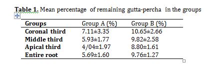
The mean percentage of remaining gutta-percha in the two groups is shown in Table 1. The amount of residual filling material in the coronal, middle, and apical thirds and the entire root canal was significantly lower in XEFR group (P=0.0001). In both groups, the amount of residual gutta-percha in the apical (P=0.000) and middle (P=0.009) thirds was significantly lower than that in the coronal (P<0.05) third; but there was no significant difference between the amount of gutta-percha remaining in the middle and apical thirds (P=0.069).
Table 1. Mean percentage of remaining gutta-percha in the groups
Discussion
This study assessed the effect of XEFR file on the amount of gutta-percha remaining after root canal retreatment. The results showed significantly lower amount of residual filling material in use of XEFR file compared with no use of this file.
Retreatment is a procedure that is required in cases of unsuccessful root canal therapy or persistent apical lesions [5], and requires removing previous infected root filling material, reshaping, cleaning, and refilling of the root canal [5,9].
The use of nickel-titanium files in root canal treatment is increasing. Most studies showed that retreatment with such rotary instruments is more effective, safer and significantly less time consuming compared with manual files [10,11]. D-RaCe system is among such files that is used to remove
gutta-percha during root canal retreatment, which comprises of 2 files [DR1 and DR2]. DR1 has an active tip which enables its easy and rapid penetration into the gutta-percha [12].
Micro-tomography [8], sectioning [2], digital radiography [13], and cone-beam computed tomography [14] have been used for evaluation of the remaining root filling material. In the current study, a stereomicroscope was used similar to a study conducted by Karamifar et al [1]. In longitudinal sectioning method, splitting may cause loss of some gutta-percha but it enables direct visualization of the filling material [15]. Digital radiography has its known limitations such as overlapping and providing a 2D view [15,16]. Micro computed tomography is nondestructive, and highly accurate for evaluation of root canal filling, and provides 3D views [6].
During the retreatment process, there is no doubt that root filling remnants cannot be completely removed regardless of the technique used, being either the rotary system or the manual file system [3,17,18]. Therefore, this situation necessitates the development of new protocols and tools.
XEFR was exclusively designed for retreatment procedure, and is used after initial removal of the filling material.
Because of special properties of the Max-Wire alloy and its expanding ability along with the small size of this file, its adaptation to the canal anatomy has improved such that it can access hard-to-reach areas. As a result, improvement of cleaning must be expected [6,8]. In this file, the M-phase can transform to A-phase when exposed to body temperature [5]. The A-phase enables the file to access unreachable areas so this file could
possibly have a better performance in the oral environment compared with the conventional laboratory environment.
To this date, performance of XEFR instruments has been limitedly evaluated. The current results confirmed the optimal efficacy of XEFR file. However, it cannot completely remove gutta-percha and other filling materials. Controversial results of studies can be due to morphological differences. For instance, Machado et al. [6] used the mesial canals of mandibular first molars with curvatures <25 degrees.
De-Deus et al. [8] evaluated the effect of XEFR on the amount of residual filling material in teeth with oval canals; whereas straight root canals were used in the present study.
The current results cannot be generalized to all teeth, especially teeth with greater curvature.
Conclusion
Within the limitations of this in vitro study, it seems that XEFR file significantly decreased the remaining root filling material in the root canals.
Full-Text: (356 Views)
| Abstract
Background and Aim: Complete removal of gutta-percha from the root canal system and its remnants is a challenge in endodontic retreatment. This ex-vivo study aimed to assess the efficacy of XP-Endo Finisher R (XEFR) in removing root filling remnants from straight canals of mandibular premolars in root canal retreatment. Materials and Methods: In this ex-vivo study, 30 single-rooted mandibular premolars with straight roots were selected. The root canals were instrumented and filled. Retreatment was then performed using D RaCe system. Teeth were then randomized into two groups of A and B (n=15). In group A, root canal retreatment was completed with XEFR. In group B, XEFR was not used. To evaluate the filling material remnants, the roots were split into halves and assessed under a stereomicroscope and then photographed. The amount of remaining gutta-percha in the coronal, middle and apical thirds of the roots was measured by AutoCAD software and analyzed with two-way ANOVA. Results: The amount of residual filling material was significantly lower in the coronal, middle and apical thirds and the entire root length in XEFR group (P<0.001). Conclusion: Use of XEFR significantly decreased the residual root filling material in the root canals. Key Words: Gutta-Percha; Root Canal Filling Material; Root Canal Therapy |
Introduction
Root canal retreatment refers to removal of infected root filling material to regain access to the root canal followed by efficient cleaning and shaping as well as subsequent re-filling of the canals [1]. Incomplete removal of gutta-percha can lead to incomplete cleaning and shaping [2]. There are several techniques for gutta-percha removal, such as rotary instruments, hand files, heat, ultrasonic tips, and laser [3,4]. However, most of the abovementioned techniques cannot completely eliminate the gutta-percha from the root canal system [1,5]. XP-Endo Finisher R (XEFR; FKG Dentaire SA, La Chaux-de-Fonds, Switzerland) was recently developed to access inaccessible parts of the canal. It is made of a specific alloy (MaxWire; Martensite-Austenite Electropolish Flex, FKG Dentaire) and can be used after the initial step of root canal retreatment to improve cleaning of the root canal system and removal of residual filling material. The tip size is #30, and non-tapered. The shape of the tip changes with temperature. At room temperature, the file is straight but it becomes spoon-shaped at body temperature when used in the root canal system.
The aim of this study was to assess the efficacy of XEFR for gutta-percha removal from straight canals of mandibular premolars in root canal retreatment.
Materials and Methods
This in vitro, experimental study was conducted on 30 single-rooted mandibular premolars after gaining approval from the Ethics Committee of Dental School, Tehran Branch, Islamic Azad University (approval ID: IR.IAU.DENTAL.REC.1399.131). The canal curvature was measured by the Schneider’s technique [6]. Teeth with less than 10-degree curvature were selected. Teeth with cracks, internal and external root resorption, calcification, caries, and open apices were excluded.
The teeth were kept in 0.5% chloramine T (Merck, Darmstadt, Germany) for 48 hours and stored in distilled water. The external surface of the teeth was mechanically cleaned with a periodontal scalpel to remove debris, calculus, and soft tissue remnants. After preparation of the access cavity, apical patency was ensured and a #15 K-file (Dentsply-Maillefer, Switzerland) was used to determine the working length 1 mm shorter than the apex. To standardize the teeth, the crowns were cut to have 15-mm roots. All root canals were prepared by one operator [1].
In all teeth, canal preparation was performed with Bio-RaCe (FKG, La-Chaux De Fonds, Switzerland) and the crown-down technique according to the manufacturer’s instructions. The files were discarded after being used in five canals. After using each file, the canal was rinsed with 2 mL of 2.5% sodium hypochlorite (Cerkamed, Poland) by a 27-gauge needle. Final irrigation was performed with 5 mL of distilled water (Faraz dental, Iran). Finally, the root canals were dried with paper points (GAPADENT Co. Tianjin, China) and obturated with gutta-percha (GAPADENT Co. Tianjin, China) and AH Plus sealer (Dentsply-Maillefer, Switzerland) by the lateral compaction technique. Coltosol (Asia Chemi Teb. Co., Tehran, Iran) temporary restorative material was used to seal the coronal access cavity, and the teeth were incubated at 37°C and 100% humidity for 2 weeks for complete setting of the sealer [7].
Next, temporary fillings were removed and coronal gutta-percha was eliminated using a #2 Gates-Glidden drill (Mani, Matsutain Seisakusho Co., Tochigi-Ken, Japan). In the process of retreatment, irrigation of root canals with 2 mL of 2.5% sodium hypochlorite was carried out after using each file. Gutta-percha was removed by the sequential use of D-RaCe retreatment files (FKG Dentaire). Accordingly, DR1 (size 30, .10 taper) was used for the coronal region, and DR2 (size 25, .04 taper) was used to the working length. Irrigation with 0.5 mL of 2.5% sodium hypochlorite was also performed before using DR1. Next, one drop of chloroform was applied into the canal, and DR2 was used after 1 minute.
Next, the root canals were randomly divided into two experimental groups of A and B (n=15). In group A, root canal retreatment was completed with XEFR file and a contra-angle handpiece (VDW Silver, Germany). Each root canal was filled with 0.5 mL of 5.25% sodium hypochlorite, and the file was introduced into the canal without rotation, and operated at 800 rpm and 1 N.cm torque with in-and-out motion and an amplitude of 7–8 mm up to the working length for 30 seconds. The canal was rinsed again with 1 mL of 2.5% sodium hypochlorite. The file was then used again for 30 seconds. The canal walls were brushed with the instrument during the procedure. A final irrigation with 5 mL of distilled water was then performed. Each file was used in two teeth and then discarded. In group B, this file was not used after the retreatment procedure [8].
To evaluate the amount of remaining filling material, a diamond disc (KG Sorensen, Barueri, Brazil) was used to create two longitudinal grooves in the buccal and lingual surfaces of the roots. The teeth were then sectioned longitudinally with an Ochsenbein chisel. The root halves were photographed under a stereomicroscope (Olympus, SZX9, Tokyo, Japan) equipped with a digital camera (Nikon D90, Nikon Corporation, Tokyo, Japan) at x20 and x6 magnifications. The photographs were transferred to a computer (Figures 1 and 2) and analyzed with AutoCAD 2020 software.
Figure 1. Stereomicroscopic images of the remaining filling material in different parts of the canal in group A (a: coronal third, b: middle third, c: apical third) (×20)
Figure 2. Stereomicroscopic images of the remaining filling material in different parts of the canal in group B (a: coronal third, b: middle third, c: apical third) (×20)
The percentage of remaining filling material on the root canal walls in the cervical, middle and apical thirds was calculated using the below-mentioned equation:
remnant surface area × 100/root canal surface area[1].
Statistical analysis:
The mean percentage of residual gutta-percha was quantified and the comparison between groups was carried out using repeated measure ANOVA, considering the file type as within subject factor (alpha=0.05).
Results
The mean percentage of remaining gutta-percha in the two groups is shown in Table 1. The amount of residual filling material in the coronal, middle, and apical thirds and the entire root canal was significantly lower in XEFR group (P=0.0001). In both groups, the amount of residual gutta-percha in the apical (P=0.000) and middle (P=0.009) thirds was significantly lower than that in the coronal (P<0.05) third; but there was no significant difference between the amount of gutta-percha remaining in the middle and apical thirds (P=0.069).
Table 1. Mean percentage of remaining gutta-percha in the groups
Discussion
This study assessed the effect of XEFR file on the amount of gutta-percha remaining after root canal retreatment. The results showed significantly lower amount of residual filling material in use of XEFR file compared with no use of this file.
Retreatment is a procedure that is required in cases of unsuccessful root canal therapy or persistent apical lesions [5], and requires removing previous infected root filling material, reshaping, cleaning, and refilling of the root canal [5,9].
The use of nickel-titanium files in root canal treatment is increasing. Most studies showed that retreatment with such rotary instruments is more effective, safer and significantly less time consuming compared with manual files [10,11]. D-RaCe system is among such files that is used to remove
gutta-percha during root canal retreatment, which comprises of 2 files [DR1 and DR2]. DR1 has an active tip which enables its easy and rapid penetration into the gutta-percha [12].
Micro-tomography [8], sectioning [2], digital radiography [13], and cone-beam computed tomography [14] have been used for evaluation of the remaining root filling material. In the current study, a stereomicroscope was used similar to a study conducted by Karamifar et al [1]. In longitudinal sectioning method, splitting may cause loss of some gutta-percha but it enables direct visualization of the filling material [15]. Digital radiography has its known limitations such as overlapping and providing a 2D view [15,16]. Micro computed tomography is nondestructive, and highly accurate for evaluation of root canal filling, and provides 3D views [6].
During the retreatment process, there is no doubt that root filling remnants cannot be completely removed regardless of the technique used, being either the rotary system or the manual file system [3,17,18]. Therefore, this situation necessitates the development of new protocols and tools.
XEFR was exclusively designed for retreatment procedure, and is used after initial removal of the filling material.
Because of special properties of the Max-Wire alloy and its expanding ability along with the small size of this file, its adaptation to the canal anatomy has improved such that it can access hard-to-reach areas. As a result, improvement of cleaning must be expected [6,8]. In this file, the M-phase can transform to A-phase when exposed to body temperature [5]. The A-phase enables the file to access unreachable areas so this file could
possibly have a better performance in the oral environment compared with the conventional laboratory environment.
To this date, performance of XEFR instruments has been limitedly evaluated. The current results confirmed the optimal efficacy of XEFR file. However, it cannot completely remove gutta-percha and other filling materials. Controversial results of studies can be due to morphological differences. For instance, Machado et al. [6] used the mesial canals of mandibular first molars with curvatures <25 degrees.
De-Deus et al. [8] evaluated the effect of XEFR on the amount of residual filling material in teeth with oval canals; whereas straight root canals were used in the present study.
The current results cannot be generalized to all teeth, especially teeth with greater curvature.
Conclusion
Within the limitations of this in vitro study, it seems that XEFR file significantly decreased the remaining root filling material in the root canals.
Type of Study: Original article |
Subject:
Oral medicine
References
1. Karamifar K, Mehrasa N, Pardis P, Saghiri MA. Cleanliness of Canal Walls following Gutta-Percha Removal with Hand Files, RaCe and RaCe plus XP-Endo Finisher Instruments: A Photographic in Vitro Analysis. Iran Endod J. 2017; 12(2): 242-7.
2. Saad AY, Al-Hadlaq SM, Al-Katheeri NH. Efficacy of two rotary NiTi instruments in the removal of Gutta-Percha during root canal retreatment. J Endod. 2007;33(1):38-41. [DOI:10.1016/j.joen.2006.08.012] [PMID]
3. Kasam S, Mariswamy AB. Efficacy of Different Methods for Removing Root Canal Filling Material in Retreatment - An In-vitro Study. J Clin Diagn Res. 2016;10(6):ZC06-10. [DOI:10.7860/JCDR/2016/17395.7904] [PMID] [PMCID]
4. Ersev H, Yilmaz B, Dincol ME, Daglaroglu R. The efficacy of ProTaper Universal rotary retreatment instrumentation to remove single gutta-percha cones cemented with several endodontic sealers. Int Endod J. 2012;45(8):756-62. [DOI:10.1111/j.1365-2591.2012.02032.x] [PMID]
5. Silva E, Belladonna FG, Zuolo AS, Rodrigues E, Ehrhardt IC, Souza EM, et al. Effectiveness of XP-endo Finisher and XP-endo Finisher R in removing root filling remnants: a micro-CT study. Int Endod J. 2018;51(1):86-91. [DOI:10.1111/iej.12788] [PMID]
6. Machado AG, Guilherme BPS, Provenzano JC, Marceliano-Alves MF, Goncalves LS, Siqueira JF, Jr., et al. Effects of preparation with the Self-Adjusting File, TRUShape and XP-endo Shaper systems, and a supplementary step with XP-endo Finisher R on filling material removal during retreatment of mandibular molar canals. Int Endod J. 2019; 52(5):709-15. [DOI:10.1111/iej.13039] [PMID]
7. Pirani C, Pelliccioni GA, Marchionni S, Montebugnoli L, Piana G, Prati C. Effectiveness of three different retreatment techniques in canals filled with compacted gutta-percha or Thermafil: a scanning electron microscope study. J Endod. 2009;35(10):1433-40. [DOI:10.1016/j.joen.2009.06.002] [PMID]
8. De-Deus G, Belladonna FG, Zuolo AS, Cavalcante DM, Carvalhal JCA, Simoes-Carvalho M, et al. XP-endo Finisher R instrument optimizes the removal of root filling remnants in oval-shaped canals. Int Endod J. 2019;52(6):899-907. [DOI:10.1111/iej.13077] [PMID]
9. Zmener O, Pameijer CH, Banegas G. Retreatment efficacy of hand versus automated instrumentation in oval-shaped root canals: an ex vivo study. Int Endod J. 2006;39(7):521-6. [DOI:10.1111/j.1365-2591.2006.01100.x] [PMID]
10. Das S, De Ida A, Das S, Nair V, Saha N, Chattopadhyay S. Comparative evaluation of three different rotary instrumentation systems for removal of gutta-percha from root canal during endodontic retreatment: An in vitro study. Journal of conservative dentistry: JCD. 2017;20(5):311-6. [DOI:10.4103/JCD.JCD_132_17] [PMID] [PMCID]
11. Joseph M, Ahlawat J, Malhotra A, Rao M, Sharma A, Talwar S. In vitro evaluation of efficacy of different rotary instrument systems for gutta percha removal during root canal retreatment. J Clin Exp Dent. 2016;8(4):e355-e60. [DOI:10.4317/jced.52488] [PMID] [PMCID]
12. Simsek N, Ahmetoglu F, Keles A, Bulut ET, Er K. 3D analysis of D-RaCe and self-adjusting file in removing filling materials from curved root canals instrumented and filled with different techniques. TheScientificWorldJournal. 2014; 2014:836513. [DOI:10.1155/2014/836513] [PMID] [PMCID]
13. Marfisi K, Mercadé M, Plotino G, Clavel T, Duran-Sindreu F, Roig M. Efficacy of Reciproc(®) and Profile(®) Instruments in the Removal of Gutta-Percha from Straight and Curved Root Canals ex Vivo. Journal of oral & maxillofacial research. 2015;6(3):e1.
14. Marfisi K, Mercade M, Plotino G, Duran-Sindreu F, Bueno R, Roig M. Efficacy of three different rotary files to remove gutta-percha and Resilon from root canals. Int Endod J. 2010;43(11):1022-8. [DOI:10.1111/j.1365-2591.2010.01758.x] [PMID]
15. Alakabani TF, Faus-Llácer V, Faus-Matoses I, Ruiz-Sánchez C, Zubizarreta-Macho Á, Sauro S, et al. The Efficacy of Rotary, Reciprocating, and Combined Non-Surgical Endodontic Retreatment Techniques in Removing a Carrier-Based Root Canal Filling Material from Straight Root Canal Systems: A Micro-Computed Tomography Analysis. Journal of clinical medicine. 2020;9(6). [DOI:10.3390/jcm9061989] [PMID] [PMCID]
16. Garg A, Nagpal A, Shetty S, Kumar S, Singh KK, Garg A. Comparison of Time Required by D-RaCe, R-Endo and Mtwo Instruments for Retreatment: An in vitro Study. J Clin Diagn Res. 2015;9(2):Zc47-9. [DOI:10.7860/JCDR/2015/11100.5596] [PMID] [PMCID]
17. Khedmat S, Azari A, Shamshiri AR, Fadae M, Bashizadeh Fakhar H. Efficacy of ProTaper and Mtwo Retreatment Files in Removal of Gutta-percha and GuttaFlow from Root Canals. Iran Endod J. 2016;11(3):184-7.
18. Gu LS, Ling JQ, Wei X, Huang XY. Efficacy of ProTaper Universal rotary retreatment system for gutta-percha removal from root canals. Int Endod J. 2008;41(4):288-95. [DOI:10.1111/j.1365-2591.2007.01350.x] [PMID]
Send email to the article author
| Rights and permissions | |
 |
This work is licensed under a Creative Commons Attribution-NonCommercial 4.0 International License. |
