

Volume 7, Issue 2 (3-2022)
J Res Dent Maxillofac Sci 2022, 7(2): 91-97 |
Back to browse issues page


Download citation:
BibTeX | RIS | EndNote | Medlars | ProCite | Reference Manager | RefWorks
Send citation to:


BibTeX | RIS | EndNote | Medlars | ProCite | Reference Manager | RefWorks
Send citation to:
Moshari A, Esnaashari E, Eskandarion S, Safari B. Comparison of Cytotoxicity of AH Plus and DC Canal SE Sealer After Final Setting. J Res Dent Maxillofac Sci 2022; 7 (2) :91-97
URL: http://jrdms.dentaliau.ac.ir/article-1-315-en.html
URL: http://jrdms.dentaliau.ac.ir/article-1-315-en.html


1- Department of Endodontics, Dental Material Research Center, Faculty of Dentistry, Tehran Medical Sciences, Islamic Azad University, Tehran, Iran , Amirabbas.moshari@gmail.com
2- Department of Endodontics, Faculty of Dentistry, Tehran Medical Sciences, Islamic Azad University, Tehran, Iran
3- Dental Material Research Center, Faculty of Dentistry, Tehran Medical Sciences, Islamic Azad University, Tehran, Iran
4- Private Dentistry Practice, Tehran, Iran
2- Department of Endodontics, Faculty of Dentistry, Tehran Medical Sciences, Islamic Azad University, Tehran, Iran
3- Dental Material Research Center, Faculty of Dentistry, Tehran Medical Sciences, Islamic Azad University, Tehran, Iran
4- Private Dentistry Practice, Tehran, Iran
Full-Text [PDF 785 kb]
(479 Downloads)
| Abstract (HTML) (1160 Views)
Full-Text: (370 Views)
| Abstract
Background and Aim: This study aimed to compare the cytotoxicity of AH Plus and DC Canal SE sealer after final setting. Materials and Methods: In this in vitro, experimental study, human gingival fibroblasts (HGFs) were cultured in Dulbecco’s modified Eagle’s medium supplemented with 10% fetal bovine serum, 100 U/mL penicillin, and 100 µg/mL streptomycin, and incubated at 37°C and 5% CO2. Cells at a density of 5000 cells/well were seeded in a 96-well plate for the methyl thiazolyl tetrazolium (MTT) assay. The cells were incubated with AH Plus and DC Canal SE sealers. Specimens of freshly mixed sealers were fabricated with 4 mm height and 10 mm diameter, and incubated at 37°C for 24 hours for setting. Each specimen was incubated with 10 mL of diluted culture medium at 37°C and 5% CO2 for 24 hours. Finally, the solution was filtered using a 0.22-µm filter. Different dilutions (1, 1/2, 1/4, 1/8) were prepared, and cell viability was assessed at 24 and 72 hours by measuring the optical density of the solutions spectrophotometrically. Data were analyzed by ANOVA and Tukey’s test. Results: Cell viability in presence of all concentrations of AH Plus and DC Canal SE was significantly lower at 72 hours compared with 24 hours (P<0.001). Cell viability in presence of AH Plus was significantly higher compared with DC Canal SE at all concentrations and time points. Conclusion: Cell viability was higher in presence of AH Plus compared with DC Canal SE sealer in all concentrations and time points. Key Words: Endodontics; Root Canal Obturation; Root Canal Filling Materials |
Introduction
Cytotoxicity of dental materials is a common concern for dental clinicians since dental materials are in close contact with the periodontium and vital tissues, and can lead to inflammatory reactions, if they are not biocompatible.[1] Endodontic materials should not prevent tissue healing. Instead, they should preferably enhance tissue healing.[2] Also, they should not elicit inflammatory reactions in the periapical tissue.[3] However, the majority of endodontic sealers have some levels of toxicity.[3,4] Sealers, particularly during their setting reaction, can trigger an inflammatory response in the periapical tissue, and can also affect the viability of cells in the periapical region such as fibroblasts.[5] Among the known sealers, methacrylate-based sealers (especially in high concentrations) have the highest level of cytotoxicity followed by resin-based and bio-ceramic sealers and then silicone-based sealers, which have shown lower cytotoxicity.[6,7] The materials used for root canal filling may have toxic effects on the periapical tissue. Thus, an ideal sealer should have minimum cytotoxic effects.[8]
Accordingly, it is important to find a sealer with minimum cytotoxicity when in contact with the periapical tissue. AH Plus (Dentsply Sirona, Berlin, Germany) is a commonly applied endodontic sealer, which is also used for the purpose of comparison with other sealers in endodontic research.[9] Studies comparing the cytotoxicity of AH Plus with other sealers reported that it had minimum cytotoxic effects on human periodontal ligament cells, osteoblasts, and L929 murine fibroblasts.[10-12] This sealer is an epoxy resin-based sealer. A recent study on the cytotoxicity of other resinous dental materials such as composite resins confirmed their optimal biocompatibility and insignificant cytotoxicity.[13] AH Plus sealer has favorable physicochemical, biological, and antimicrobial properties, and minimum adverse effect on cell viability and proliferation.[14-20]
Recently, some new root canal filling materials were introduced to the market such as DC Canal SE sealer (S & C polymer GmbH, Elmshorn, Germany), which is a methacrylate-based self-etch sealer. Advances in bonding technology aim to minimize apical and coronal leakage by enhancement of the bond strength of sealers to root dentin, and subsequent mono-block obturation. Methacrylate-based sealers were introduced to achieve this goal, and include urethane dimethacrylate, bisphenol-A-glycidyl dimethacrylate, pigments, and radiopaque materials. These sealers also have an etching primer.[21,22] The results regarding the cytotoxicity of these sealers have been controversial.[23] Thus, this study aimed to compare the cytotoxicity of AH Plus, and DC Canal SE sealer after final setting.
Materials and Methods
This in vitro experimental study was approved by the ethics committee of Islamic Azad University. For assessment of cytotoxicity, Human Gingival Fibroblasts (HGFs) were purchased from the Pasteur Institute of Iran and cultured in Dulbecco’s modified Eagle’s medium (Gibco, Munich, Germany) supplemented with 10% fetal bovine serum, 100 U/mL penicillin, and 100 µg/mL streptomycin. They were then incubated at 37°C and 5% CO2. Second passage cells were plated with 5000 cells/well density in a 96-well plate for the methyl thiazolyl tetrazolium (MTT) assay.[24] The cells were then incubated with AH Plus resin-based sealer and DC Canal SE sealer that were prepared as follows: Freshly applied sealers were mixed according to the manufacturers’ instructions for 30 seconds, and discs with 4 mm height and 10 mm diameter were fabricated. After UV radiation for 24 hours, they were incubated in a humidified incubator at 37°C. Each specimen was immersed in 10 mL of culture medium at 37°C and 5% CO2 for 24 hours. Next, the solution was collected by a sterile syringe and filtered using a 0.22-µm filter. The solution was then serially diluted to prepare 1, 1/2, 1/4, and 1/8 concentrations; for this purpose, 100 mL of the sealer solution was mixed with 100 mL of the medium to obtain 1/2 concentration. The same process was repeated to obtain 1/4 and 1/8 concentrations.[24,25]
Cell viability was assessed by measuring the optical density of the solutions at 24 and 72 hours, and untreated tissue culture was used as the control group throughout the experiment. For this purpose, the color change following the addition of yellow MTT salt and formation of purple formazan crystals by the activity of mitochondrial dehydrogenase enzyme in metabolically active cells was measured by a spectrophotometer. For this purpose, the MTT salt was dissolved in phosphate buffered saline (Zist Mavad Pharmed, Tehran, Iran) and added to the culture medium in final concentration of 0.5 mg/mL. After 2 hours of incubation at 37°C, the overlaying medium was carefully collected, and 100 mL of dimethyl sulfoxide was added to each well. The optical density was read by a spectrophotometer (ELX808 Microplate Reader, BioTek Instruments Inc., VT, USA) at 490 nm wavelength. The mean optical density measured for each concentration was divided by the optical density of the control wells and multiplied by 100. The obtained value indicated the percentage of cell viability. The results were interpreted based on the drawn cell viability curves.[26] Normal distribution of data was evaluated by the Kolmogorov-Smirnov test. Data were analyzed by ANOVA followed by the Tukey’s test.
Results
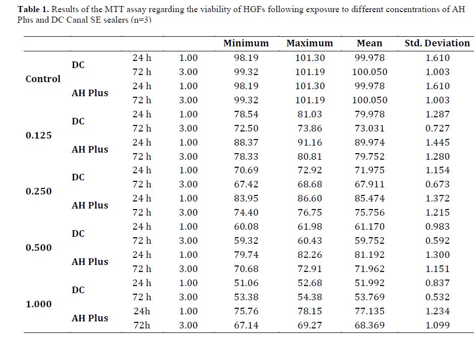
Table 1 presents the results of the MTT assay regarding the viability of HGFs following exposure to different concentrations of AH Plus and DC Canal SE sealers at 24 and 72 hours.
Three-way ANOVA showed significant interaction effect of concentration, time, and type of sealer. The Tukey’s test was applied to assess the individual effect of sealer type, concentration, and time.
Effect of sealer type:
DC Canal SE:
A significant difference was noted in cell viability among all concentrations of this sealer at 24 and 72 hours (P<0.05). In all groups, biocompatibility was significantly lower than that of the control group (P<0.001). Significant differences were noted when comparing different concentrations of the sealer with each other (P<0.001). Lower concentrations had higher biocompatibility and cell viability.
Accordingly, maximum cell viability was noted in 1/8 concentration of DC Canal SE sealer at both 24 and 72 hours.
AH Plus:
A significant difference was noted in cell viability among all concentrations at 24 and 72 hours (P<0.05). In all groups, biocompatibility was lower than that of the control group.
Significant differences were noted when comparing different concentrations of the sealer with each other (P<0.001). Lower concentrations had higher biocompatibility and cell viability. Accordingly, maximum cell viability was noted in 1/8 concentration of AH Plus sealer at both 24 and 72 hours.
Cell viability at 24 and 72 hours:
24 hours:
Cell viability of HGFs was significantly higher in presence of 1, 1/2, 1/4, and 1/8 concentrations of AH Plus compared with similar concentrations of DC Canal SE (P<0.001).
72 hours:
Cell viability of HGFs was significantly higher in presence of 1, 1/2, 1/4, and 1/8 concentrations of AH Plus compared with similar concentrations of DC Canal SE (P<0.001).
In general, the viability of HGFs in presence of AH Plus was significantly higher than that of DC Canal SE sealer in all concentrations and time points (P<0.001).
Based on concentration, comparison of cell viability at 24 and 72 hours revealed that cell viability at 72 hours was significantly lower than that at 24 hours in presence of all concentrations of AH Plus (P<0.001).
Cell viability at 72 hours was significantly lower than that at 24 hours in presence of 1/4 and 1/8 concentrations of DC Canal SE (P<0.001). However, this difference was not significant in presence of 1 and 1/2 concentrations (P>0.05). Therefore, it may be stated that irrespective of type of sealer, lower concentration of sealer was associated with higher cell viability. Also, cell viability decreased following prolonged exposure to sealer. AH Plus was more biocompatible than DC Canal SE in all concentrations and time points. Also, according to ISO 10993-5, reduction over 30% in cell viability indicates cytotoxicity of an agent. Accordingly, AH Plus was not cytotoxic in any concentration or time point. However, DC Canal SE was only non-toxic and biocompatible in 1/8 concentration at 24 and 72 hours, and was toxic in higher concentrations.
Figure 1 shows the viability of HGFs exposed to different concentrations of AH Plus and DC Canal SE sealers.
Table 1. Results of the MTT assay regarding the viability of HGFs following exposure to different concentrations of AH Plus and DC Canal SE sealers (n=3)
Discussion
This study assessed the cytotoxicity of two resin-based sealers namely AH Plus and DC
Figure 1. Viability of HGFs exposed to different concentrations of AH Plus and DC Canal SE sealers
Canal SE (which is a self-etch epoxy resin sealer) against HGFs. The cytotoxicity of both sealers in 1, 1/2, 1/4, and 1/8 concentrations was evaluated at 24 and 72 hours using the MTT assay. In general, AH Plus showed significantly lower cytotoxicity than DC Canal SE sealer, and the only non-toxic concentration of DC Canal SE was its 1/8 concentration. It showed cytotoxic effects in all other tested concentrations. This group of resin sealers bond to thermoplastic root filling materials as well as root dentin by the formation of hybrid layers. They are also used along with Resilon points or as a conventional sealer along with gutta-percha with the lateral compaction or warm vertical condensation techniques.
Assessment of different properties of newly introduced sealers in comparison with commonly used sealers such as AH Plus is imperative. The MTT assay is often used for assessment of cytotoxicity of biomaterials. In the MTT assay, absence of a significant difference between the test and control wells would indicate that the tested sealer has no cytotoxic effect. However, a significant reduction in viability of cells treated with the sealer would indicate cytotoxic effect of the sealer.
HGFs were used in this study due to their high sensitivity to toxic agents, similar to the study by Parirokh study.[25] Assessment of the cytotoxicity of freshly mixed sealers is clinically appropriate since sealers in the root canal system are not completely set after application, and may leak into the periapical tissue.
The present results showed significantly higher viability of HGFs in presence of AH Plus compared with DC Canal SE at all concentrations and time points. Irrespective of sealer type, lower concentrations of sealer were associated with higher cell viability. Also, cell viability decreased following prolonged exposure to sealers. It has been demonstrated that cytotoxic effects on cell culture may be due to monomer release. Since curing of resin-based sealers is not often complete, non-polymerized monomers can leak out from resin into the adjacent aqueous phase, and diffuse into the periapical space through dentin. Thus, prior to complete setting, the cytotoxicity of monomer may affect the periapical tissue. However, after setting, the effect of residual monomers on the tissues depends on sealer washout over time.[25] Consistent with the present results, several studies have confirmed the high biocompatibility and low cytotoxicity of AH Plus.[25,27,28] In line with present results regarding decreased biocompatibility of AH Plus sealer after 72 hours, Cotti et al.[3] showed that after completion of setting time, AH Plus still decreased the viability of cells, probably due to the toxicity of epoxy resin component of this sealer, which still remains after setting. Unlike the present study, Parirokh et al.[25] reported decreased cytotoxicity of AH Plus after 24 hours, and explained the reason to be the decreased concentration of the leached toxic resin component. However, they did not assess the cytotoxicity after 72 hours. In contrast to the present study, Bin et al. [29] found that 1, 1/2 and 1/4 concentrations of AH Plus significantly decreased the biocompatibility of hamster fibroblasts at 48 hours, and were reported to be toxic according to ISO 10993-5. Also, they found no significant correlation between the concentration of this sealer and cytotoxicity rate. However, in line with the present results, they demonstrated that cytotoxicity increased at 48 hours, compared with 12 hours. They explained that the epoxy resin component of AH Plus was responsible for cytotoxicity of this sealer as well as the presence of small amount of amine to enhance the curing of epoxy component. Variations in the results regarding the cytotoxicity of AH Plus can be due to differences in setting time as a result of variations in temperature and humidity.[29]
Consistent with the present results regarding the inverse correlation of concentration and biocompatibility of sealers, Silva et al.[28] showed that by an increase in concentration of AH Plus and a methacrylate-based sealer, cell viability decreased. Also, similar to the present study, Parirokh et al.[25] indicated that by an increase in concentration of AH Plus, viability of fibroblasts decreased, and sealer cytotoxicity increased. Karapınar-Kazandag et al. demonstrated that cytotoxicity of resin sealers had a direct correlation with time and concentration.[26]
Consistent with the present results regarding the cytotoxicity of SC Canal SE sealer (which has a methacrylate base) in all concentrations, Silva et al.[28] reported that methacrylate-based sealers showed high toxicity compared with AH Plus due to the presence of urethane dimethacrylate in their composition, which increases the intracellular free oxygen radicals and decreases the concentration of intracellular glutathione, and leads the cells towards apoptosis.[23] Reduction in concentration of glutathione initiates intracellular cytotoxicity and cell death, because it decreases the self-protective property of target cells.[30] In contrast to the present results regarding no cytotoxicity of methacrylate-based sealers, Oztan et al.
reported that AH Plus had a higher cytotoxicity than methacrylate-based sealers. This controversy can be attributed to different in vitro conditions, biological mechanisms, cell type, method of contact of material with cells, preparation of extracts, and duration of exposure.[31]
Conclusion
The results of current study showed lower cytotoxicity of AH Plus than DC Canal SE sealer. DC Canal SE sealer was more cytotoxic at 72 hours compared with 24 hours at all concentrations.
Cytotoxicity of dental materials is a common concern for dental clinicians since dental materials are in close contact with the periodontium and vital tissues, and can lead to inflammatory reactions, if they are not biocompatible.[1] Endodontic materials should not prevent tissue healing. Instead, they should preferably enhance tissue healing.[2] Also, they should not elicit inflammatory reactions in the periapical tissue.[3] However, the majority of endodontic sealers have some levels of toxicity.[3,4] Sealers, particularly during their setting reaction, can trigger an inflammatory response in the periapical tissue, and can also affect the viability of cells in the periapical region such as fibroblasts.[5] Among the known sealers, methacrylate-based sealers (especially in high concentrations) have the highest level of cytotoxicity followed by resin-based and bio-ceramic sealers and then silicone-based sealers, which have shown lower cytotoxicity.[6,7] The materials used for root canal filling may have toxic effects on the periapical tissue. Thus, an ideal sealer should have minimum cytotoxic effects.[8]
Accordingly, it is important to find a sealer with minimum cytotoxicity when in contact with the periapical tissue. AH Plus (Dentsply Sirona, Berlin, Germany) is a commonly applied endodontic sealer, which is also used for the purpose of comparison with other sealers in endodontic research.[9] Studies comparing the cytotoxicity of AH Plus with other sealers reported that it had minimum cytotoxic effects on human periodontal ligament cells, osteoblasts, and L929 murine fibroblasts.[10-12] This sealer is an epoxy resin-based sealer. A recent study on the cytotoxicity of other resinous dental materials such as composite resins confirmed their optimal biocompatibility and insignificant cytotoxicity.[13] AH Plus sealer has favorable physicochemical, biological, and antimicrobial properties, and minimum adverse effect on cell viability and proliferation.[14-20]
Recently, some new root canal filling materials were introduced to the market such as DC Canal SE sealer (S & C polymer GmbH, Elmshorn, Germany), which is a methacrylate-based self-etch sealer. Advances in bonding technology aim to minimize apical and coronal leakage by enhancement of the bond strength of sealers to root dentin, and subsequent mono-block obturation. Methacrylate-based sealers were introduced to achieve this goal, and include urethane dimethacrylate, bisphenol-A-glycidyl dimethacrylate, pigments, and radiopaque materials. These sealers also have an etching primer.[21,22] The results regarding the cytotoxicity of these sealers have been controversial.[23] Thus, this study aimed to compare the cytotoxicity of AH Plus, and DC Canal SE sealer after final setting.
Materials and Methods
This in vitro experimental study was approved by the ethics committee of Islamic Azad University. For assessment of cytotoxicity, Human Gingival Fibroblasts (HGFs) were purchased from the Pasteur Institute of Iran and cultured in Dulbecco’s modified Eagle’s medium (Gibco, Munich, Germany) supplemented with 10% fetal bovine serum, 100 U/mL penicillin, and 100 µg/mL streptomycin. They were then incubated at 37°C and 5% CO2. Second passage cells were plated with 5000 cells/well density in a 96-well plate for the methyl thiazolyl tetrazolium (MTT) assay.[24] The cells were then incubated with AH Plus resin-based sealer and DC Canal SE sealer that were prepared as follows: Freshly applied sealers were mixed according to the manufacturers’ instructions for 30 seconds, and discs with 4 mm height and 10 mm diameter were fabricated. After UV radiation for 24 hours, they were incubated in a humidified incubator at 37°C. Each specimen was immersed in 10 mL of culture medium at 37°C and 5% CO2 for 24 hours. Next, the solution was collected by a sterile syringe and filtered using a 0.22-µm filter. The solution was then serially diluted to prepare 1, 1/2, 1/4, and 1/8 concentrations; for this purpose, 100 mL of the sealer solution was mixed with 100 mL of the medium to obtain 1/2 concentration. The same process was repeated to obtain 1/4 and 1/8 concentrations.[24,25]
Cell viability was assessed by measuring the optical density of the solutions at 24 and 72 hours, and untreated tissue culture was used as the control group throughout the experiment. For this purpose, the color change following the addition of yellow MTT salt and formation of purple formazan crystals by the activity of mitochondrial dehydrogenase enzyme in metabolically active cells was measured by a spectrophotometer. For this purpose, the MTT salt was dissolved in phosphate buffered saline (Zist Mavad Pharmed, Tehran, Iran) and added to the culture medium in final concentration of 0.5 mg/mL. After 2 hours of incubation at 37°C, the overlaying medium was carefully collected, and 100 mL of dimethyl sulfoxide was added to each well. The optical density was read by a spectrophotometer (ELX808 Microplate Reader, BioTek Instruments Inc., VT, USA) at 490 nm wavelength. The mean optical density measured for each concentration was divided by the optical density of the control wells and multiplied by 100. The obtained value indicated the percentage of cell viability. The results were interpreted based on the drawn cell viability curves.[26] Normal distribution of data was evaluated by the Kolmogorov-Smirnov test. Data were analyzed by ANOVA followed by the Tukey’s test.
Results
Table 1 presents the results of the MTT assay regarding the viability of HGFs following exposure to different concentrations of AH Plus and DC Canal SE sealers at 24 and 72 hours.
Three-way ANOVA showed significant interaction effect of concentration, time, and type of sealer. The Tukey’s test was applied to assess the individual effect of sealer type, concentration, and time.
Effect of sealer type:
DC Canal SE:
A significant difference was noted in cell viability among all concentrations of this sealer at 24 and 72 hours (P<0.05). In all groups, biocompatibility was significantly lower than that of the control group (P<0.001). Significant differences were noted when comparing different concentrations of the sealer with each other (P<0.001). Lower concentrations had higher biocompatibility and cell viability.
Accordingly, maximum cell viability was noted in 1/8 concentration of DC Canal SE sealer at both 24 and 72 hours.
AH Plus:
A significant difference was noted in cell viability among all concentrations at 24 and 72 hours (P<0.05). In all groups, biocompatibility was lower than that of the control group.
Significant differences were noted when comparing different concentrations of the sealer with each other (P<0.001). Lower concentrations had higher biocompatibility and cell viability. Accordingly, maximum cell viability was noted in 1/8 concentration of AH Plus sealer at both 24 and 72 hours.
Cell viability at 24 and 72 hours:
24 hours:
Cell viability of HGFs was significantly higher in presence of 1, 1/2, 1/4, and 1/8 concentrations of AH Plus compared with similar concentrations of DC Canal SE (P<0.001).
72 hours:
Cell viability of HGFs was significantly higher in presence of 1, 1/2, 1/4, and 1/8 concentrations of AH Plus compared with similar concentrations of DC Canal SE (P<0.001).
In general, the viability of HGFs in presence of AH Plus was significantly higher than that of DC Canal SE sealer in all concentrations and time points (P<0.001).
Based on concentration, comparison of cell viability at 24 and 72 hours revealed that cell viability at 72 hours was significantly lower than that at 24 hours in presence of all concentrations of AH Plus (P<0.001).
Cell viability at 72 hours was significantly lower than that at 24 hours in presence of 1/4 and 1/8 concentrations of DC Canal SE (P<0.001). However, this difference was not significant in presence of 1 and 1/2 concentrations (P>0.05). Therefore, it may be stated that irrespective of type of sealer, lower concentration of sealer was associated with higher cell viability. Also, cell viability decreased following prolonged exposure to sealer. AH Plus was more biocompatible than DC Canal SE in all concentrations and time points. Also, according to ISO 10993-5, reduction over 30% in cell viability indicates cytotoxicity of an agent. Accordingly, AH Plus was not cytotoxic in any concentration or time point. However, DC Canal SE was only non-toxic and biocompatible in 1/8 concentration at 24 and 72 hours, and was toxic in higher concentrations.
Figure 1 shows the viability of HGFs exposed to different concentrations of AH Plus and DC Canal SE sealers.
Table 1. Results of the MTT assay regarding the viability of HGFs following exposure to different concentrations of AH Plus and DC Canal SE sealers (n=3)
Discussion
This study assessed the cytotoxicity of two resin-based sealers namely AH Plus and DC
Figure 1. Viability of HGFs exposed to different concentrations of AH Plus and DC Canal SE sealers
Canal SE (which is a self-etch epoxy resin sealer) against HGFs. The cytotoxicity of both sealers in 1, 1/2, 1/4, and 1/8 concentrations was evaluated at 24 and 72 hours using the MTT assay. In general, AH Plus showed significantly lower cytotoxicity than DC Canal SE sealer, and the only non-toxic concentration of DC Canal SE was its 1/8 concentration. It showed cytotoxic effects in all other tested concentrations. This group of resin sealers bond to thermoplastic root filling materials as well as root dentin by the formation of hybrid layers. They are also used along with Resilon points or as a conventional sealer along with gutta-percha with the lateral compaction or warm vertical condensation techniques.
Assessment of different properties of newly introduced sealers in comparison with commonly used sealers such as AH Plus is imperative. The MTT assay is often used for assessment of cytotoxicity of biomaterials. In the MTT assay, absence of a significant difference between the test and control wells would indicate that the tested sealer has no cytotoxic effect. However, a significant reduction in viability of cells treated with the sealer would indicate cytotoxic effect of the sealer.
HGFs were used in this study due to their high sensitivity to toxic agents, similar to the study by Parirokh study.[25] Assessment of the cytotoxicity of freshly mixed sealers is clinically appropriate since sealers in the root canal system are not completely set after application, and may leak into the periapical tissue.
The present results showed significantly higher viability of HGFs in presence of AH Plus compared with DC Canal SE at all concentrations and time points. Irrespective of sealer type, lower concentrations of sealer were associated with higher cell viability. Also, cell viability decreased following prolonged exposure to sealers. It has been demonstrated that cytotoxic effects on cell culture may be due to monomer release. Since curing of resin-based sealers is not often complete, non-polymerized monomers can leak out from resin into the adjacent aqueous phase, and diffuse into the periapical space through dentin. Thus, prior to complete setting, the cytotoxicity of monomer may affect the periapical tissue. However, after setting, the effect of residual monomers on the tissues depends on sealer washout over time.[25] Consistent with the present results, several studies have confirmed the high biocompatibility and low cytotoxicity of AH Plus.[25,27,28] In line with present results regarding decreased biocompatibility of AH Plus sealer after 72 hours, Cotti et al.[3] showed that after completion of setting time, AH Plus still decreased the viability of cells, probably due to the toxicity of epoxy resin component of this sealer, which still remains after setting. Unlike the present study, Parirokh et al.[25] reported decreased cytotoxicity of AH Plus after 24 hours, and explained the reason to be the decreased concentration of the leached toxic resin component. However, they did not assess the cytotoxicity after 72 hours. In contrast to the present study, Bin et al. [29] found that 1, 1/2 and 1/4 concentrations of AH Plus significantly decreased the biocompatibility of hamster fibroblasts at 48 hours, and were reported to be toxic according to ISO 10993-5. Also, they found no significant correlation between the concentration of this sealer and cytotoxicity rate. However, in line with the present results, they demonstrated that cytotoxicity increased at 48 hours, compared with 12 hours. They explained that the epoxy resin component of AH Plus was responsible for cytotoxicity of this sealer as well as the presence of small amount of amine to enhance the curing of epoxy component. Variations in the results regarding the cytotoxicity of AH Plus can be due to differences in setting time as a result of variations in temperature and humidity.[29]
Consistent with the present results regarding the inverse correlation of concentration and biocompatibility of sealers, Silva et al.[28] showed that by an increase in concentration of AH Plus and a methacrylate-based sealer, cell viability decreased. Also, similar to the present study, Parirokh et al.[25] indicated that by an increase in concentration of AH Plus, viability of fibroblasts decreased, and sealer cytotoxicity increased. Karapınar-Kazandag et al. demonstrated that cytotoxicity of resin sealers had a direct correlation with time and concentration.[26]
Consistent with the present results regarding the cytotoxicity of SC Canal SE sealer (which has a methacrylate base) in all concentrations, Silva et al.[28] reported that methacrylate-based sealers showed high toxicity compared with AH Plus due to the presence of urethane dimethacrylate in their composition, which increases the intracellular free oxygen radicals and decreases the concentration of intracellular glutathione, and leads the cells towards apoptosis.[23] Reduction in concentration of glutathione initiates intracellular cytotoxicity and cell death, because it decreases the self-protective property of target cells.[30] In contrast to the present results regarding no cytotoxicity of methacrylate-based sealers, Oztan et al.
reported that AH Plus had a higher cytotoxicity than methacrylate-based sealers. This controversy can be attributed to different in vitro conditions, biological mechanisms, cell type, method of contact of material with cells, preparation of extracts, and duration of exposure.[31]
Conclusion
The results of current study showed lower cytotoxicity of AH Plus than DC Canal SE sealer. DC Canal SE sealer was more cytotoxic at 72 hours compared with 24 hours at all concentrations.
Type of Study: Original article |
Subject:
Oral medicine
References
1. Keiser K, Johnson CC, Tipton DA. Cytotoxicity of mineral trioxide aggregate using human periodontal ligament fibroblasts. J Endod. 2000 May;26(5):288-91. [DOI:10.1097/00004770-200005000-00010] [PMID]
2. Jung S, Sielker S, Hanisch MR, Libricht V, Schäfer E, Dammaschke T. Cytotoxic effects of four different root canal sealers on human osteoblasts. PLoS One. 2018 Mar 26; 13 (3):e0194467. [DOI:10.1371/journal.pone.0194467] [PMID] [PMCID]
3. Cotti E, Petreucic V, Re D, Simbula G. Cytotoxicity evaluation of a new resin-based hybrid root canal sealer: an in vitro study. J Endod. 2014 Jan;40(1):124-8. [DOI:10.1016/j.joen.2013.09.038] [PMID]
4. Martinho FC, Camargo SEA, Fernandes AMM, Campos MS, Prado RF, Camargo CHR, Valera MC. Comparison of cytotoxicity, genotoxicity and immunological inflammatory biomarker activity of several endodontic sealers against immortalized human pulp cells. Int Endod J. 2018 Jan; 51 (1):41-57. [DOI:10.1111/iej.12785] [PMID]
5. Kim TG, Lee YH, Lee NH, Bhattarai G, Lee IK, Yun BS, Yi HK. The antioxidant property of pachymic acid improves bone disturbance against AH plus-induced inflammation in MC-3T3 E1 cells. J Endod. 2013 Apr;39(4):461-6. [DOI:10.1016/j.joen.2012.11.022] [PMID]
6. Benetti F, de Azevedo Queiroz ÍO, Oliveira PHC, Conti LC, Azuma MM, Oliveira SHP, Cintra LTA. Cytotoxicity and biocompatibility of a new bioceramic endodontic sealer containing calcium hydroxide. Braz Oral Res. 2019;33:e042. [DOI:10.1590/1807-3107bor-2019.vol33.0042] [PMID]
7. Camps J, About I. Cytotoxicity testing of endodontic sealers: a new method. J Endod. 2003 Sep;29(9):583-6. [DOI:10.1097/00004770-200309000-00010] [PMID]
8. Vouzara T, Dimosiari G, Koulaouzidou EA, Economides N. Cytotoxicity of a New Calcium Silicate Endodontic Sealer. J Endod. 2018 May;44(5):849-52. [DOI:10.1016/j.joen.2018.01.015] [PMID]
9. Silva EJ, Perez R, Valentim RM, Belladonna FG, De-Deus GA, Lima IC, Neves AA. Dissolution, dislocation and dimensional changes of endodontic sealers after a solubility challenge: a micro-CT approach. Int Endod J. 2017 Apr; 50 (4):407-14. [DOI:10.1111/iej.12636] [PMID]
10. Araki K, Suda H, Spångberg LS. Indirect longitudinal cytotoxicity of root canal sealers on L929 cells and human periodontal ligament fibroblasts. J Endod. 1994 Feb;20(2): 67-70. [DOI:10.1016/S0099-2399(06)81183-0]
11. Huang FM, Tai KW, Chou MY, Chang YC. Cytotoxicity of resin-, zinc oxide-eugenol-, and calcium hydroxide-based root canal sealers on human periodontal ligament cells and permanent V79 cells. Int Endod J. 2002 Feb;35(2):153-8. [DOI:10.1046/j.1365-2591.2002.00459.x] [PMID]
12. Lee DH, Lim BS, Lee YK, Yang HC. Mechanisms of root canal sealers cytotoxicity on osteoblastic cell line MC3T3-E1. Oral Surg Oral Med Oral Pathol Oral Radiol Endod. 2007 Nov;104(5):717-21. [DOI:10.1016/j.tripleo.2007.05.018] [PMID]
13. Kamalak H, Kamalak A, Taghizadehghalehjoughi A. Cytotoxic effects of new-generation bulk-fill composites on human dental pulp stem cells. Cell Mol Biol (Noisy-le-grand). 2018 Feb 28;64(3):62-71. [DOI:10.14715/cmb/2018.64.3.11] [PMID]
14. Heyder M, Kranz S, Völpel A, Pfister W, Watts DC, Jandt KD, Sigusch BW. Antibacterial effect of different root canal sealers on three bacterial species. Dent Mater. 2013 May; 29 (5):542-9. [DOI:10.1016/j.dental.2013.02.007] [PMID]
15. Leonardo MR, da Silva LA, Almeida WA, Utrilla LS. Tissue response to an epoxy resin-based root canal sealer. Endod Dent Traumatol. 1999 Feb;15(1):28-32. [DOI:10.1111/j.1600-9657.1999.tb00745.x] [PMID]
16. Mutoh N, Satoh T, Watabe H, Tani-Ishii N. Evaluation of the biocompatibility of resin-based root canal sealers in rat periapical tissue. Dent Mater J. 2013;32(3):413-9. [DOI:10.4012/dmj.2012-218] [PMID]
17. Saleh IM, Ruyter IE, Haapasalo M, Ørstavik D. Survival of Enterococcus faecalis in infected dentinal tubules after root canal filling with different root canal sealers in vitro. Int Endod J. 2004 Mar;37(3):193-8. [DOI:10.1111/j.0143-2885.2004.00785.x] [PMID]
18. Schäfer E, Bering N, Bürklein S. Selected physicochemical properties of AH Plus, EndoREZ and RealSeal SE root canal sealers. Odontology. 2015 Jan;103(1):61-5. [DOI:10.1007/s10266-013-0137-y] [PMID]
19. Tanomaru-Filho M, Tanomaru JM, Leonardo MR, da Silva LA. Periapical repair after root canal filling with different root canal sealers. Braz Dent J. 2009;20(5):389-95. [DOI:10.1590/S0103-64402009000500006] [PMID]
20. Wang Z, Shen Y, Haapasalo M. Dentin extends the antibacterial effect of endodontic sealers against Enterococcus faecalis biofilms. J Endod. 2014 Apr;40(4):505-8. [DOI:10.1016/j.joen.2013.10.042] [PMID]
21. Costa JA, Rached-Júnior FA, Souza-Gabriel AE, Silva-Sousa YT, Sousa-Neto MD. Push-out strength of methacrylate resin-based sealers to root canal walls. Int Endod J. 2010 Aug;43(8):698-706. [DOI:10.1111/j.1365-2591.2010.01766.x] [PMID]
22. Scelza MZ, Linhares AB, da Silva LE, Granjeiro JM, Alves GG. A multiparametric assay to compare the cytotoxicity of endodontic sealers with primary human osteoblasts. Int Endod J. 2012 Jan;45(1):12-8. [DOI:10.1111/j.1365-2591.2011.01941.x] [PMID]
23. Graunaite I, Lodiene G, Arandarcikaite O, Pukalskas A, Machiulskiene V. Leachables and cytotoxicity of root canal sealers. J Oral Sci. 2018 Sep 23;60(3):381-7. [DOI:10.2334/josnusd.17-0173] [PMID]
24. van Meerloo J, Kaspers GJ, Cloos J. Cell sensitivity assays: the MTT assay. Methods Mol Biol. 2011;731:237-45. [DOI:10.1007/978-1-61779-080-5_20] [PMID]
25. Parirokh M, Forghani FR, Paseban H, Asgary S, Askarifard S, Esmaeeli Mahani S. Cytotoxicity of two resin-based sealers and a fluoride varnish on human gingival fibroblasts. Iran Endod J. 2015;10(2):89-92.
26. Karapınar-Kazandağ M, Bayrak OF, Yalvaç ME, Ersev H, Tanalp J, Sahin F, Bayırlı G. Cytotoxicity of 5 endodontic sealers on L929 cell line and human dental pulp cells. Int Endod J. 2011 Jul;44(7):626-34. [DOI:10.1111/j.1365-2591.2011.01863.x] [PMID]
27. Silva EJ, Rosa TP, Herrera DR, Jacinto RC, Gomes BP, Zaia AA. Evaluation of cytotoxicity and physicochemical properties of calcium silicate-based endodontic sealer MTA Fillapex. J Endod. 2013 Feb;39(2):274-7. [DOI:10.1016/j.joen.2012.06.030] [PMID]
28. Silva GO, Cavalcanti BN, Oliveira TR, Bin CV, Camargo SE, Camargo CH. Cytotoxicity and genotoxicity of natural resin-based experimental endodontic sealers. Clin Oral Investig. 2016 May;20(4):815-9. [DOI:10.1007/s00784-015-1567-4] [PMID]
29. Bin CV, Valera MC, Camargo SE, Rabelo SB, Silva GO, Balducci I, Camargo CH. Cytotoxicity and genotoxicity of root canal sealers based on mineral trioxide aggregate. J Endod. 2012 Apr;38(4):495-500. [DOI:10.1016/j.joen.2011.11.003] [PMID]
30. Lodienė G, Kopperud HM, Ørstavik D, Bruzell EM. Detection of leachables and cytotoxicity after exposure to methacrylate- and epoxy-based root canal sealers in vitro. Eur J Oral Sci. 2013 Oct;121(5):488-96. [DOI:10.1111/eos.12065] [PMID]
31. Oztan MD, Yilmaz S, Kalayci A, Zaimoğlu L. A comparison of the in vitro cytotoxicity of two root canal sealers. J Oral Rehabil. 2003 Apr;30(4):426-9. [DOI:10.1046/j.1365-2842.2003.01053.x] [PMID]
Send email to the article author
| Rights and permissions | |
 |
This work is licensed under a Creative Commons Attribution-NonCommercial 4.0 International License. |
