

Volume 6, Issue 1 (3-2021)
J Res Dent Maxillofac Sci 2021, 6(1): 4-13 |
Back to browse issues page

Download citation:
BibTeX | RIS | EndNote | Medlars | ProCite | Reference Manager | RefWorks
Send citation to:


BibTeX | RIS | EndNote | Medlars | ProCite | Reference Manager | RefWorks
Send citation to:
KHodarahmi E, Salari M, Azizi A, Lawaf S. Discoloration of Vita Classical Shade Guide by Glutaraldehyde Disinfectant. J Res Dent Maxillofac Sci 2021; 6 (1) :4-13
URL: http://jrdms.dentaliau.ac.ir/article-1-293-en.html
URL: http://jrdms.dentaliau.ac.ir/article-1-293-en.html


1- Dentist
2- Assistant professor, Prosthodontics Dept, Faculty of Dentistry, Tehran Medical Sciences
3- Professor of oral medicine Dept, Faculty of Dentistry, Tehran Medical Sciences,
4- Associate Professor, Prosthodontics Dept, Member ship of Dental Material Research Center, Faculty of Dentistry, Tehran Medical Sciences, Islamic Azad University , Tehran, Iran. , Drshlawaf@yahoo.com
Keywords: Color, Colorimetry, Dental Disinfectants, Discoloration, Dental Restorations, Spectrophotometry, Time Factors
Full-Text [PDF 530 kb]
(1002 Downloads)
| Abstract (HTML) (2198 Views)
Full-Text: (585 Views)
Abstract
Background and Aim: Shade guide discoloration after disinfection can interfere with the appropriate color selection for dental restorations. Since one of the most important issues for patients is the color of the final restoration, the discoloration of shade guides due to disinfectants will be important. Infection control is a definite and important matter in dentistry. Due to the contradictory results of studies on the effect of disinfectants on shade guide discoloration, this study aimed to examine the discoloration of the Vita classical shade guide by glutaraldehyde disinfectant.
Materials and Methods: In this experimental study, samples of A4, B4, C4, and D4 colors were selected from the Vitapan classical shade guide, 10 pieces each (40 samples in total). Three samples of each color were immersed in distilled water as a control while the other seven were immersed in a 2% glutaraldehyde disinfectant solution. The shade pilot spectrophotometer was used for colorimetry, which was performed at baseline and 24, 48, and 72 hours after the immersion. The color of the samples was evaluated based on the CIE Lab system. The data were analyzed with two-way analysis of variance (ANOVA) and Tukey's test.
Results: The rate of color change (ΔE) of the samples was higher in the glutaraldehyde group than in distilled water (P<0.05). In addition, color change in both groups showed a significant difference at different times (P<0.05).
Conclusion: The Vitapan classical color samples discolor by immersion in 2% glutaraldehyde disinfectant after 24, 48, and 72 hours, but this color change is not clinically detectable (ΔE<1).
Keywords: Color, Colorimetry, Dental Disinfectants, Discoloration, Dental Restorations, Spectrophotometry, Time Factors
Introduction
The discoloration of shade guides after disinfection can interfere with appropriate color selection for dental restorations. (1) Shade guides are used to match the color between the veneer and the natural tooth; they require disinfection to control the infection. (2) The use of disinfectants can eventually cause changes in the color of the equipment. (2)
Any difference in the color of the restoration compared to natural teeth will be significant as it is noticeable to the patient. (3) Dental veneers should be made similar in color to natural teeth. (4) Various studies have shown that disinfectants affect the physical and mechanical properties of resin teeth; these substances may affect the color and surface properties of cast restorations. (4) Glutaraldehyde, as a sterilizing substance, destroys all pathogens and even the endospore; it is a strong disinfectant. (5)
The color matching process must be in accordance with shade guides. Several shade guides are used in restorative dentistry; one of the most widely used types is the Vitapan classical shade guide (VITA Zahnfabrik, Bad Säckingen, Germany) with 16 colors, which has been selected for use in this study. (1) Few studies have been performed to determine whether surface disinfectants affect the appearance of shade guides. (1)
In 1999, Tsun et al conducted a study to investigate the effect of immersion of pressable ceramics and ceramo-metal porcelain in various surface disinfectants and did not report any noticeable discoloration, (4) whereas Koosha et al proved that disinfectants have a significant effect on shade guide discoloration. (2)
Due to inconsistencies and insufficient information in this field, this study aimed to investigate the color change of the Vita classical shade guide after immersion in 2% glutaraldehyde disinfectant at the Faculty of Dentistry of Islamic Azad University of Medical Sciences, Tehran, Iran. The results of this study can be very useful for improving clinical conditions and raising the awareness of dentists.
Materials and Methods
In this experimental study, A4, B4, C4, and D4 color samples were selected from the Vitapan classical shade guide (VITA Zahnfabrik, Bad Säckingen, Germany), 10 pieces each (40 samples in total). (1) Then, the metal handle of each shade tab was separated, and the porcelain of that shade tab was coded from 1 to 40 for performing the test steps. Next, all the tabs were assessed using the shade pilot spectrophotometer (Dentsply Sirona, Bensheim, Germany) for measuring color in a single-blind manner. Then, three random tabs of each color were immersed in distilled water as a control, and the other seven tabs of each color were immersed in 2% glutaraldehyde surface disinfectant (Behsa Razi Co., Tehran, Iran) for 24 hours. (2) The samples were immersed three times a week for 10 minutes each time to simulate a one-year use. (2) After 24 hours of immersion in the disinfectant, all tabs were washed with distilled water for 5 minutes in an ultrasonic device, were kept under running water for 30 seconds, (2) and examined again by the shade pilot spectrophotometer. In addition to 24 hours (the first year), this process was repeated for 48 hours (the second year) and 72 hours (the third year; Figures 1 to 6). (1) The color of the samples was evaluated based on the CIE Lab system. (4) Data were analyzed using two-way repeated-measures analysis of variance (ANOVA) and Tukey’s test.
The research project was performed by immersing A4, B4, C4, and D4 color samples in glutaraldehyde and distilled water at three different times (24, 48, and 72 hours) and colorimetry at four different times (0, 24, 48, and 72 hours). The following results were obtained:
Table 1: Comparison of discoloration rate (ΔE) of the samples at different immersion times in the two studied solutions
According to the results of Table 1, it was shown that the type of solution is effective in discoloration of all the samples, regardless of the type of the color sample (P=0.006<0.05).
Also, color change in glutaraldehyde solution (P=0.0001) and distilled water (P=0.0001) showed a significant difference at different times (P<0.05). It was also shown that time change (regardless of the type of solution) is effective in ΔE of all samples (P=0.0001<0.05).
Table 2: Comparison of discoloration rate (ΔE) at different immersion times in the two types of solution studied for the A4 color sample
According to the results of Table 2, it was shown that the type of solution is effective in the color change of the A4 color sample (P=0.024<0.05). Also, color change in glutaraldehyde solution (P=0.041<0.05) showed a significant difference at different times, while the color change in distilled water (P=0.119>0.05) did not show any significant difference at different times. Time change (regardless of the type of solution) was also effective in ΔE changes (P=0.007<0.05).
The result of the color change statistical test according to each of the times studied in the A4 color sample is as follows:
At 24 and 72 hours, the difference in discoloration was not significant between the two solutions (P=0.357,0.201). However, at 48 hours, the difference in discoloration was significant between the two solutions (P=0.026).
Table 3: Comparison of discoloration rate (ΔE) at different immersion times in the two types of solution studied for the B4 color sample
According to the results of Table 3, it was shown that the type of solution was not effective in the discoloration of the B4 color sample (P=0.775>0.05). Also, color change in glutaraldehyde solution (P=0.006) showed a significant difference at different times, while the color change in distilled water (P=0.296) did not show any significant difference at different times. Time change (regardless of the type of solution) was also effective in changes of ΔE (P=0.0001<0.05).
The result of the statistical test of the color change for the B4 color sample according to each of the times studied is as follows:
At 24, 48, and 72 hours, the difference in the color change was not significant between the two solutions (P>0.05).
Table 4: Comparison of discoloration rate (ΔE) at different immersion times in the two types of solution studied for the C4 color sample
According to the results of Table 4, it was shown that the type of solution was effective in discoloration of the C4 color sample (P=0.008<0.05). Also, the color change in glutaraldehyde solution (P=0.013) showed a significant difference at different times, while the color change in distilled water (P=0.096) did not show any significant difference at different times. Time change (regardless of the type of solution) was effective in ΔE changes (P=0.002).
The result of the statistical test of the color change of the C4 color sample according to each of the times studied is as follows:
At 24 hours, the difference in color change was significant between the two solutions (P=0.001). But at 48 and 72 hours, the difference in color change was not significant between the two solutions (P=0.37,0.251).
Table 5: Comparison of discoloration rate (ΔE) at different immersion times in the two types of solution studied for the D4 color sample
According to the results of Table 5, it was shown that the type of solution was not effective in the color change of the D4 color sample (P=0.368>0.05). Also, color change in glutaraldehyde solution (P=0.004) showed a significant difference at different times, while the color change in distilled water (P=0.347) did not show any significant difference at different times. Time change (regardless of the type of solution) was effective in the changes of ΔE (P=0.001<0.05).
The result of the statistical test of the color change of the D4 color sample according to each of the times studied is as follows:
At 24, 48, and 72 hours, the difference in discoloration was not significant between the two solutions (P=0.892,0.536,0.172).
Table 6: Comparison of discoloration rate (ΔE) at different immersion times in the color groups studied
According to the results of Table 6, discoloration rates in the studied color samples (regardless of the type of solution and immersion time) were significantly different from each other (P=0.007).
Also, color change at different times of immersion (regardless of the type of solution and color samples studied) showed a significant difference (P=0.006).
The result of the statistical test of the color change of different color samples according to each of the studied times is as follows:
At 48 hours, the difference in color change was significant among the color samples (P=0.001), but at the other two times, the difference in color change was not significant among the color samples (P=0.254,0.402).
Table 7: Tukey’s statistical test results in the color groups studied (A4, B4, C4, and D4)
According to Table 7, the mean E∆ in the color groups studied was not significantly different.
To investigate the relationship between the amount of color change recorded by the spectrophotometer and that in the clinical environment, the information was converted to the National Bureau Standards (NBS) units using the following equation:
NBS unit = ΔE × 0.92.
According to the NBS units, the color changes are as follows:
0.0-0.5: Very slight changes
0.5-1.5: Slight changes
1.5-3.0: Noticeable changes
3.0-6.0: Appreciable changes
6.0-12.0: Very appreciable changes
12 or more: Conversion to another color.
In this study, color changes in the color groups were in the range of 0.5-1.5 i.e. slight changes.
Table 8: Mean (L a b) of the A4 color sample at baseline, 24, 48, and 72 hours
Table 9: Pairwise comparison of the mean (L a b) at different times for the A4 color sample (P<0.05) Pair 2 L( base) - L( 72h) -.4285714 .2627691 -4.315 .005
Pair 3 L( 24h) - L( 48h) -.1142857 .1214986 -2.489 .047
Pair 4 L( 24h) - L( 72h) -.4000000 .2309401 -4.583 .004
Pair 5 L( 48h) - L( 72h) -.2857143 .2115701 -3.573 .012
Pair 6 a( base) - a( 48h) .2857143 .2734262 2.765 .033
Pair 7 a( base) - a( 72h) .5285714 .1976047 7. 077 .000
Pair 8 a( 24h) - a( 48h) .2714286 .0487950 14.717 .000
Pair 9 a( 24h) - a( 72h) .5142857 .2035401 6.685 .001
Pair 10 a( 48h) - a( 72h) .2428571 .2070197 3.104 .021
Pair 11 b( base) - b( 24h) .2143 .1345 4.215 .006
Pair 12 b( 24h) - b( 48h) -.1143 .1215 -2.489 .047
Pair 13 b( 24h) - b( 72h) -.2143 .1864 -3.041 .023
The pairwise comparison of the mean L a b (Table 8) at different times for the A4 color sample is shown in Table 9.
The “L” index indicates the degree of brightness; the more positive the index, the brighter the color becomes, and if the index becomes negative, the darker will be the color. For the A4 color sample, the ΔL was in the positive direction in the case group, which indicates brightening. Also, for the A4 color sample, a significant difference was observed in years 2 and 3 compared to baseline, in years 2 and 3 compared to the first year, and in year 3 compared to the second year (P<0.05).
Component “a” (which indicates redness and greenness with positive changes towards redness and negative changes towards green) was negative for the A4 color sample, indicating greening of the samples. Also, there was a significant difference in component “a” in years 2 and 3 compared to baseline and in years 2 and 3 compared to the first year (P<0.05).
Component “b”, the positive of which indicates yellowing, and the negative of which indicates bluing of the sample, was positive for the A4 color sample, indicating the yellowing of the samples. Also, a significant difference was observed in component “b” in year 1 compared to baseline and in years 2 and 3 compared to the first year (P<0.05; Table 10).
Table 10: Mean (L a b) of the B4 color sample at baseline, 24, 48, and 72 hours
Discussion
Shade guides are mainly used to achieve a suitable color matching. To prevent the transmission of diseases and infectious agents, it is necessary to disinfect shade guides after each use. (2)
The 2005 OSHA guidelines for disinfection of shade guides have categorized them as semicritical items, which can be disinfected with an intermediate-level disinfectant approved by the Environmental Protection Agency (EPA). (1)
Some offices and clinics use autoclaves to disinfect shade guides, but the plastic tab holder cannot withstand the flow of autoclave vapors and will degrade over time. Also, the use of autoclaves in many offices is too time-consuming. (1)
Centers such as dental schools use sterilization (gas) to disinfect shade guides due to the volume of items and the availability of this sterilization method. Whereas in most offices, surface disinfectants are used between patients. (1)
The present study was performed to investigate the effect of glutaraldehyde disinfectant on the rate of the color change of the Vita classical shade guide when it is immersed in the solution at least 3 times a week for 10 minutes each time. (6) In this study, the simulation of use for one year was with 24 hours of immersion in the disinfectant while the simulation of use for 2 and 3 years was with 48 and 72 hours of immersion in the disinfectant. (6) The A4, B4, C4, and D4 colors were selected to cover all the different pigments (A: brown, B: yellow, C: gray, D: red-brown).
The shade pilot spectrophotometer was used to record the before and after values of (L a b) of the Vita classical shade guide in the middle third of the color sample. This device is a digital color analyzer spectrophotometer that is more accurate and reproducible compared to the human eye.
The results of this study showed that color change occurs in both glutaraldehyde and distilled water groups over time, but color changes are more significant in the glutaraldehyde group (P<0.006).
In similar studies, Hombesh et al, Brandt et al, Huang et al, and Öngül et al have also shown that color changes increase over time. (7-10)
In this study, it was found that the colors of the samples become brighter and the “L” component increases over time. These changes can be attributed to the solubility or abrasion of surface properties by glutaraldehyde and distilled water. (1) With glutaraldehyde, this color change and brightening occur more probably due to its unique chemical compounds.
The results of the present study showed that the mean ΔE of the color groups studied (A4, B4, C4, and D4) was less than one. The results of the amount of ΔE have been reported differently in different studies.
In studies by Hombesh et al, Brandt et al, and Öngül et al, disinfectants produced significant color changes (ΔE>1). (7,8,10). In the study by Huang et al, disinfectants produced minimal and minor changes (ΔE<1). (9)
According to the results, it is clear that the type of substance and its chemical composition has a significant effect on the rate of change of ΔE, L, a, and b. Disinfectants can change the parameters (L, a, b) through two mechanisms: abrasion of surface properties and residue deposition. (1)
Tsun et al examined the effect of 2% alkaline glutaraldehyde on porcelain samples and showed that this material could be used for a long time without significant discoloration of the samples. (4) However, in the present study, after 24 hours of immersion in glutaraldehyde solution, ΔE more than one was reported. The reason for this discrepancy can be attributed to the metal handle of the color sample and the effect of metal corrosion and deposition of corrosive materials on porcelain.
However, in the study conducted by Tsun et al on color changes of porcelain, glutaraldehyde was the only substance that could be used for long-term immersion (7 days) without significant color changes. (4)
According to the results of the current study, glutaraldehyde can be used to disinfect the Vita classical shade guide without worrying about significant color changes.
According to the results of this study, all the studied color samples (A4, B4, C4, and D4) showed increased “L” (brightening), decreased “a” (greening), and increased “b” (yellowing) during the time of immersion in glutaraldehyde. Changes of L, a, b in the control group of the A4, B4, C4, and D4 color samples were very insignificant and had a greater effect on decreasing component “a” and then on increasing component “L”.
In general, in both the case and control groups, the color changes were towards an increase in “L”, decrease in “a”, and increase in “b”, which in the case group (glutaraldehyde), these changes were more significant compared to the control group (distilled water; P<0.05).
The solution type (glutaraldehyde and distilled water) was effective in color change (ΔE) of the A4 and C4 color samples but not for the B4 and D4 samples (Tables 3 to 6).
These findings indicate that the behavior of color samples can defer with various disinfectants, which can be due to the type of pigment, their solubility, and the chemical properties of the compounds that produce these pigments.
However, despite the difference in the discoloration behavior of the samples, ΔE of all A4, B4, C4, and D4 color samples was less than one with glutaraldehyde disinfectant, indicating that these changes are small and imperceptible and cannot be seen with the naked eye.
One of the positive points of this research was that we removed the metal handle of the shade tabs to prevent surface or subsurface adsorption of corrosive products on porcelain (11) to check only the color change of the porcelain itself without the intervention of an interfering agent. However, these changes may become more clinically evident in the clinical setting and over time; this can be due to the abrasion of surface properties and deposition of surface residues. (1)
In this study, the shade pilot spectrophotometer was used, and only the color of the middle one-third area of the facial surface of the tooth was examined. This area is the best for choosing tooth color. (1)
How the manufacturer makes the shade guide can also affect its discoloration after disinfection. Shade guides may be made of several layers of compacted porcelain, or they may be porcelain samples whose surface is stained and glazed. Samples made of several layers of porcelain, such as Vitapan classical shade guide, are more resistant to discoloration. (1)
Conclusion
Based on the results of the present study, it can be concluded that:
1. If we follow the instructions provided by the manufacturers of shade guides, as well as the time and manner of use of disinfectants recommended by their manufacturer, there would be no noticeable (clinically visible) changes in ΔE (ΔE<1).
2. Color changes following immersion of A4, B4, C4, and D4 color samples in glutaraldehyde solution for 24, 48, and 72 hours show a significant difference at different times, i.e. time change is effective in changes of ΔE (P<0.05 and P=0.0001). Nevertheless, this color change is insignificant, meaning it causes small changes in ΔE that are not visually obvious to observers (ΔE<1).
3. The process of color change after 72 hours of immersion in glutaraldehyde is towards lightening, greening, and yellowing of color samples.
4. Color change (ΔE) of shade guides should be evaluated occasionally (a standard color sample should be set aside for comparison with the types used in the clinic).
5. The type of disinfectant and its interaction with the surface of color samples has a direct effect on the color change process.
Background and Aim: Shade guide discoloration after disinfection can interfere with the appropriate color selection for dental restorations. Since one of the most important issues for patients is the color of the final restoration, the discoloration of shade guides due to disinfectants will be important. Infection control is a definite and important matter in dentistry. Due to the contradictory results of studies on the effect of disinfectants on shade guide discoloration, this study aimed to examine the discoloration of the Vita classical shade guide by glutaraldehyde disinfectant.
Materials and Methods: In this experimental study, samples of A4, B4, C4, and D4 colors were selected from the Vitapan classical shade guide, 10 pieces each (40 samples in total). Three samples of each color were immersed in distilled water as a control while the other seven were immersed in a 2% glutaraldehyde disinfectant solution. The shade pilot spectrophotometer was used for colorimetry, which was performed at baseline and 24, 48, and 72 hours after the immersion. The color of the samples was evaluated based on the CIE Lab system. The data were analyzed with two-way analysis of variance (ANOVA) and Tukey's test.
Results: The rate of color change (ΔE) of the samples was higher in the glutaraldehyde group than in distilled water (P<0.05). In addition, color change in both groups showed a significant difference at different times (P<0.05).
Conclusion: The Vitapan classical color samples discolor by immersion in 2% glutaraldehyde disinfectant after 24, 48, and 72 hours, but this color change is not clinically detectable (ΔE<1).
Keywords: Color, Colorimetry, Dental Disinfectants, Discoloration, Dental Restorations, Spectrophotometry, Time Factors
Introduction
The discoloration of shade guides after disinfection can interfere with appropriate color selection for dental restorations. (1) Shade guides are used to match the color between the veneer and the natural tooth; they require disinfection to control the infection. (2) The use of disinfectants can eventually cause changes in the color of the equipment. (2)
Any difference in the color of the restoration compared to natural teeth will be significant as it is noticeable to the patient. (3) Dental veneers should be made similar in color to natural teeth. (4) Various studies have shown that disinfectants affect the physical and mechanical properties of resin teeth; these substances may affect the color and surface properties of cast restorations. (4) Glutaraldehyde, as a sterilizing substance, destroys all pathogens and even the endospore; it is a strong disinfectant. (5)
The color matching process must be in accordance with shade guides. Several shade guides are used in restorative dentistry; one of the most widely used types is the Vitapan classical shade guide (VITA Zahnfabrik, Bad Säckingen, Germany) with 16 colors, which has been selected for use in this study. (1) Few studies have been performed to determine whether surface disinfectants affect the appearance of shade guides. (1)
In 1999, Tsun et al conducted a study to investigate the effect of immersion of pressable ceramics and ceramo-metal porcelain in various surface disinfectants and did not report any noticeable discoloration, (4) whereas Koosha et al proved that disinfectants have a significant effect on shade guide discoloration. (2)
Due to inconsistencies and insufficient information in this field, this study aimed to investigate the color change of the Vita classical shade guide after immersion in 2% glutaraldehyde disinfectant at the Faculty of Dentistry of Islamic Azad University of Medical Sciences, Tehran, Iran. The results of this study can be very useful for improving clinical conditions and raising the awareness of dentists.
Materials and Methods
In this experimental study, A4, B4, C4, and D4 color samples were selected from the Vitapan classical shade guide (VITA Zahnfabrik, Bad Säckingen, Germany), 10 pieces each (40 samples in total). (1) Then, the metal handle of each shade tab was separated, and the porcelain of that shade tab was coded from 1 to 40 for performing the test steps. Next, all the tabs were assessed using the shade pilot spectrophotometer (Dentsply Sirona, Bensheim, Germany) for measuring color in a single-blind manner. Then, three random tabs of each color were immersed in distilled water as a control, and the other seven tabs of each color were immersed in 2% glutaraldehyde surface disinfectant (Behsa Razi Co., Tehran, Iran) for 24 hours. (2) The samples were immersed three times a week for 10 minutes each time to simulate a one-year use. (2) After 24 hours of immersion in the disinfectant, all tabs were washed with distilled water for 5 minutes in an ultrasonic device, were kept under running water for 30 seconds, (2) and examined again by the shade pilot spectrophotometer. In addition to 24 hours (the first year), this process was repeated for 48 hours (the second year) and 72 hours (the third year; Figures 1 to 6). (1) The color of the samples was evaluated based on the CIE Lab system. (4) Data were analyzed using two-way repeated-measures analysis of variance (ANOVA) and Tukey’s test.
The research project was performed by immersing A4, B4, C4, and D4 color samples in glutaraldehyde and distilled water at three different times (24, 48, and 72 hours) and colorimetry at four different times (0, 24, 48, and 72 hours). The following results were obtained:
Table 1: Comparison of discoloration rate (ΔE) of the samples at different immersion times in the two studied solutions
According to the results of Table 1, it was shown that the type of solution is effective in discoloration of all the samples, regardless of the type of the color sample (P=0.006<0.05).
Also, color change in glutaraldehyde solution (P=0.0001) and distilled water (P=0.0001) showed a significant difference at different times (P<0.05). It was also shown that time change (regardless of the type of solution) is effective in ΔE of all samples (P=0.0001<0.05).
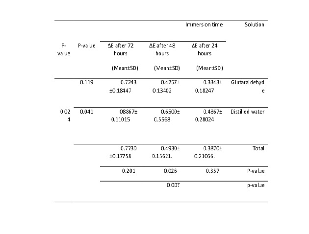
Table 2: Comparison of discoloration rate (ΔE) at different immersion times in the two types of solution studied for the A4 color sample
According to the results of Table 2, it was shown that the type of solution is effective in the color change of the A4 color sample (P=0.024<0.05). Also, color change in glutaraldehyde solution (P=0.041<0.05) showed a significant difference at different times, while the color change in distilled water (P=0.119>0.05) did not show any significant difference at different times. Time change (regardless of the type of solution) was also effective in ΔE changes (P=0.007<0.05).
The result of the color change statistical test according to each of the times studied in the A4 color sample is as follows:
At 24 and 72 hours, the difference in discoloration was not significant between the two solutions (P=0.357,0.201). However, at 48 hours, the difference in discoloration was significant between the two solutions (P=0.026).
Table 3: Comparison of discoloration rate (ΔE) at different immersion times in the two types of solution studied for the B4 color sample
According to the results of Table 3, it was shown that the type of solution was not effective in the discoloration of the B4 color sample (P=0.775>0.05). Also, color change in glutaraldehyde solution (P=0.006) showed a significant difference at different times, while the color change in distilled water (P=0.296) did not show any significant difference at different times. Time change (regardless of the type of solution) was also effective in changes of ΔE (P=0.0001<0.05).
The result of the statistical test of the color change for the B4 color sample according to each of the times studied is as follows:
At 24, 48, and 72 hours, the difference in the color change was not significant between the two solutions (P>0.05).
Table 4: Comparison of discoloration rate (ΔE) at different immersion times in the two types of solution studied for the C4 color sample
According to the results of Table 4, it was shown that the type of solution was effective in discoloration of the C4 color sample (P=0.008<0.05). Also, the color change in glutaraldehyde solution (P=0.013) showed a significant difference at different times, while the color change in distilled water (P=0.096) did not show any significant difference at different times. Time change (regardless of the type of solution) was effective in ΔE changes (P=0.002).
The result of the statistical test of the color change of the C4 color sample according to each of the times studied is as follows:
At 24 hours, the difference in color change was significant between the two solutions (P=0.001). But at 48 and 72 hours, the difference in color change was not significant between the two solutions (P=0.37,0.251).
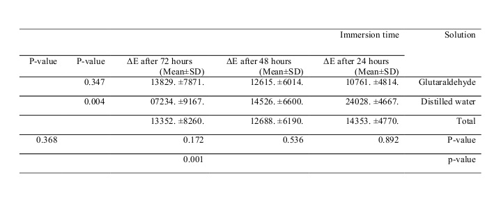
Table 5: Comparison of discoloration rate (ΔE) at different immersion times in the two types of solution studied for the D4 color sample
According to the results of Table 5, it was shown that the type of solution was not effective in the color change of the D4 color sample (P=0.368>0.05). Also, color change in glutaraldehyde solution (P=0.004) showed a significant difference at different times, while the color change in distilled water (P=0.347) did not show any significant difference at different times. Time change (regardless of the type of solution) was effective in the changes of ΔE (P=0.001<0.05).
The result of the statistical test of the color change of the D4 color sample according to each of the times studied is as follows:
At 24, 48, and 72 hours, the difference in discoloration was not significant between the two solutions (P=0.892,0.536,0.172).
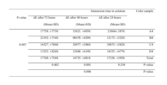
Table 6: Comparison of discoloration rate (ΔE) at different immersion times in the color groups studied
According to the results of Table 6, discoloration rates in the studied color samples (regardless of the type of solution and immersion time) were significantly different from each other (P=0.007).
Also, color change at different times of immersion (regardless of the type of solution and color samples studied) showed a significant difference (P=0.006).
The result of the statistical test of the color change of different color samples according to each of the studied times is as follows:
At 48 hours, the difference in color change was significant among the color samples (P=0.001), but at the other two times, the difference in color change was not significant among the color samples (P=0.254,0.402).
Table 7: Tukey’s statistical test results in the color groups studied (A4, B4, C4, and D4)
According to Table 7, the mean E∆ in the color groups studied was not significantly different.
To investigate the relationship between the amount of color change recorded by the spectrophotometer and that in the clinical environment, the information was converted to the National Bureau Standards (NBS) units using the following equation:
NBS unit = ΔE × 0.92.
According to the NBS units, the color changes are as follows:
0.0-0.5: Very slight changes
0.5-1.5: Slight changes
1.5-3.0: Noticeable changes
3.0-6.0: Appreciable changes
6.0-12.0: Very appreciable changes
12 or more: Conversion to another color.
In this study, color changes in the color groups were in the range of 0.5-1.5 i.e. slight changes.
Table 8: Mean (L a b) of the A4 color sample at baseline, 24, 48, and 72 hours
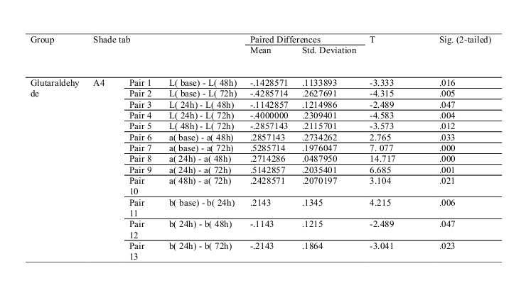
Table 9: Pairwise comparison of the mean (L a b) at different times for the A4 color sample (P<0.05) Pair 2 L( base) - L( 72h) -.4285714 .2627691 -4.315 .005
Pair 3 L( 24h) - L( 48h) -.1142857 .1214986 -2.489 .047
Pair 4 L( 24h) - L( 72h) -.4000000 .2309401 -4.583 .004
Pair 5 L( 48h) - L( 72h) -.2857143 .2115701 -3.573 .012
Pair 6 a( base) - a( 48h) .2857143 .2734262 2.765 .033
Pair 7 a( base) - a( 72h) .5285714 .1976047 7. 077 .000
Pair 8 a( 24h) - a( 48h) .2714286 .0487950 14.717 .000
Pair 9 a( 24h) - a( 72h) .5142857 .2035401 6.685 .001
Pair 10 a( 48h) - a( 72h) .2428571 .2070197 3.104 .021
Pair 11 b( base) - b( 24h) .2143 .1345 4.215 .006
Pair 12 b( 24h) - b( 48h) -.1143 .1215 -2.489 .047
Pair 13 b( 24h) - b( 72h) -.2143 .1864 -3.041 .023
The pairwise comparison of the mean L a b (Table 8) at different times for the A4 color sample is shown in Table 9.
The “L” index indicates the degree of brightness; the more positive the index, the brighter the color becomes, and if the index becomes negative, the darker will be the color. For the A4 color sample, the ΔL was in the positive direction in the case group, which indicates brightening. Also, for the A4 color sample, a significant difference was observed in years 2 and 3 compared to baseline, in years 2 and 3 compared to the first year, and in year 3 compared to the second year (P<0.05).
Component “a” (which indicates redness and greenness with positive changes towards redness and negative changes towards green) was negative for the A4 color sample, indicating greening of the samples. Also, there was a significant difference in component “a” in years 2 and 3 compared to baseline and in years 2 and 3 compared to the first year (P<0.05).
Component “b”, the positive of which indicates yellowing, and the negative of which indicates bluing of the sample, was positive for the A4 color sample, indicating the yellowing of the samples. Also, a significant difference was observed in component “b” in year 1 compared to baseline and in years 2 and 3 compared to the first year (P<0.05; Table 10).
Table 10: Mean (L a b) of the B4 color sample at baseline, 24, 48, and 72 hours
Discussion
Shade guides are mainly used to achieve a suitable color matching. To prevent the transmission of diseases and infectious agents, it is necessary to disinfect shade guides after each use. (2)
The 2005 OSHA guidelines for disinfection of shade guides have categorized them as semicritical items, which can be disinfected with an intermediate-level disinfectant approved by the Environmental Protection Agency (EPA). (1)
Some offices and clinics use autoclaves to disinfect shade guides, but the plastic tab holder cannot withstand the flow of autoclave vapors and will degrade over time. Also, the use of autoclaves in many offices is too time-consuming. (1)
Centers such as dental schools use sterilization (gas) to disinfect shade guides due to the volume of items and the availability of this sterilization method. Whereas in most offices, surface disinfectants are used between patients. (1)
The present study was performed to investigate the effect of glutaraldehyde disinfectant on the rate of the color change of the Vita classical shade guide when it is immersed in the solution at least 3 times a week for 10 minutes each time. (6) In this study, the simulation of use for one year was with 24 hours of immersion in the disinfectant while the simulation of use for 2 and 3 years was with 48 and 72 hours of immersion in the disinfectant. (6) The A4, B4, C4, and D4 colors were selected to cover all the different pigments (A: brown, B: yellow, C: gray, D: red-brown).
The shade pilot spectrophotometer was used to record the before and after values of (L a b) of the Vita classical shade guide in the middle third of the color sample. This device is a digital color analyzer spectrophotometer that is more accurate and reproducible compared to the human eye.
The results of this study showed that color change occurs in both glutaraldehyde and distilled water groups over time, but color changes are more significant in the glutaraldehyde group (P<0.006).
In similar studies, Hombesh et al, Brandt et al, Huang et al, and Öngül et al have also shown that color changes increase over time. (7-10)
In this study, it was found that the colors of the samples become brighter and the “L” component increases over time. These changes can be attributed to the solubility or abrasion of surface properties by glutaraldehyde and distilled water. (1) With glutaraldehyde, this color change and brightening occur more probably due to its unique chemical compounds.
The results of the present study showed that the mean ΔE of the color groups studied (A4, B4, C4, and D4) was less than one. The results of the amount of ΔE have been reported differently in different studies.
In studies by Hombesh et al, Brandt et al, and Öngül et al, disinfectants produced significant color changes (ΔE>1). (7,8,10). In the study by Huang et al, disinfectants produced minimal and minor changes (ΔE<1). (9)
According to the results, it is clear that the type of substance and its chemical composition has a significant effect on the rate of change of ΔE, L, a, and b. Disinfectants can change the parameters (L, a, b) through two mechanisms: abrasion of surface properties and residue deposition. (1)
Tsun et al examined the effect of 2% alkaline glutaraldehyde on porcelain samples and showed that this material could be used for a long time without significant discoloration of the samples. (4) However, in the present study, after 24 hours of immersion in glutaraldehyde solution, ΔE more than one was reported. The reason for this discrepancy can be attributed to the metal handle of the color sample and the effect of metal corrosion and deposition of corrosive materials on porcelain.
However, in the study conducted by Tsun et al on color changes of porcelain, glutaraldehyde was the only substance that could be used for long-term immersion (7 days) without significant color changes. (4)
According to the results of the current study, glutaraldehyde can be used to disinfect the Vita classical shade guide without worrying about significant color changes.
According to the results of this study, all the studied color samples (A4, B4, C4, and D4) showed increased “L” (brightening), decreased “a” (greening), and increased “b” (yellowing) during the time of immersion in glutaraldehyde. Changes of L, a, b in the control group of the A4, B4, C4, and D4 color samples were very insignificant and had a greater effect on decreasing component “a” and then on increasing component “L”.
In general, in both the case and control groups, the color changes were towards an increase in “L”, decrease in “a”, and increase in “b”, which in the case group (glutaraldehyde), these changes were more significant compared to the control group (distilled water; P<0.05).
The solution type (glutaraldehyde and distilled water) was effective in color change (ΔE) of the A4 and C4 color samples but not for the B4 and D4 samples (Tables 3 to 6).
These findings indicate that the behavior of color samples can defer with various disinfectants, which can be due to the type of pigment, their solubility, and the chemical properties of the compounds that produce these pigments.
However, despite the difference in the discoloration behavior of the samples, ΔE of all A4, B4, C4, and D4 color samples was less than one with glutaraldehyde disinfectant, indicating that these changes are small and imperceptible and cannot be seen with the naked eye.
One of the positive points of this research was that we removed the metal handle of the shade tabs to prevent surface or subsurface adsorption of corrosive products on porcelain (11) to check only the color change of the porcelain itself without the intervention of an interfering agent. However, these changes may become more clinically evident in the clinical setting and over time; this can be due to the abrasion of surface properties and deposition of surface residues. (1)
In this study, the shade pilot spectrophotometer was used, and only the color of the middle one-third area of the facial surface of the tooth was examined. This area is the best for choosing tooth color. (1)
How the manufacturer makes the shade guide can also affect its discoloration after disinfection. Shade guides may be made of several layers of compacted porcelain, or they may be porcelain samples whose surface is stained and glazed. Samples made of several layers of porcelain, such as Vitapan classical shade guide, are more resistant to discoloration. (1)
Conclusion
Based on the results of the present study, it can be concluded that:
1. If we follow the instructions provided by the manufacturers of shade guides, as well as the time and manner of use of disinfectants recommended by their manufacturer, there would be no noticeable (clinically visible) changes in ΔE (ΔE<1).
2. Color changes following immersion of A4, B4, C4, and D4 color samples in glutaraldehyde solution for 24, 48, and 72 hours show a significant difference at different times, i.e. time change is effective in changes of ΔE (P<0.05 and P=0.0001). Nevertheless, this color change is insignificant, meaning it causes small changes in ΔE that are not visually obvious to observers (ΔE<1).
3. The process of color change after 72 hours of immersion in glutaraldehyde is towards lightening, greening, and yellowing of color samples.
4. Color change (ΔE) of shade guides should be evaluated occasionally (a standard color sample should be set aside for comparison with the types used in the clinic).
5. The type of disinfectant and its interaction with the surface of color samples has a direct effect on the color change process.
Type of Study: Original article |
Subject:
Dental implant
References
1. Pohjola RM, Hackman ST, Browning WD. Evaluation of a standard shade guide for color change after disinfection. Quintessence Int. 2007 Sep;38(8):671-6.
2. Koosha S, Pourmahdi Borujeni M, Amraii H. Evaluation shade guide color changes after application of disinfectant materials. Jentashapir. 2012;3(4):505-11.
3. Browning WD. Use of shade guides for color measurement in tooth-bleaching studies. J Esthet Restor Dent. 2003;15 Suppl 1:S13-20. [DOI:10.1111/j.1708-8240.2003.tb00314.x] [PMID]
4. Ma T, Johnson GH, Gordon GE. Effects of chemical disinfectants on surface characteristics and color of three fixed prosthodontic crown materials. J Prosthet Dent. 1999 Nov;82(5):600-7. [DOI:10.1016/S0022-3913(99)70061-2]
5. Paul SJ, Peter A, Rodoni L, Pietrobon N. Conventional visual vs spectrophotometric shade taking for porcelain-fused-to-metal crowns: a clinical comparison. Int J Periodontics Restorative Dent. 2004 Jun;24(3):222-31.
6. Swepston JH, Miller AW 3rd. Esthetic matching. J Prosthet Dent. 1985 Nov;54(5):623-5. [DOI:10.1016/0022-3913(85)90234-3]
7. Hombesh MN, Praveen B, Sinha HV, Prasanna BG, Sachin B, Chandrashekar S. Two years survivability of VITA 3D master shade matching guides after disinfection with isopropyl alcohol: An in vitro study. J Conserv Dent. 2019 May-Jun;22(3):275-80. [DOI:10.4103/JCD.JCD_573_18] [PMID] [PMCID]
8. Brandt J, Nelson S, Lauer HC, von Hehn U, Brandt S. In vivo study for tooth colour determination-visual versus digital. Clin Oral Investig. 2017 Dec;21(9):2863-71. [DOI:10.1007/s00784-017-2088-0] [PMID]
9. Huang PY, Masri R, Romberg E, Driscoll CF. The effect of various disinfectants on dental shade guides. J Prosthet Dent. 2014 Sep;112(3):613-7. [DOI:10.1016/j.prosdent.2014.04.006] [PMID]
10. Öngül D, Şermet B, Balkaya MC. Visual and instrumental evaluation of color match ability of 2 shade guides on a ceramic system. J Prosthet Dent. 2012 Jul;108(1):9-14. [DOI:10.1016/S0022-3913(12)60102-4]
11. Mostafavi A, Koosha S, Amjad M. Effect of Disinfection on the Surface Roughness of Dental Casts Retrieved from Addition Silicone Impressions. J Res Dentomaxillofac Sci. 2018;3(1):27-33. [DOI:10.29252/jrdms.3.1.27]
12. Aguilar FG, Roberti Garcia Lda F, Cruvinel DR, Sousa AB, de Carvalho Panzeri Pires-de-Souza F. Color and opacity of composites protected with surface sealants and submitted to artificial accelerated aging. Eur J Dent. 2012 Jan;6(1):24-33.
Send email to the article author
| Rights and permissions | |
 |
This work is licensed under a Creative Commons Attribution-NonCommercial 4.0 International License. |
