


BibTeX | RIS | EndNote | Medlars | ProCite | Reference Manager | RefWorks
Send citation to:
URL: http://jrdms.dentaliau.ac.ir/article-1-201-en.html


2- Assistant professor, Oral and Maxillofacial Pathology Dept, Faculty of Dentistry,Tehran Medical Sciences
3- Assistant professor, Oral and Maxillofacial Surgery Dept, Faculty of Dentistry,Tehran Medical Sciences
4- Assistant professor, Pediatric Dentistry Dept, Faculty of Dentistry,Tehran Medical Sciences, Islamic Azad University, Tehran, Iran , tavasolisara@yahoo.com
5- Associate professor, Laser Research Center of Dentistry, Department of Pediatric Dentistry
Abstract
Background: Radicular cysts are the most frequent odontogenic cysts of the jaws. Radicular cysts arising from the primary teeth are very rare and comprise 0.5% to 3.3% of radicular cysts in both primary and permanent dentitions.
Case Presentation: In this paper, we report the clinical, radiographical, and histological characteristics of a radicular cyst associated with a maxillary deciduous first molar. The treatment plan included extraction and enucleation of the cyst under local anesthesia after the elevation of a mucoperiosteal flap, which led to satisfactory and uneventful healing.
Conclusion: Early diagnosis of radicular cysts associated with the primary dentition allows for a less aggressive treatment plan and prevents adverse effects on the permanent successor.
Keywords: Radicular Cyst, Primary Molar, Deciduous, Periapical Cyst
Introduction
Periapical cysts, which are associated with non-vital teeth, are the most common inflammatory odontogenic cysts. The associated tooth may be deeply carious, traumatized, or improperly restored. (1) The usual cause of periapical cysts is an infected tooth with a necrotic pulp. Toxins that exit from the apex of the tooth cause periapical inflammation, which stimulates the epithelial rests of Malassez in the apical periodontal ligament (PDL) and causes the formation of a periapical granuloma. Finally, this epithelium undergoes necrosis due to the lack of blood supply, and the granuloma turns into a cyst. (2) Periapical cysts comprise about 52.3% of all jaw cysts. (3) Periapical cysts are rarely observed in the deciduous dentition. Based on the estimations, only 0.5-3.3% of radicular cysts involve the deciduous dentition and the majority of them are associated with mandibular molars. It has been observed that cysts associated with the primary teeth are present in the interradicular region not close to the apex of the teeth; therefore, the term ‘periradicular cyst’ is more appropriate. (1) These lesions can lead to bony expansion and resorption. Furthermore, radicular cysts may also lead to displacement and damage to the developing permanent dentition. (4) It seems that the most applicable treatment option is complete enucleation of the cyst with the extraction of the associated primary teeth and preservation of the permanent teeth, after which the permanent teeth spontaneously align in their normal position. (5) Some periapical cysts have been reported in association with the primary teeth but those arising from untreated maxillary primary teeth are very rare. (5,6)
This case report aimed to present a periapical cyst associated with an untreated carious maxillary primary molar that can be confused with a dentigerous cyst.
Case presentation
A healthy eight-year-old girl was referred to the Department of Pediatric Dentistry of the Dental Faculty of Islamic Azad University of Medical Sciences, Tehran, Iran, with the chief complaint of a painless swelling located at the maxillary right buccal region (Figure 1).
Figure 1. A painless swelling at the maxillary right buccal region
The extraoral examination indicated a painless bony hard swelling on the upper right side of the maxilla. According to the intraoral examination, a well-circumscribed swelling was noticed in the right upper buccal sulcus, extending from the distal aspect of the primary canine to the mesial aspect of the second primary molar. The size of the circumscribed swelling was approximately 2×1 cm2. The periapical radiograph revealed a well-defined unilocular radiolucency involving the periradicular area of the primary canine and the first primary molar as well as the developing first premolar (Figure 2).
Figure 2. A well-defined unilocular radiolucency involving the periradicular area of the primary canine and the first primary molar as well as the developing first premolar
The occlusal radiograph indicated that the first bicuspid was buccally displaced because of the expansion of the lesion (Figure 3).
Figure 3. Occlusal radiograph indicating the buccal displacement of the first bicuspid
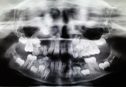
A round radiolucent unilocular lesion with smooth and well-defined borders in the periapical area of the maxillary right primary canine and first primary molar was observed in the panoramic radiograph (Figure 4).
Figure 4. Panoramic radiograph showing a round radiolucent unilocular lesion with smooth and well-defined borders in the periapical area of the maxillary right primary canine and first primary molar
A needle biopsy was taken from the lesion, and the result of aspiration was a light yellow odorless liquid. According to the patient’s records and clinical and radiographical examinations, the differential diagnoses of the lesion included radicular cyst and dentigerous cyst. Surgical enucleation of the lesion was the treatment plan for the case. The patient was scheduled for the surgery after the patient’s parents signed the consent form. The standard sterilization and disinfection procedures were followed, and the area was anesthetized by infraorbital block and infiltration injections of 2% lidocaine hydrochloride with epinephrine 1:100000. A crevicular incision was made from the distal surface of the maxillary lateral incisor to the mesial surface of the first molar. The cystic site was exposed after mucoperiosteal flap elevation, which revealed expansion, thinning, and perforation of the buccal cortical plate (Figure 5).
Figure 5. The cystic site was exposed after the elevation of the mucoperiosteal flap
The primary canine and the roots of the primary molars were extracted, and the cyst was enucleated along with the developing first premolar bud (Figure 6).
Figure 6. The primary canine and the roots of the primary molars were extracted
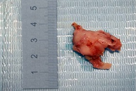
A decision was made by the surgeon and the pediatric dentist during surgery to extract the impacted first premolar because it was involved with cystic lining with a very poor prognosis. The flaps were repositioned and sutured. Then, the tissue specimens were sent for histopathological examinations (Figure 7).
Figure 7. The enucleated cyst
The primary closure was done following debridement with normal saline and hemostasis.
The patient was medicated with 200mg Ibuprofen every six hours until pain relief and with 250 mg/5ml Amoxicillin every eight hours for a week.
Seven days later, the patient referred for post-surgical examination and suture removal. Post-surgical healing was uneventful.
The histopathologic examination revealed a lesion composed of fibrous connective tissue infiltrated in several parts by chronic inflammatory cells. The cystic cavity was lined with a thin stratified squamous epithelium. There were no signs of malignancy (Figure 8).
Figure 8. The epithelial lining and cystic structure as well as chronic infiltration of lymphocytes and plasma cells
The case was diagnosed with a radicular cyst, and the patient was scheduled for the remaining required dental treatments. The patient also was referred for an orthodontic evaluation. At the 20-month recall session, the radiographical and clinical evaluations proved a successful treatment (Figure 9).
Figure 9. The 20-month recall panoramic radiograph showing a successful treatment outcome
Discussion
Periapical cysts are the most widely recognized inflammatory odontogenic cysts that associate with non-vital teeth. Periapical cysts originating from the primary teeth involve the mixed dentition with a rate of 0.5-3.3%. (1) The majority of periapical cysts are symptomless and are found during routine radiographical examinations. (1) Very few cases of periapical cysts are found in the first decade after which there is a steep rise with a peak occurrence in the third decade. Nagata et al reported 112 cases of periapical cysts in 2004. (7) The most commonly involved primary teeth were the mandibular molars (67%), maxillary molars (17%), maxillary anterior teeth (13%), and mandibular anterior teeth (3%). (7) Periapical cysts are frequently detected in the mandibular primary dentition because these teeth are frequently affected by caries, which is the most well-known etiological factor for the development of these cysts. In the permanent teeth, the maxillary incisors are mostly involved because of trauma, caries, and old silicate restorations. (3)
Periapical radiolucencies associated with the primary teeth may be mistaken for a periapical granuloma or a dentigerous cyst associated with the corresponding permanent tooth. Dentigerous cysts are characterized by a well-defined unilocular radiolucency in the pericoronal region of an unerupted permanent tooth. Their cortical margins are continuous with the follicle at the cementoenamel junction (CEJ) of the permanent tooth. (8) Pulpal and periapical infections in the primary dentition drain more quickly compared to the permanent dentition; the antigenic stimuli, which elicit the changes leading to the development of periapical cysts, may be distinctive. (9,10) Also, these lesions tend to obviate on their own following the extraction or exfoliation of the related tooth and are usually not submitted for histopathological investigation. (11) Histologically, there is no distinction between cysts associated with the primary teeth and those of the permanent teeth except for the scarcity of cholesterol crystal slits in cysts associated with the primary teeth. This is because the lesion associated with the primary teeth is of a shorter duration before removal compared to the cysts associated with the permanent teeth. (3)
In the present case, the treatment plan comprised the extraction of the involved primary tooth followed by the enucleation of the cyst. The other conservative and effective alternative treatment option is marsupialization of the lesion with a fixed resin tube placed in the alveolar socket after the extraction of the involved deciduous tooth. (12) In a similar case, the treatment included extraction of the associated primary teeth followed by marsupialization. A removable appliance with a resin extension into the cystic cavity was used to decompress the lesion. This treatment permitted fast healing of the lesion and eruption of the permanent tooth. (13) The consequences of an untreated or undiagnosed periapical cyst could jeopardize the patient’s dental development. (13) An untreated periapical cyst causes swelling, tenderness, tooth mobility, and a bluish color after cortical plate expansion. (14) Moreover, the successor tooth may be displaced or lose its vitality. However, early diagnosis through regular radiographical monitoring of these teeth may prove profitable in preventing the above-mentioned issues and avoiding aggressive surgeries. (14,15) Postsurgical osseous defects heal properly in children as they have a high natural tendency for bone regeneration; this makes post-surgical healing uncomplicated. (11,15,16)
Conclusion
This article introduced an uncommon case of a periapical cyst associated with primary maxillary molars. Diagnosis of the sequelae of an untreated or undiscovered periapical cyst is vital for preventing adverse effects on the permanent successor and the need for invasive surgical treatment. Moreover, regular clinical and radiographical follow-ups for pulp-treated primary teeth are highly recommended.
| Rights and permissions | |
 |
This work is licensed under a Creative Commons Attribution-NonCommercial 4.0 International License. |
