

Volume 1, Issue 4 (12-2016)
J Res Dent Maxillofac Sci 2016, 1(4): 32-38 |
Back to browse issues page


Download citation:
BibTeX | RIS | EndNote | Medlars | ProCite | Reference Manager | RefWorks
Send citation to:


BibTeX | RIS | EndNote | Medlars | ProCite | Reference Manager | RefWorks
Send citation to:
Talayi Pour A, Hafezi L, Yarahmadi A, Ghaznavi A, Iranparvar A, Sahabi L. Comparison of the Diagnostic Accuracy of Digital Intraoral Radiography with PSP and CBCT in the Detection of Horizontal and Vertical Dental Root Fractures. J Res Dent Maxillofac Sci 2016; 1 (4) :32-38
URL: http://jrdms.dentaliau.ac.ir/article-1-132-en.html
URL: http://jrdms.dentaliau.ac.ir/article-1-132-en.html


1- professor,Oral and maxilofacial Radiology Dept , Dental Branch, Ialamic azad university tehran,Iran
2- Associated professor, Oral and maxilofacial Radiology Dept , Dental Branch, Ialamic azad university tehran,Iran
3- Postgraduate Student, Oral and maxilofacial Radiology Dept , Dental Branch, Ialamic azad university tehran,Iran , dr.yarahmadi169@yahoo .com
4- Assistant professor, Oral and maxilofacial Radiology Dept, oromeyeh dental branch university
5- Dentist
Keywords: Intraoral digital radiography, Photostimulable Phosphor Plate, Cone Beam Computed Tomography, Horizontal root fracture, Vertical root fracture
Full-Text [PDF 329 kb]
(1648 Downloads)
| Abstract (HTML) (6196 Views)
Abstract
Background and Aim: Clinical and radiographic diagnoses of dental root fractures have always been difficult and require high accuracy in dental care and treatment. The aim of this study was to compare the diagnostic accuracy of intraoral digital radiography (PSP) and CBCT in the detection of horizontal and vertical root fractures (HRF and VRF).
Materials and Methods: For this experimental study, 60 human mandibular teeth (30 anterior and 30 posterior multi-rooted teeth) were selected. Thirty randomly-selected teeth were fractured horizontally while the next 30 randomly-selected teeth were fractured vertically by use of a hammer and then the pieces were glued back together and were placed in a sheep mandible. Radiographic images of all the teeth were taken using intraoral digital radiography (PSP) and CBCT methods. Afterwards, two oral and maxillofacial radiologists assessed the images separately. The data were subjected to diagnostic analytic tests.
Results: There were significant differences in specificity, sensitivity, positive predictive value and negative predictive value between digital intraoral radiography (PSP) and CBCT in the detection of HRF and VRF. Kappa value for inter-observer and intra-observer agreement in VRF equaled 73.3% for CBCT and 54.2% for PSP, while in HRF it equaled 63.3% for CBCT and 55.4% for PSP.
Conclusion: CBCT method has higher specificity and sensitivity in the detection of HRF and VRF compared with intraoral digital radiography.
Keywords: Intraoral digital radiography, Photostimulable Phosphor Plate, Cone Beam Computed Tomography, Horizontal root fracture, Vertical root fracture
Introduction
Clinical and radiographic diagnoses of dental root fractures have always been difficult. Radiographic diagnosis of dental root fracture requires high accuracy in dental care and treatments. Overall, dental root fractures comprise 0.5 to 7% of the injuries that inflict the permanent dentition. (1) Horizontal and vertical root fractures (HRF and VRF) are difficult to detect due to the challenges in diagnosis and tracing on intraoral radiographs, except when definite clinical findings exist. This difficulty in diagnosis leads to unnecessary tooth extractions, poor long-term prognosis and extensive bone loss. (1, 2) Conventional and digital intraoral radiography are the most common techniques for tracing dental root fractures. Root fractures can also be assessed by routine dental examinations. (1,3)
Dental root fractures have become detectable since two-dimensional radiography was implemented in dentistry in 1896. (4,5) Nowadays, intraoral radiography with Photostimulable Phosphor Plate (PSP) is being used for detection of root fracture as a digital intraoral radiography method. (6) Conventional two-dimensional radiography can be completed with a third view (the orofacial view). Cone Beam Computed Tomography (CBCT) was introduced in dento-alveolar imaging in 1998. (7) CBCT has been implemented as a valuable imaging modality in different dentistry fields such as surgery and orthodontics. Nevertheless, the advantages and limitations of CBCT in dental traumatology, especially in the diagnosis of teeth with fractured roots have remained indefinite.(8)
The present study aimed to compare the diagnostic accuracy of digital intraoral radiography (two-dimensional) and CBCT (three-dimensional) in the detection of the presence or absence of HRF and VRF at the radiology department of the dental school of Islamic Azad University of Tehran during 2013-2014.
Materials and methods
In this in vitro diagnostic study, 60 anterior and multi-rooted posterior teeth without fractures, cracks or root fillings were selected and coded. Dental sockets were formed with a bur in a sheep mandible to hold and stabilize the teeth. Thirty randomly-selected teeth were fractured horizontally, while the next 30 randomly- selected teeth were fractured vertically by a hammer, and then the pieces were glued back together and the teeth were placed inside the dental sockets. Afterwards, the mandible was radiographed with PSP (using parallel method with the aid of a film holder and using DIGORA OPTIME device (Soredex, Helsinki, Finland) with radiographic exposure parameters of t=0.1 s, kVp=70, mA=8 and resolution= 3/14 lp/mm) and the digital images were saved. Afterwards, CBCT images were obtained from the mandible in axial and coronal views using NEWTOM VGI CBCT (QR SRL Company, Verona, Italy) with an 8*8 cm field of view in high resolution mode and the images were saved.(Fig1,2)
Fig 1-Digital image and CBCTimag coronal view
Fig 2- Digital image and CBCTimag Axial view
The images were evaluated separately by two oral and maxillofacial radiologists. Each observer evaluated the images of each fracture type obtained by each method independently in a time interval separated by three weeks (intra-observer and inter-observer reliability). The results were compared regarding the reliability and were registered in designated lists. The data were statistically analyzed by use of statistical indices (specificity, sensitivity, positive predictive value, negative predictive value, and kappa coefficient) using SPSS software version 22 (SPSS Inc., Chicago IL, USA).
Results
After analyzing 120 radiographic images obtained by digital intraoral radiography (PSP) and CBCT, the following results were achieved:
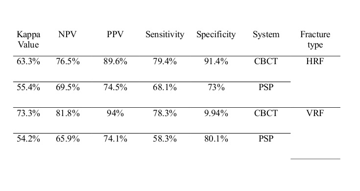
The results are presented in tables 1 to 4. According to tables 1 and 2, CBCT has the highest sensitivity and specificity in the diagnosis of HRF and VRF. CBCT has higher sensitivity (79.4%) for detection of HRF compared with digital intraoral radiography with PSP (68.1%). Also, CBCT has higher specificity (91.4%) in the detection of intact samples compared with digital intraoral radiography with PSP (73%). (Table 3) CBCT has higher sensitivity (78.3%) in the detection of VRF compared with digital intraoral radiography with PSP (58.3%). CBCT has higher specificity (94.9%) in the detection of intact samples compared with digital intraoral radiography with PSP (80.1%). (Table 3)
Positive predictive value (PPV) is significantly higher in CBCT compared with digital intraoral radiography with PSP in the detection of HRF. This means that the observers had 89.6% correct diagnoses of HRF with CBCT system. While PPV equaled 74.5% in PSP system, which means that the observers had 74.5% cases of correct diagnoses of HRF with PSP system. Negative predictive value (NPV) (correct diagnosis of intact samples) equaled 76.5% in CBCT and 69.5% in intraoral radiography with PSP. (Table 3)
Likewise, PPV is significantly higher in CBCT compared with digital intraoral radiography with PSP in the detection of VRF, while PPV equaled 74.1% in PSP system. NPV equaled 81.8% in CBCT and 65.9% in digital intraoral radiography with PSP. (Table 3)
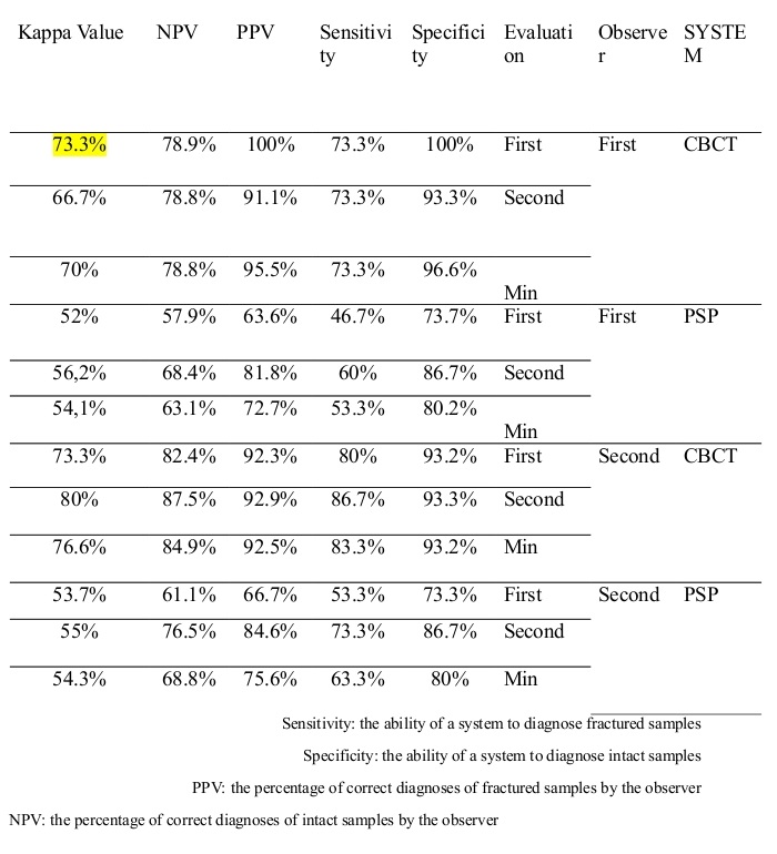
Intra-observer and inter-observer agreement between the two observers in the detection of HRF were higher in CBCT compared with digital radiography with PSP. Kappa value for inter-observer and intra-observer agreement equaled 63.3% for CBCT and equaled 55.4% for PSP. (Table 4)
Likewise, Intra-observer and inter-observer agreement between the two observers in the detection of VRF were higher in CBCT compared with digital intraoral radiography with PSP. Kappa value for inter-observer and intra-observer agreement equaled 73.3% for CBCT and equaled 54.2% for PSP. (Table 4)
Table 1- Diagnostic results regarding HRF based on the evaluated radiographic modality
Table 2- Diagnostic results regarding VRF based on the evaluated radiographic modality
Table 3- Diagnostic results of the two observers regarding HRF and VRF based on the evaluated radiographic modality
Table 4- Results of inter-observer and intra-observer agreement regarding HRF and VRF based on the evaluated radiographic modality
Discussion
HRF and VRF have always been difficult to diagnose and trace on intraoral radiographs. This difficulty in diagnosis leads to unnecessary tooth extractions, poor long-term prognosis and extensive bone loss. (1,2)
In this in vitro study, the diagnostic accuracy of digital intraoral radiography with PSP and CBCT in the detection of HRF and VRF was assessed.
60 human mandibular anterior and multi-rooted posterior teeth were selected for this study. A sheep mandible was used for stabilizing the teeth. Dental roots were fractured randomly by use of a hammer, (similar to the method used by Avsever et al and da Silveira et al) (9,10) and then the fractured pieces were glued back together and were placed inside the dental sockets. Afterwards, the mandible was radiographed with PSP and CBCT similar to the method used by Kambungton et al. (11)
The results showed that CBCT has higher diagnostic accuracy than digital intraoral radiography with PSP in the detection of HRF and VRF. Moreover, inter-observer and intra-observer agreement between the observers were higher in CBCT system compared to digital intraoral radiography with PSP. Considering the results, the accuracy of CBCT was higher in the detection of intact and fractured samples, which can be attributed to three-dimensional evaluation of the presence or absence of fracture line in different sections of the teeth. Also, many radiographic errors of two-dimensional radiography techniques (like digital intraoral radiography) such as overlapping of adjacent teeth and superimposition of different structures are absent in CBCT method due to the ability of visualization of each tooth at different angles and views.
Bornstein et al reported that the diagnosis of the location and angle of root fracture is significantly different in CBCT method compared with the diagnoses based merely on intraoral radiography. (8) This finding is in line with the results of the present study.
Kambungton et al showed that no statistically significant differences exist among CBCT, digital and conventional intraoral radiography in the detection of VRF in single-rooted mandibular teeth. (11) This difference with our results can be attributed to the use of various types of teeth (anterior and multi-rooted posterior teeth) in the present study.
Wang et al concluded that CBCT is significantly more accurate and valid in the diagnosis of root fractures in comparison with intraoral radiography, (12) which is in line with the results of the present research.
Avsever et al showed that the diagnostic accuracy of CBCT is significantly higher for tracing HRF compared with digital intraoral radiography with CCD and stated that CBCT should be selected as the most reliable imaging modality in diagnosis of HRF. (9) This finding is in accordance with the results of our study.
A report by Brady et al showed that periapical radiography and CBCT were unreliable in the detection of partial fractures and that the width of the fracture can influence the accuracy of CBCT. Also, they found that complete fractures can be traced more easily than partial fractures by all radiographic modalities. (13) In the present study, which only evaluated complete fracture lines, specificity and PPV of CBCT was extremely high and reliable in tracing the mentioned fractures.
Edlund et al reported PPV of 92%, sensitivity of 88% and specificity of 95% for CBCT and stated that CBCT has high accuracy in the detection of VRF, (14) which confirms the results of the present study.
After the evaluation of the conventional intraoral radiographs and CBCT images, da Silveira et al stated that the results of the diagnostic tests have shown similar capability of the detection of VRF for periapical radiography and CBCT scans in non-endodontically treated teeth and after metallic post insertion, and they also stated that radiographic evaluation at multiple horizontal angles should be considered as the first complementary modality in the detection of VRF. They concluded that when conventional radiography is incapable of rendering adequate information, CBCT can be used. (10) This finding contradicts our results. The difference can be attributed to the use of single-rooted teeth and multiple exposures at different horizontal angles in the mentioned study.
Considering the expenses and facilities, when definite clinical signs of root fracture are present, digital intraoral radiography can be used as the first step but if CBCT is available, it is strongly recommended to use this imaging modality as the first evaluative step.
Conclusion
CBCT has higher sensitivity and specificity in the detection of horizontal and vertical root fractures compared with digital intraoral radiography.
Full-Text: (810 Views)
Abstract
Background and Aim: Clinical and radiographic diagnoses of dental root fractures have always been difficult and require high accuracy in dental care and treatment. The aim of this study was to compare the diagnostic accuracy of intraoral digital radiography (PSP) and CBCT in the detection of horizontal and vertical root fractures (HRF and VRF).
Materials and Methods: For this experimental study, 60 human mandibular teeth (30 anterior and 30 posterior multi-rooted teeth) were selected. Thirty randomly-selected teeth were fractured horizontally while the next 30 randomly-selected teeth were fractured vertically by use of a hammer and then the pieces were glued back together and were placed in a sheep mandible. Radiographic images of all the teeth were taken using intraoral digital radiography (PSP) and CBCT methods. Afterwards, two oral and maxillofacial radiologists assessed the images separately. The data were subjected to diagnostic analytic tests.
Results: There were significant differences in specificity, sensitivity, positive predictive value and negative predictive value between digital intraoral radiography (PSP) and CBCT in the detection of HRF and VRF. Kappa value for inter-observer and intra-observer agreement in VRF equaled 73.3% for CBCT and 54.2% for PSP, while in HRF it equaled 63.3% for CBCT and 55.4% for PSP.
Conclusion: CBCT method has higher specificity and sensitivity in the detection of HRF and VRF compared with intraoral digital radiography.
Keywords: Intraoral digital radiography, Photostimulable Phosphor Plate, Cone Beam Computed Tomography, Horizontal root fracture, Vertical root fracture
Introduction
Clinical and radiographic diagnoses of dental root fractures have always been difficult. Radiographic diagnosis of dental root fracture requires high accuracy in dental care and treatments. Overall, dental root fractures comprise 0.5 to 7% of the injuries that inflict the permanent dentition. (1) Horizontal and vertical root fractures (HRF and VRF) are difficult to detect due to the challenges in diagnosis and tracing on intraoral radiographs, except when definite clinical findings exist. This difficulty in diagnosis leads to unnecessary tooth extractions, poor long-term prognosis and extensive bone loss. (1, 2) Conventional and digital intraoral radiography are the most common techniques for tracing dental root fractures. Root fractures can also be assessed by routine dental examinations. (1,3)
Dental root fractures have become detectable since two-dimensional radiography was implemented in dentistry in 1896. (4,5) Nowadays, intraoral radiography with Photostimulable Phosphor Plate (PSP) is being used for detection of root fracture as a digital intraoral radiography method. (6) Conventional two-dimensional radiography can be completed with a third view (the orofacial view). Cone Beam Computed Tomography (CBCT) was introduced in dento-alveolar imaging in 1998. (7) CBCT has been implemented as a valuable imaging modality in different dentistry fields such as surgery and orthodontics. Nevertheless, the advantages and limitations of CBCT in dental traumatology, especially in the diagnosis of teeth with fractured roots have remained indefinite.(8)
The present study aimed to compare the diagnostic accuracy of digital intraoral radiography (two-dimensional) and CBCT (three-dimensional) in the detection of the presence or absence of HRF and VRF at the radiology department of the dental school of Islamic Azad University of Tehran during 2013-2014.
Materials and methods
In this in vitro diagnostic study, 60 anterior and multi-rooted posterior teeth without fractures, cracks or root fillings were selected and coded. Dental sockets were formed with a bur in a sheep mandible to hold and stabilize the teeth. Thirty randomly-selected teeth were fractured horizontally, while the next 30 randomly- selected teeth were fractured vertically by a hammer, and then the pieces were glued back together and the teeth were placed inside the dental sockets. Afterwards, the mandible was radiographed with PSP (using parallel method with the aid of a film holder and using DIGORA OPTIME device (Soredex, Helsinki, Finland) with radiographic exposure parameters of t=0.1 s, kVp=70, mA=8 and resolution= 3/14 lp/mm) and the digital images were saved. Afterwards, CBCT images were obtained from the mandible in axial and coronal views using NEWTOM VGI CBCT (QR SRL Company, Verona, Italy) with an 8*8 cm field of view in high resolution mode and the images were saved.(Fig1,2)
Fig 1-Digital image and CBCTimag coronal view
Fig 2- Digital image and CBCTimag Axial view
The images were evaluated separately by two oral and maxillofacial radiologists. Each observer evaluated the images of each fracture type obtained by each method independently in a time interval separated by three weeks (intra-observer and inter-observer reliability). The results were compared regarding the reliability and were registered in designated lists. The data were statistically analyzed by use of statistical indices (specificity, sensitivity, positive predictive value, negative predictive value, and kappa coefficient) using SPSS software version 22 (SPSS Inc., Chicago IL, USA).
Results
After analyzing 120 radiographic images obtained by digital intraoral radiography (PSP) and CBCT, the following results were achieved:
The results are presented in tables 1 to 4. According to tables 1 and 2, CBCT has the highest sensitivity and specificity in the diagnosis of HRF and VRF. CBCT has higher sensitivity (79.4%) for detection of HRF compared with digital intraoral radiography with PSP (68.1%). Also, CBCT has higher specificity (91.4%) in the detection of intact samples compared with digital intraoral radiography with PSP (73%). (Table 3) CBCT has higher sensitivity (78.3%) in the detection of VRF compared with digital intraoral radiography with PSP (58.3%). CBCT has higher specificity (94.9%) in the detection of intact samples compared with digital intraoral radiography with PSP (80.1%). (Table 3)
Positive predictive value (PPV) is significantly higher in CBCT compared with digital intraoral radiography with PSP in the detection of HRF. This means that the observers had 89.6% correct diagnoses of HRF with CBCT system. While PPV equaled 74.5% in PSP system, which means that the observers had 74.5% cases of correct diagnoses of HRF with PSP system. Negative predictive value (NPV) (correct diagnosis of intact samples) equaled 76.5% in CBCT and 69.5% in intraoral radiography with PSP. (Table 3)
Likewise, PPV is significantly higher in CBCT compared with digital intraoral radiography with PSP in the detection of VRF, while PPV equaled 74.1% in PSP system. NPV equaled 81.8% in CBCT and 65.9% in digital intraoral radiography with PSP. (Table 3)
Intra-observer and inter-observer agreement between the two observers in the detection of HRF were higher in CBCT compared with digital radiography with PSP. Kappa value for inter-observer and intra-observer agreement equaled 63.3% for CBCT and equaled 55.4% for PSP. (Table 4)
Likewise, Intra-observer and inter-observer agreement between the two observers in the detection of VRF were higher in CBCT compared with digital intraoral radiography with PSP. Kappa value for inter-observer and intra-observer agreement equaled 73.3% for CBCT and equaled 54.2% for PSP. (Table 4)
Table 1- Diagnostic results regarding HRF based on the evaluated radiographic modality
Table 2- Diagnostic results regarding VRF based on the evaluated radiographic modality
Table 3- Diagnostic results of the two observers regarding HRF and VRF based on the evaluated radiographic modality
Table 4- Results of inter-observer and intra-observer agreement regarding HRF and VRF based on the evaluated radiographic modality
Discussion
HRF and VRF have always been difficult to diagnose and trace on intraoral radiographs. This difficulty in diagnosis leads to unnecessary tooth extractions, poor long-term prognosis and extensive bone loss. (1,2)
In this in vitro study, the diagnostic accuracy of digital intraoral radiography with PSP and CBCT in the detection of HRF and VRF was assessed.
60 human mandibular anterior and multi-rooted posterior teeth were selected for this study. A sheep mandible was used for stabilizing the teeth. Dental roots were fractured randomly by use of a hammer, (similar to the method used by Avsever et al and da Silveira et al) (9,10) and then the fractured pieces were glued back together and were placed inside the dental sockets. Afterwards, the mandible was radiographed with PSP and CBCT similar to the method used by Kambungton et al. (11)
The results showed that CBCT has higher diagnostic accuracy than digital intraoral radiography with PSP in the detection of HRF and VRF. Moreover, inter-observer and intra-observer agreement between the observers were higher in CBCT system compared to digital intraoral radiography with PSP. Considering the results, the accuracy of CBCT was higher in the detection of intact and fractured samples, which can be attributed to three-dimensional evaluation of the presence or absence of fracture line in different sections of the teeth. Also, many radiographic errors of two-dimensional radiography techniques (like digital intraoral radiography) such as overlapping of adjacent teeth and superimposition of different structures are absent in CBCT method due to the ability of visualization of each tooth at different angles and views.
Bornstein et al reported that the diagnosis of the location and angle of root fracture is significantly different in CBCT method compared with the diagnoses based merely on intraoral radiography. (8) This finding is in line with the results of the present study.
Kambungton et al showed that no statistically significant differences exist among CBCT, digital and conventional intraoral radiography in the detection of VRF in single-rooted mandibular teeth. (11) This difference with our results can be attributed to the use of various types of teeth (anterior and multi-rooted posterior teeth) in the present study.
Wang et al concluded that CBCT is significantly more accurate and valid in the diagnosis of root fractures in comparison with intraoral radiography, (12) which is in line with the results of the present research.
Avsever et al showed that the diagnostic accuracy of CBCT is significantly higher for tracing HRF compared with digital intraoral radiography with CCD and stated that CBCT should be selected as the most reliable imaging modality in diagnosis of HRF. (9) This finding is in accordance with the results of our study.
A report by Brady et al showed that periapical radiography and CBCT were unreliable in the detection of partial fractures and that the width of the fracture can influence the accuracy of CBCT. Also, they found that complete fractures can be traced more easily than partial fractures by all radiographic modalities. (13) In the present study, which only evaluated complete fracture lines, specificity and PPV of CBCT was extremely high and reliable in tracing the mentioned fractures.
Edlund et al reported PPV of 92%, sensitivity of 88% and specificity of 95% for CBCT and stated that CBCT has high accuracy in the detection of VRF, (14) which confirms the results of the present study.
After the evaluation of the conventional intraoral radiographs and CBCT images, da Silveira et al stated that the results of the diagnostic tests have shown similar capability of the detection of VRF for periapical radiography and CBCT scans in non-endodontically treated teeth and after metallic post insertion, and they also stated that radiographic evaluation at multiple horizontal angles should be considered as the first complementary modality in the detection of VRF. They concluded that when conventional radiography is incapable of rendering adequate information, CBCT can be used. (10) This finding contradicts our results. The difference can be attributed to the use of single-rooted teeth and multiple exposures at different horizontal angles in the mentioned study.
Considering the expenses and facilities, when definite clinical signs of root fracture are present, digital intraoral radiography can be used as the first step but if CBCT is available, it is strongly recommended to use this imaging modality as the first evaluative step.
Conclusion
CBCT has higher sensitivity and specificity in the detection of horizontal and vertical root fractures compared with digital intraoral radiography.
Type of Study: Original article |
Subject:
Radiology
References
1. Martins JNR, Canta JP, Coelho A, Baharestani M. Vertical root fracture diagnosis of crowned premolars with root canal treatment – Two case reports. Rev Port Estomatol Med Dent Cir Maxilofac 2014;55(1):60–64.
2. Edlund M, Nair MK, Nair UP. Detection of vertical root fractures by using cone- beam computed tomography: A clinical study. J Endod 2011;37(6):768-72.
3. Farman AG, Farman TT. A comparison of 18 different x-ray detectors currently used in dentistry. Oral Surg Oral Med Oral Pathol Oral Radiol Endod 2005;99(4):485-9.
4. Dehghani M, Montazer lotf elahi H, Moeini M, Bardal R. Comparing the
5. accuracy of cone beam computed tomography, digital intraoral radiography and conventional intraoral radiography in the measurement of periodontal bone defects . J Res Dentomaxillofac Sci 2016;1(1):34-39.
6. De Vos W, Casselman J, Swennen GR. Cone beam computerized tomography (CBCT) imaging of the oral and maxillofacial region: A systematic review of the literature. Int J Oral Maxillofac Surg 2009;38(6):609-25.
7. Lofthag-Hansen S. Cone beam computed tomography radiation dose and image quality assessments. Swed Dent J Suppl 2010;(209):4-55.
8. Mehralizade S, Nemati Anaraki S, Sakhdari S, Miraba S, Amiri Siavashani M Bayat S. Comparing the diagnostic accuracy of two different resolution radiographs captured with PSP digital intraoral receptors in detection of secondary caries (In Vitro). J Res Dentomaxillofac Sci 2016;1(1):40-4
9. Alamri HM, Sadrameli M, Alshalhoob MA, Sadrameli M, Alshehri MA. Applications of CBCT in dental practice: A review of the literature. Gen Dent 2012;60(5):390-400.
10. Bornstein MM, Wölner-Hanssen AB, Sendi P, von Arx T. Comparison of intra oral radiography and limited cone beam computed tomography for the assessment of root-fractured permanent teeth. Dent Traumatol 2009;25(6):571-7.
11. Avsever H, Gunduz K, Orhan K, Uzun I, Ozmen B, Egrioglu E, Midilli M. Comparison of Intraoral radiography and cone -beam computed tomography for the detection of horizontal root fractures: an in vitro study. Clin Oral Investig 2014;18(1):285-92.
12. da Silveira PF, Vizzotto MB, Liedke GS, da Silveira HL, Montagner F, da Silveira HE. Detection of vertical root fractures by conventional radiographic examination and cone beam computed tomography- an in vitro analysis. Dent Traumatol 2013;29(1):41-6.
13. Kambungton J, Janhom A, Prapayasatok S, Pongsiriwet S. Assessment of vertical root fractures using three imaging modalities: cone beam CT, intraoral digital radiography and film. Dentomaxillofac Radiol 2012;41(2):91-5.
14. Wang P, Yan XB, Lui DG, Zhang WL, Zhang Y, Ma XC. Detection of dental root fractures by using cone-beam computed tomography. Dentomaxillofac Radiol 2011;40(5):290-8.
15. Brady E, Mannocci F, Brown J, Wilson R, Patel S. A comparison of cone beam computed tomography and periapical radiography for the detection of vertical root fractures in nonendodontically treated teeth. Int Endod J 2014;47(8):735-46.
16. Edlund M, Nair MK, Nair UP. Detection of vertical root fractures by using cone-beam computed tomography: a clinical study. J Endod 2011;37(6):768-72.
17. Bechara B, McMahan CA, Noujeim M, Faddoul T, Moore WS, Teixeira FB,et al. Comparison of cone beam CT scans with enhanced photostimulated phosphor plate images in the detection of root fracture of endodontically treated teeth. Dentomaxillofac Radiol 2013;42(7):20120404.
Send email to the article author
| Rights and permissions | |
 |
This work is licensed under a Creative Commons Attribution-NonCommercial 4.0 International License. |
