

Volume 1, Issue 3 (7-2016)
J Res Dent Maxillofac Sci 2016, 1(3): 1-7 |
Back to browse issues page


Download citation:
BibTeX | RIS | EndNote | Medlars | ProCite | Reference Manager | RefWorks
Send citation to:


BibTeX | RIS | EndNote | Medlars | ProCite | Reference Manager | RefWorks
Send citation to:
Vatanpour M, Falah Doust A, Shojaee G, Jamshidian A, Farasat A. In Vitro Comparison of the Effect of Ledermix® Paste and Green Tea Extract on the Concentration of Inflammatory Mediators. J Res Dent Maxillofac Sci 2016; 1 (3) :1-7
URL: http://jrdms.dentaliau.ac.ir/article-1-110-en.html
URL: http://jrdms.dentaliau.ac.ir/article-1-110-en.html


1- Assistant Professor, Endodontics Dept, Dental Branch of Tehran, Islamic Azad University, Tehran, Iran
2- Post Graduate student, Endodontics Dept, Dental faculty,Qom university of medical sciences,Qom,Iran
3- Post Graduate student,Endodontics Dept, Dental Branch of Tehran, Islamic Azad University, Tehran, Iran. , jmdn.ali@gmail.com
4- Pharmaceutical Sciences Branch,Islamic Azad University,Tehran,Iran
Full-Text [PDF 325 kb]
(2266 Downloads)
| Abstract (HTML) (5911 Views)
Abstract
Background and aim: The purpose of this study is to compare the effect of Ledermix® paste and green tea extract on the concentration of inflammatory mediators.
Materials and methods: In this in-vitro experimental study, first, the noncytotoxic concentrations of Ledermix® medicament and green tea extract were determined by MTT assay. Afterwards, the concentration./files/site1/images/awt_thumbnails/2019.4(1)/1.jpgs of IL-1β, IL-6, and TNF-α inflammatory mediators were assessed by ELISA test in the four experimental groups and also in the control group at different time intervals of 2, 24, and 48 hours and after 7 days. One-way analysis of variance (ANOVA) was used for statistical analysis of the obtained raw data followed by Tukey's post-hoc test for pair comparisons.
Results: the noncytotoxic concentration of green tea extract and Ledermix® paste equaled 12 µg/ml and 0.25 µg/ml, respectively. Time interval had no significant effect on the concentration of the mediators. Generally, both substances significantly decreased the concentration of IL-1β and IL-6 inflammatory mediators in comparison with the control group.
Conclusions: Green tea extract is less cytotoxic and more biocompatible than Ledermix® paste. Both Ledermix® paste and green tea extract expressed anti-inflammatory effects.
Keywords: Cytotoxicity, Green tea extract, Inflammatory mediators, Ledermix® paste
Introduction
Progression of pulpitis to the periapical area and accumulation of microorganisms in the root canal system trigger innate and acquired immune responses and consequently, inflammatory mediators such as IL-1β, IL-6, and TNF-α are released in the periapical area (1). The release of these cytokines can lead to alveolar bone destruction and formation of periapical lesion (2).
The basis of a successful endodontic treatment which leads to ideal reparation is the identification and elimination of factors that induce apical periodontitis. Generally, mechanical and chemical debridement of the root canal and local application of intracanal medicaments (such as calcium hydroxide, Ledermix® paste and antibiotics) help eliminate the inflammation and repair the periapical tissues (1). Considering the controversies, none of these methods seems ideal and there is no consensus regarding their use. Nevertheless, reducing the inflammation to the minimum level is necessary to ensure successful treatment outcomes (1, 2).
Studies have displayed that intracanal medicaments such as calcium hydroxide and Ledermix® paste can have side effects on vital tissues and since these substances are in direct contact with periapical tissues, it is important to consider the biocompatibility of intracanal medicaments upon selection (3). Various studies have evaluated the antibacterial and bactericidal properties of green tea, as an intracanal medicament, on different bacterial species. The results proved that green tea extracts are probably beneficial as medicaments in infected root canals (4). But the anti-inflammatory effects of Ledermix® and green tea have not been compared. Therefore, in the present study MTT assay was conducted to determine the noncytotoxic concentration of Ledermix® paste and green tea extract in monocyte cell culture medium. Also, the effects of Ledermix® paste and green tea extract on the level of IL-1β, IL-6, and TNF-α inflammatory mediators in the cell culture medium were compared.
Materials and Methods
In this in vitro experimental study, U937 monocyte cell line culture media in 96-well plates were assessed (96 samples including 3 samples for each inflammatory mediator and one sample as the control), and purposive sampling method was used. In addition, the concentrations of different inflammatory mediators (IL-1β, IL-6, and TNF-α) were measured at different time intervals of 2, 24, and 48 hours and after 7 days in the presence of Ledermix® paste (Sigma, Australia) and green tea medicament (Soha Jissa, Iran).
All the experimental steps were performed by one operator after comprehensive training. All the samples were incubated at 37°C and 95% relative humidity in compliance with ISO standard 6876 (This international standard specifies the requirements and test methods for root canal (endodontic) sealing materials which set with or without moisture, and are used for permanent obturation of the root canal with or without the aid of obturating points/cones. It only covers the sealers that are intended for orthograde use i.e. a root filling placed from the coronal aspect of a tooth)(5)
In the first phase of the study, MTT assay was repeated three times to evaluate the effect of Ledermix® paste and green tea extract on monocyte viability. U937 monocyte cell lines obtained from the cell bank of Pasteur institute of Iran were cultured in Dulbecco's Modified Eagle's Medium (DMEM) containing 10% FBS (Fetal Bovine Serum), 2 mmol/l of L-glutamine and 1,000,000 U/L penicillin/streptomycin at 37°C, 5% Co2 and 95% humidity. After 24 hours of incubation with green tea extract at the concentrations of 12, 25, 50 and 100 µg/ml and Ledermix® at the concentrations of 0.25, 0.5, 1 and 2 µg/ml, the cells were washed twice with Phosphate Buffered Saline (PBS) according to the study by Taylor et al (6), and were incubated for one hour in fresh MMT culture medium at a dilution of 1 gr /l at 37°C. Afterwards, an equal volume of Lysis Buffer (pH=4.7) containing 200 gsDs/L, 50 %(v/v) N,N-Dimethylformamide was added to the culture medium and incubated for 24 hours.
Finally, the amount of formazan absorbance was measured by spectrophotometer (SHIMADZU, UV 2100) at 570 nm wavelength, and the noncytotoxic concentrations of green tea extract and Ledermix® were determined. The highest concentration of the medicament with an optical density (OD) similar to that of the control group was determined as the final concentration, which indicated the maximum cell activity unaffected by the medicaments in the culture medium.
In the second phase of the study, U937 monocyte cell lines were cultured in DMEM culture medium inside 96-well plates, containing 10% FBS and 2 mmol/l of L-glutamine and 1,000,000 U/L penicillin/streptomycin and were incubated at 37°C, 10% Co2, and 95% humidity. Four to five cell passages were transferred to fresh culture medium in 96-well plates with the capacity of 20000 cell/well.
In the first group (control), lipopolysaccharide (LPS) from E.coli K12 bacteria procured from Sigma-Aldrich Company (St. Louis, MO, USA) at the concentration of 1µg/ml was added to the monocyte cell culture medium and incubated (7). In the second group (case), the same concentration of LPS from E.coli K12 bacteria was placed on monocyte cell culture medium and after one hour of incubation, 1ml of green tea extract at the concentration of 12 µg/ml was supplemented (0.25 µg/ml concentration of Ledermix® and 12 µg/ml concentration of green tea were selected due to the similarity of the optical density with that of the control group). In the third group (case), the same concentration of LPS from E.coli K-12 bacteria was placed on the culture medium and after one hour of incubation, 1ml of Ledermix® at a concentration of 0.25 µg/ml was added. In the fourth group (case), initially 1ml of green tea extract at a concentration of 12 µg/ml was added to the culture medium and incubated. After one hour (8), 1µg/ml LPS from E.coli K12 bacteria was added. In the fifth group (case), 1ml of Ledermix® at a concentration of 12 µg/ml was added to the culture medium and incubated and one hour later, 1µg/ml LPS from E.coli K12 bacteria was supplemented. The samples were incubated and subsequently, the floating liquid surface over the culture media of the five experimental groups was collected at the time intervals of 2,24, and 48 hours and after 7days, and the concentrations of IL-1β, IL-6, and TNF-α inflammatory mediators were measured by ELISA kit ( Minneapolis, R&D systems, MN,USA). One-way analysis of variance (ANOVA) was used for statistical analysis of the obtained raw data followed by Tukey's post-hoc test for pair comparisons.
Results
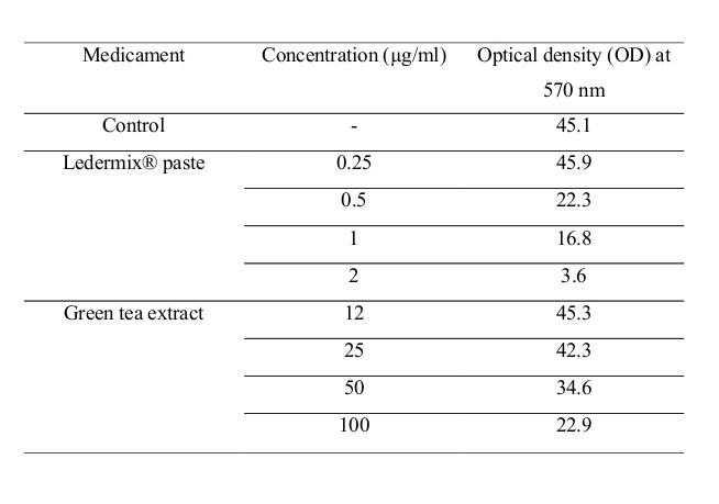
The basic objective of this experiment was determination of the proper concentration of the medicaments to be used in the second phase of the study. Therefore, MTT Cell Viability Assay was performed on U937 monocyte cell line. The results of the MTT Cell Viability Assay are summarized in Table 1. The final concentrations of green tea extract were 12, 25, 50 and 100 µg/ml while for Ledermix® paste the concentrations equaled 0.25, 0.5, 1 and 2 µg/ml. It was shown that green tea extract at the concentration of 100µg/ml and Ledermix® at the concentration of 0.5 µg/ml have the ability to destroy half the cells in the culture medium, and can cause 50% reduction in the formation of formazan and 50% reduction in the optical density (OD) at 570 nm (measured by spectrophotometer) compared with the control culture medium. These values represent the cytotoxic concentration (Inhibitory Concentration50 or IC50) of these medicaments. 0.25 µg/ml of Ledermix® paste and 12 µg/ml of green tea extract are the noncytotoxic concentrations, which were used in the subsequent phase of the experiment.
Table 1- Results of the MTT Cell Viability Assay performed on Ledermix® paste and green tea extract
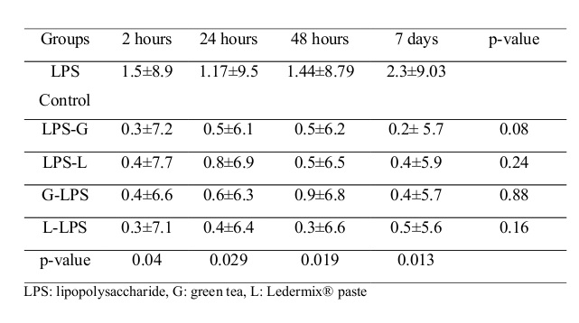
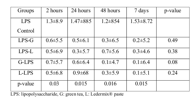
In the second phase of the study, the concentrations of IL-1β, IL-6, and TNF-α inflammatory mediators at the time intervals of 2, 24, and 48 hours and also after 7 days were assessed by ELISA kit in the four case groups and also in the control group. The concentrations of IL-1β, IL-6, and TNF-α inflammatory mediators are shown in Tables 2, 3 and 4, respectively. The levels of significance (p-value) of the results of one-way ANOVA in comparing the concentrations of IL-1β, IL-6, and TNF-α inflammatory mediators in the control group and the four study groups at the time intervals of 2, 24, and 48 hours and after 7 days are also presented in these tables.
Table 2- Average concentrations of IL-1β inflammatory mediator at different time intervals
Table 3- Average concentrations of IL-6 inflammatory mediator at different time intervals
Table 4- Average concentrations of TNF-α inflammatory mediator at different time intervals
Discussion
MTT Cell Viability Assay is a customary technique for assessing the biocompatibility of medications and different materials used in various medical fields (9), and also in endodontics (10, 11). Various researches have estimated the cytotoxic effect of green tea and its polyphenol derivatives such as Catechin on different cell lineages including carcinogenic cells (12). Studies have shown that intracanal medicaments such as calcium hydroxide and Ledermix® can have side effects on vital tissues and since these substances are in direct contact with periapical tissues, it is important to consider the biocompatibility of the intracanal medicaments upon selection (3, 13).
Currently, Ledermix® paste is a combination of 9% demeclocycline hydrochloride (antibiotic), and 1% and 2% triamcinolone acetonide (corticosteroid) in a polyethylene glycol base. The corticosteroid ingredient is used for mediating the pain and inflammation and suppressing the local immune responses, while the antibiotic part is used for preventing the bacterial growth. The therapeutic ingredients of Ledermix® have the ability to permeate through the dentinal tubules and cementum to reach periodontal and peri-radicular tissues (14).
Catechins (flavan-3-ols) are the main polyphenols of green tea. The four main catechins are: epigallocatechin-3-gallate (EGCG) (59% of total catechins), epigallocatechin (EGC) (19%), epicatechin-3-gallate (ECG) (13.6%) and epicatechin (EC) (6.4%) (15). Beside their direct role as antioxidants, the polyphenols have supplementary mechanisms by which they reduce oxidation level: 1- Binding of metal ions such as iron and copper and preventing their participation in the oxidation reactions (leading to the formation of hydroxyl radical). 2- Preventing the activation of redox-sensitive transcription factors that serve as mediators of inflammatory reactions. 3- Suppressing the oxidation stimulants such as induced nitric oxide synthase (iNOS), cyclooxygenase 2 (COX-2), lipoxygenase 2 (LOX-2) and xanthine oxidase. 4- Inducing the antioxidant enzymes such as glutathione S-transferase and superoxide dismutase (16).
In the first phase of this study, MTT assay was performed to determine the cytotoxicity of Ledermix® medicament and green tea extract on U937 monocyte cell line. 100µg/ml green tea extract and 0.5 µg/ml Ledermix® have the ability to destroy half the cells in the culture medium, and indicate the cytotoxic concentration (Inhibitory Concentration50 or IC50) of these medicaments. Considering the obtained results, higher concentrations of green tea extract in comparison with Ledermix® are needed to destroy half the cells in the culture medium. In other words, green tea extract is less cytotoxic and more biocompatible than Ledermix®. 0.25 µg/ml of Ledermix® and 12 µg/ml of green tea extract are the noncytotoxic concentrations which had an OD similar to that of the control group, and were used in the subsequent phase of the experiment.
In 1989, Taylor and colleagues evaluated the effect of Ledermix® paste on mouse fibroblasts in vitro. Ledermix® at the concentration of 0.001 µg/ml terminated cell mitosis, while 1 µg/ml concentration caused cell death (5). In the present study, Ledermix® paste at the concentration of 1 µg/ml caused 50% cell death, while based on the study by Taylor et al, 10-4 mg/ml concentration caused more than 80% of the cells to fail mitosis (5). These differences can be attributed to the use of different cell lines and different methods for the assessment of medication's impact. In the study by Taylor et al, mouse fibroblasts were observed while in the present study, monocyte cell lineage was assessed.
Han et al in 2009 assessed the cytotoxic effect of green tea polyphenols (GTPs) by MTT Assay. IC50 of GTPs extract in hamster pulmonary cells equaled 50 µg/ml, while on human leukemic cells, this value was over 100 µg/ml, which indicates low cytotoxicity (12). Overall, these findings concur with the results of the present study.
Lee et al in 2004 assessed the impact of Epigallocatechin-3-Gallate, a type of catechin derived from green tea, on human gastric epithelial cell line by the aid of MTT Assay. The results of this research showed that Epigallocatechin-3-Gallate has low cytotoxicity and it even has cytoprotective influence on the Helicobacter pylori-induced cells (17).
In the second phase of the present study, the anti-inflammatory properties of Ledermix® and green tea extract were estimated. The concentrations of IL-1β, IL-6, and TNF-α inflammatory mediators of the control group and four study groups were measured by ELISA kit at the time intervals of 2, 24, and 48 hours and after 7 days. Overall, based on the obtained results, time interval has no significant influence on the level of the mediators released from the monocytes that have been induced by different medicaments. Generally, both substances significantly decreased the concentration of IL-1β, IL-6, and TNF-α inflammatory mediators at the mentioned time intervals in comparison with the control group. Nevertheless, the efficacy of these substances decreased gradually over time, and showed no significant difference from the control group after one week.
The anti-inflammatory effect of these two substances has been assessed in previous studies, especially in the field of endodontics. The findings are generally in line with our results. The impact of Ledermix® has not been directly evaluated on Interleukins but Chen et al in 2008 showed that immediate application of Ledermix® intracanal medicament prevents root resorption of replanted dog teeth (18). Considering that IL-1β, IL-6 and TNF-α have a major role in hard tissue resorption (19, 20, 21), it can be presumed that Ledermix® may affect the released concentration of these interleukins.
A research by Thong et al in 2001 revealed that Ledermix® prevented inflammatory root resorption of replanted monkey teeth, and that its ability to induce healing in periodontal tissues is slightly higher than that of calcium hydroxide (22).
In an article published by Nakanishi and colleagues in 2010, the anti-inflammatory effect of catechin on human dental pulp cells was studied. They exposed the cells to bacteria-derived factors such as LPS, and afterwards the effect of EGCG and ECG (which are the main ingredients of green tea catechins) on the expression of pre-inflammatory cytokines was evaluated. The expression of IL-6 and IL-8 in the dental pulp cells exposed to LPS or PG was significantly decreased in the presence of EGCG and ECG (8).
In 2009, Lee and colleagues stated that a green tea extract, epigallocatechin-3-gallate, reduces the size of periapical lesions by inhibiting the expression of cysteine-rich61 in osteoblasts, and decreases the infiltration of macrophages into the lesions and prevents the development of apical periodontitis (23). Since the studied Interleukins can induce bone resorption, green tea extract may be useful in healing the periapical lesions according to the study by lee et al (17).
A research by Hirao et al in 2010 showed that tea catechins reduce inflammatory reactions in stimulated dental pulp cells (24). They also showed that the expression of IL-8 and PGE2 in the fibroblasts of stimulated human dental pulp cells is blocked by catechins (24).
In the present study, both Ledermix® and green tea extract showed distinctive anti-inflammatory effects compared with the control group. However, no significant differences were identified between these two substances.
Ledermix® paste inhibits phospholipase and PGE2 pathway and consequently impedes the production of other cytokines, probably due to its corticosteroid ingredient (triamcinolone) (25). Ledermix® reduces the activity of clast cells and ends root resorption process (3). Likewise, green tea contains active ingredients such as variety of catechin polyphenols which can probably block Cyclooxigenase-2 pathway and discontinue the production of inflammatory mediators especially TNF-α. It can also reduce the activity of osteoclasts (26). Accordingly, it seems that in addition to controlling the secretion of IL-1β, IL-6, and TNF-α mediators and hard tissue resorption, green tea can also mediate pain by blocking the byproducts of Cyclooxygenase-2.
Contemporary studies have proven that Ledermix® can induce tooth discoloration due to its demeclocycline ingredient, but no tooth discoloration induced by the regular concentrations of green tea extract has been reported. Perhaps, green tea lacks some of the disadvantages of Ledermix® paste such as higher cytotoxicity and probable tooth discoloration (27, 28). Therefore, green tea extract can be proposed as a suitable and highly biocompatible intracanal medicament to reduce inflammation. Nevertheless, more comprehensive studies of the other important characteristics of intracanal medicaments should be performed on green tea extract.
Conclusions
The results of the present research indicate that green tea extract is less cytotoxic and more biocompatible than Ledermix® paste. Moreover, both Ledermix® paste and green tea extract show anti-inflammatory effects. It is noteworthy that in particular cases, green tea extract shows higher anti-inflammatory effects in comparison with Ledermix®.
Full-Text: (838 Views)
Abstract
Background and aim: The purpose of this study is to compare the effect of Ledermix® paste and green tea extract on the concentration of inflammatory mediators.
Materials and methods: In this in-vitro experimental study, first, the noncytotoxic concentrations of Ledermix® medicament and green tea extract were determined by MTT assay. Afterwards, the concentration./files/site1/images/awt_thumbnails/2019.4(1)/1.jpgs of IL-1β, IL-6, and TNF-α inflammatory mediators were assessed by ELISA test in the four experimental groups and also in the control group at different time intervals of 2, 24, and 48 hours and after 7 days. One-way analysis of variance (ANOVA) was used for statistical analysis of the obtained raw data followed by Tukey's post-hoc test for pair comparisons.
/1.jpg)
Results: the noncytotoxic concentration of green tea extract and Ledermix® paste equaled 12 µg/ml and 0.25 µg/ml, respectively. Time interval had no significant effect on the concentration of the mediators. Generally, both substances significantly decreased the concentration of IL-1β and IL-6 inflammatory mediators in comparison with the control group.
Conclusions: Green tea extract is less cytotoxic and more biocompatible than Ledermix® paste. Both Ledermix® paste and green tea extract expressed anti-inflammatory effects.
Keywords: Cytotoxicity, Green tea extract, Inflammatory mediators, Ledermix® paste
Introduction
Progression of pulpitis to the periapical area and accumulation of microorganisms in the root canal system trigger innate and acquired immune responses and consequently, inflammatory mediators such as IL-1β, IL-6, and TNF-α are released in the periapical area (1). The release of these cytokines can lead to alveolar bone destruction and formation of periapical lesion (2).
The basis of a successful endodontic treatment which leads to ideal reparation is the identification and elimination of factors that induce apical periodontitis. Generally, mechanical and chemical debridement of the root canal and local application of intracanal medicaments (such as calcium hydroxide, Ledermix® paste and antibiotics) help eliminate the inflammation and repair the periapical tissues (1). Considering the controversies, none of these methods seems ideal and there is no consensus regarding their use. Nevertheless, reducing the inflammation to the minimum level is necessary to ensure successful treatment outcomes (1, 2).
Studies have displayed that intracanal medicaments such as calcium hydroxide and Ledermix® paste can have side effects on vital tissues and since these substances are in direct contact with periapical tissues, it is important to consider the biocompatibility of intracanal medicaments upon selection (3). Various studies have evaluated the antibacterial and bactericidal properties of green tea, as an intracanal medicament, on different bacterial species. The results proved that green tea extracts are probably beneficial as medicaments in infected root canals (4). But the anti-inflammatory effects of Ledermix® and green tea have not been compared. Therefore, in the present study MTT assay was conducted to determine the noncytotoxic concentration of Ledermix® paste and green tea extract in monocyte cell culture medium. Also, the effects of Ledermix® paste and green tea extract on the level of IL-1β, IL-6, and TNF-α inflammatory mediators in the cell culture medium were compared.
Materials and Methods
In this in vitro experimental study, U937 monocyte cell line culture media in 96-well plates were assessed (96 samples including 3 samples for each inflammatory mediator and one sample as the control), and purposive sampling method was used. In addition, the concentrations of different inflammatory mediators (IL-1β, IL-6, and TNF-α) were measured at different time intervals of 2, 24, and 48 hours and after 7 days in the presence of Ledermix® paste (Sigma, Australia) and green tea medicament (Soha Jissa, Iran).
All the experimental steps were performed by one operator after comprehensive training. All the samples were incubated at 37°C and 95% relative humidity in compliance with ISO standard 6876 (This international standard specifies the requirements and test methods for root canal (endodontic) sealing materials which set with or without moisture, and are used for permanent obturation of the root canal with or without the aid of obturating points/cones. It only covers the sealers that are intended for orthograde use i.e. a root filling placed from the coronal aspect of a tooth)(5)
In the first phase of the study, MTT assay was repeated three times to evaluate the effect of Ledermix® paste and green tea extract on monocyte viability. U937 monocyte cell lines obtained from the cell bank of Pasteur institute of Iran were cultured in Dulbecco's Modified Eagle's Medium (DMEM) containing 10% FBS (Fetal Bovine Serum), 2 mmol/l of L-glutamine and 1,000,000 U/L penicillin/streptomycin at 37°C, 5% Co2 and 95% humidity. After 24 hours of incubation with green tea extract at the concentrations of 12, 25, 50 and 100 µg/ml and Ledermix® at the concentrations of 0.25, 0.5, 1 and 2 µg/ml, the cells were washed twice with Phosphate Buffered Saline (PBS) according to the study by Taylor et al (6), and were incubated for one hour in fresh MMT culture medium at a dilution of 1 gr /l at 37°C. Afterwards, an equal volume of Lysis Buffer (pH=4.7) containing 200 gsDs/L, 50 %(v/v) N,N-Dimethylformamide was added to the culture medium and incubated for 24 hours.
Finally, the amount of formazan absorbance was measured by spectrophotometer (SHIMADZU, UV 2100) at 570 nm wavelength, and the noncytotoxic concentrations of green tea extract and Ledermix® were determined. The highest concentration of the medicament with an optical density (OD) similar to that of the control group was determined as the final concentration, which indicated the maximum cell activity unaffected by the medicaments in the culture medium.
In the second phase of the study, U937 monocyte cell lines were cultured in DMEM culture medium inside 96-well plates, containing 10% FBS and 2 mmol/l of L-glutamine and 1,000,000 U/L penicillin/streptomycin and were incubated at 37°C, 10% Co2, and 95% humidity. Four to five cell passages were transferred to fresh culture medium in 96-well plates with the capacity of 20000 cell/well.
In the first group (control), lipopolysaccharide (LPS) from E.coli K12 bacteria procured from Sigma-Aldrich Company (St. Louis, MO, USA) at the concentration of 1µg/ml was added to the monocyte cell culture medium and incubated (7). In the second group (case), the same concentration of LPS from E.coli K12 bacteria was placed on monocyte cell culture medium and after one hour of incubation, 1ml of green tea extract at the concentration of 12 µg/ml was supplemented (0.25 µg/ml concentration of Ledermix® and 12 µg/ml concentration of green tea were selected due to the similarity of the optical density with that of the control group). In the third group (case), the same concentration of LPS from E.coli K-12 bacteria was placed on the culture medium and after one hour of incubation, 1ml of Ledermix® at a concentration of 0.25 µg/ml was added. In the fourth group (case), initially 1ml of green tea extract at a concentration of 12 µg/ml was added to the culture medium and incubated. After one hour (8), 1µg/ml LPS from E.coli K12 bacteria was added. In the fifth group (case), 1ml of Ledermix® at a concentration of 12 µg/ml was added to the culture medium and incubated and one hour later, 1µg/ml LPS from E.coli K12 bacteria was supplemented. The samples were incubated and subsequently, the floating liquid surface over the culture media of the five experimental groups was collected at the time intervals of 2,24, and 48 hours and after 7days, and the concentrations of IL-1β, IL-6, and TNF-α inflammatory mediators were measured by ELISA kit ( Minneapolis, R&D systems, MN,USA). One-way analysis of variance (ANOVA) was used for statistical analysis of the obtained raw data followed by Tukey's post-hoc test for pair comparisons.
Results
The basic objective of this experiment was determination of the proper concentration of the medicaments to be used in the second phase of the study. Therefore, MTT Cell Viability Assay was performed on U937 monocyte cell line. The results of the MTT Cell Viability Assay are summarized in Table 1. The final concentrations of green tea extract were 12, 25, 50 and 100 µg/ml while for Ledermix® paste the concentrations equaled 0.25, 0.5, 1 and 2 µg/ml. It was shown that green tea extract at the concentration of 100µg/ml and Ledermix® at the concentration of 0.5 µg/ml have the ability to destroy half the cells in the culture medium, and can cause 50% reduction in the formation of formazan and 50% reduction in the optical density (OD) at 570 nm (measured by spectrophotometer) compared with the control culture medium. These values represent the cytotoxic concentration (Inhibitory Concentration50 or IC50) of these medicaments. 0.25 µg/ml of Ledermix® paste and 12 µg/ml of green tea extract are the noncytotoxic concentrations, which were used in the subsequent phase of the experiment.
Table 1- Results of the MTT Cell Viability Assay performed on Ledermix® paste and green tea extract
In the second phase of the study, the concentrations of IL-1β, IL-6, and TNF-α inflammatory mediators at the time intervals of 2, 24, and 48 hours and also after 7 days were assessed by ELISA kit in the four case groups and also in the control group. The concentrations of IL-1β, IL-6, and TNF-α inflammatory mediators are shown in Tables 2, 3 and 4, respectively. The levels of significance (p-value) of the results of one-way ANOVA in comparing the concentrations of IL-1β, IL-6, and TNF-α inflammatory mediators in the control group and the four study groups at the time intervals of 2, 24, and 48 hours and after 7 days are also presented in these tables.
Table 2- Average concentrations of IL-1β inflammatory mediator at different time intervals
Table 3- Average concentrations of IL-6 inflammatory mediator at different time intervals
Table 4- Average concentrations of TNF-α inflammatory mediator at different time intervals
Discussion
MTT Cell Viability Assay is a customary technique for assessing the biocompatibility of medications and different materials used in various medical fields (9), and also in endodontics (10, 11). Various researches have estimated the cytotoxic effect of green tea and its polyphenol derivatives such as Catechin on different cell lineages including carcinogenic cells (12). Studies have shown that intracanal medicaments such as calcium hydroxide and Ledermix® can have side effects on vital tissues and since these substances are in direct contact with periapical tissues, it is important to consider the biocompatibility of the intracanal medicaments upon selection (3, 13).
Currently, Ledermix® paste is a combination of 9% demeclocycline hydrochloride (antibiotic), and 1% and 2% triamcinolone acetonide (corticosteroid) in a polyethylene glycol base. The corticosteroid ingredient is used for mediating the pain and inflammation and suppressing the local immune responses, while the antibiotic part is used for preventing the bacterial growth. The therapeutic ingredients of Ledermix® have the ability to permeate through the dentinal tubules and cementum to reach periodontal and peri-radicular tissues (14).
Catechins (flavan-3-ols) are the main polyphenols of green tea. The four main catechins are: epigallocatechin-3-gallate (EGCG) (59% of total catechins), epigallocatechin (EGC) (19%), epicatechin-3-gallate (ECG) (13.6%) and epicatechin (EC) (6.4%) (15). Beside their direct role as antioxidants, the polyphenols have supplementary mechanisms by which they reduce oxidation level: 1- Binding of metal ions such as iron and copper and preventing their participation in the oxidation reactions (leading to the formation of hydroxyl radical). 2- Preventing the activation of redox-sensitive transcription factors that serve as mediators of inflammatory reactions. 3- Suppressing the oxidation stimulants such as induced nitric oxide synthase (iNOS), cyclooxygenase 2 (COX-2), lipoxygenase 2 (LOX-2) and xanthine oxidase. 4- Inducing the antioxidant enzymes such as glutathione S-transferase and superoxide dismutase (16).
In the first phase of this study, MTT assay was performed to determine the cytotoxicity of Ledermix® medicament and green tea extract on U937 monocyte cell line. 100µg/ml green tea extract and 0.5 µg/ml Ledermix® have the ability to destroy half the cells in the culture medium, and indicate the cytotoxic concentration (Inhibitory Concentration50 or IC50) of these medicaments. Considering the obtained results, higher concentrations of green tea extract in comparison with Ledermix® are needed to destroy half the cells in the culture medium. In other words, green tea extract is less cytotoxic and more biocompatible than Ledermix®. 0.25 µg/ml of Ledermix® and 12 µg/ml of green tea extract are the noncytotoxic concentrations which had an OD similar to that of the control group, and were used in the subsequent phase of the experiment.
In 1989, Taylor and colleagues evaluated the effect of Ledermix® paste on mouse fibroblasts in vitro. Ledermix® at the concentration of 0.001 µg/ml terminated cell mitosis, while 1 µg/ml concentration caused cell death (5). In the present study, Ledermix® paste at the concentration of 1 µg/ml caused 50% cell death, while based on the study by Taylor et al, 10-4 mg/ml concentration caused more than 80% of the cells to fail mitosis (5). These differences can be attributed to the use of different cell lines and different methods for the assessment of medication's impact. In the study by Taylor et al, mouse fibroblasts were observed while in the present study, monocyte cell lineage was assessed.
Han et al in 2009 assessed the cytotoxic effect of green tea polyphenols (GTPs) by MTT Assay. IC50 of GTPs extract in hamster pulmonary cells equaled 50 µg/ml, while on human leukemic cells, this value was over 100 µg/ml, which indicates low cytotoxicity (12). Overall, these findings concur with the results of the present study.
Lee et al in 2004 assessed the impact of Epigallocatechin-3-Gallate, a type of catechin derived from green tea, on human gastric epithelial cell line by the aid of MTT Assay. The results of this research showed that Epigallocatechin-3-Gallate has low cytotoxicity and it even has cytoprotective influence on the Helicobacter pylori-induced cells (17).
In the second phase of the present study, the anti-inflammatory properties of Ledermix® and green tea extract were estimated. The concentrations of IL-1β, IL-6, and TNF-α inflammatory mediators of the control group and four study groups were measured by ELISA kit at the time intervals of 2, 24, and 48 hours and after 7 days. Overall, based on the obtained results, time interval has no significant influence on the level of the mediators released from the monocytes that have been induced by different medicaments. Generally, both substances significantly decreased the concentration of IL-1β, IL-6, and TNF-α inflammatory mediators at the mentioned time intervals in comparison with the control group. Nevertheless, the efficacy of these substances decreased gradually over time, and showed no significant difference from the control group after one week.
The anti-inflammatory effect of these two substances has been assessed in previous studies, especially in the field of endodontics. The findings are generally in line with our results. The impact of Ledermix® has not been directly evaluated on Interleukins but Chen et al in 2008 showed that immediate application of Ledermix® intracanal medicament prevents root resorption of replanted dog teeth (18). Considering that IL-1β, IL-6 and TNF-α have a major role in hard tissue resorption (19, 20, 21), it can be presumed that Ledermix® may affect the released concentration of these interleukins.
A research by Thong et al in 2001 revealed that Ledermix® prevented inflammatory root resorption of replanted monkey teeth, and that its ability to induce healing in periodontal tissues is slightly higher than that of calcium hydroxide (22).
In an article published by Nakanishi and colleagues in 2010, the anti-inflammatory effect of catechin on human dental pulp cells was studied. They exposed the cells to bacteria-derived factors such as LPS, and afterwards the effect of EGCG and ECG (which are the main ingredients of green tea catechins) on the expression of pre-inflammatory cytokines was evaluated. The expression of IL-6 and IL-8 in the dental pulp cells exposed to LPS or PG was significantly decreased in the presence of EGCG and ECG (8).
In 2009, Lee and colleagues stated that a green tea extract, epigallocatechin-3-gallate, reduces the size of periapical lesions by inhibiting the expression of cysteine-rich61 in osteoblasts, and decreases the infiltration of macrophages into the lesions and prevents the development of apical periodontitis (23). Since the studied Interleukins can induce bone resorption, green tea extract may be useful in healing the periapical lesions according to the study by lee et al (17).
A research by Hirao et al in 2010 showed that tea catechins reduce inflammatory reactions in stimulated dental pulp cells (24). They also showed that the expression of IL-8 and PGE2 in the fibroblasts of stimulated human dental pulp cells is blocked by catechins (24).
In the present study, both Ledermix® and green tea extract showed distinctive anti-inflammatory effects compared with the control group. However, no significant differences were identified between these two substances.
Ledermix® paste inhibits phospholipase and PGE2 pathway and consequently impedes the production of other cytokines, probably due to its corticosteroid ingredient (triamcinolone) (25). Ledermix® reduces the activity of clast cells and ends root resorption process (3). Likewise, green tea contains active ingredients such as variety of catechin polyphenols which can probably block Cyclooxigenase-2 pathway and discontinue the production of inflammatory mediators especially TNF-α. It can also reduce the activity of osteoclasts (26). Accordingly, it seems that in addition to controlling the secretion of IL-1β, IL-6, and TNF-α mediators and hard tissue resorption, green tea can also mediate pain by blocking the byproducts of Cyclooxygenase-2.
Contemporary studies have proven that Ledermix® can induce tooth discoloration due to its demeclocycline ingredient, but no tooth discoloration induced by the regular concentrations of green tea extract has been reported. Perhaps, green tea lacks some of the disadvantages of Ledermix® paste such as higher cytotoxicity and probable tooth discoloration (27, 28). Therefore, green tea extract can be proposed as a suitable and highly biocompatible intracanal medicament to reduce inflammation. Nevertheless, more comprehensive studies of the other important characteristics of intracanal medicaments should be performed on green tea extract.
Conclusions
The results of the present research indicate that green tea extract is less cytotoxic and more biocompatible than Ledermix® paste. Moreover, both Ledermix® paste and green tea extract show anti-inflammatory effects. It is noteworthy that in particular cases, green tea extract shows higher anti-inflammatory effects in comparison with Ledermix®.
Type of Study: Original article |
Subject:
Oral & maxillofacial surgery
References
1. Law A, Messer H. An evidence-based analysis of the antibacterial effectiveness of intracanal medicaments. J Endod 2004;30(10):689.94.
2. Silva TA, Garlet GP, Fukada SY, Silva JS, Cunha FQ. Chemokines in oral inflammatory diseases: apical periodontitis and periodontal disease. J Dent Res 2007;86(4):306-19.
3. Hauman CH, Love RM. Biocompatibility of dental materials used in contemporary therapy: a review. Part1. Intracanal drugs and substances. Int Endod J 2003;36(2):75-85.
4. Narotzki B, Rezinck AZ, Aizenbud D, Levy Y. Green Tea: A promising natural product in oral health. Arch Oral biol 2001;57(5);429-35.
5. Chng HK, Islam I, Yap AU, Tong YW, Koh ET. Properties of a new root-end filling material. J Endod 2005;31(9):665-8.
6. Nakanishi T, Mukai K, Yumoto H, Hirao K, Hosokawa Y, Matsuo T. Anti-inflammatory effect of catechin on cultured human dental pulp cells affected by bacteria-derived factors. Eur J Oral Sci 2010;118(2):145-50.
7. Bilmin k, Kopczynska B, Grib P. Influence of serum and albumin on the in vitro anandamide cytotoxicity toward C6golima cells assessed by the MTT cell viability assay: implications for the methodology of the MTT tests. Folia Neuropathol 2013;51(1);44-50.
8. Osorio RM, Hefti A, Vertucci FJ, Shawley AL. Cytotoxicity of endodontic materials. J Endod 1998;24(2):91-6.
9. Day PF, Duggal MS, High AS, Robertson A, Gregg TA, Ashley PF, et al. Discoloration of teeth after avulsion and replantation: results from a multicenter randomized controlled trial. J Endod 2011;37(8):1052-7.
10. Han DH, Jeong JH, Kim JH. Anti-proliferative and apoptosis induction activity of tea polyphenols on human promyelocytic Leukemia HL-60 cells. Antic ancer Res2009;29(4):1417-21.
11. EIdeniz AU, Mustafa K, Orstavik D, Dahl JE. Cytotoxicity of new resin-calcium hydroxide and silicone-based root canal sealers on fibroblasts derived from human gingiva and L929 cell lines. Int Endod J 2007;40(5):329-37.
12. Athansseadis B, Abottot PV, Wash LJ. The use of Calcium Hydroxide , antibiotics and biocides as antimicrobical medicaments in endodontics. Aust Dent J 2007;52(1 Suppl):S64-82.
13. McKay DL, Blumberg JB. The role of tea in human health: an update. J Am Coll Nutr 2002;21(1):1-13.
14. Cabrera C, Artacho R, Giménez R. Beneficial effects of green tea--a review. J Am Coll Nutrn 2006;25(2):79-99.
15. Taylor MA, Humen WR, Heithersay GS. Some effects of Ledermix paste and Pulpdent paste on mouse fibroblasts and on bacteria in vitro. Endod Dent Traumatol 1989;5(6):266-73.
16. Lee KM, Yeo M, Chouse JS, Jin JH, Park SJ , Cheong JY, et al. Protective mechanism of epigallocatechin-3-gallate against Helicobacter pylori-induced gastric epithelial cytotoxicity via the blockage of TLR-4 signaling. Helicobacter 2004;9(6):632-42.
17. Chen H, Teixeira FB, Ritter Al, Levin L, Trope M. The effect of intracanal anti-inflammatory medicaments on external root resorption of replanted dog teeth after extended extra-oral dry time. Dent Traumatol 2008;24(1):74-8.
18. Pezelj-Ribarić S, Magasić K, Prpić J, Miletić I, Karlović Z.Tumor necrosis factor-alpha in periapical tissue exudates of teeth with a periodontitis. Mediators Inflamm 2007;2007:69416.
19. Shimauchi H, Takayama S, Imai-Tanaka T, Okada H. Balance of interleukin-1 beta and interleukin-1 receptor antagonist in human periapical lesions. J Endod 1998;24(2):116-9.
20. Balto K, Sasaki H, Stashenko P. Interleukin-6 deficiency increases inflammatory bone destruction. Infect Immun 2001;69(2):744-50.
21. Thong YL, Messer HH, Siar CH, Saw LH. Periodontal response to two intracanal medicaments in replanted monkey incisors. Dent Traumatol 2001;17(6):254_9.
22. Lee YL, Hong CY, Kok SH, Hou KL, Lin YT, Chen MH, et al. An extract of green tea, epigallocatechin-3-gallate, reduces periapical lesions by inhibiting cysteine-rich61 expression in osteoblasts. J Endod 2009;35(2):206-11.
23. Hirao K, Yumoto H, Nakanishi T, Mukai K, Takahashi K, Takegawa D, et al. Tea catechins reduce inflammatory reactions via mitogen-activated protein Kinase pathways in toll-like receptor 2 ligand-stimulated dental pulp cells. Life Sci2010 24;86(17-18):654-60.
24. Vane JR. Antiinflammatory drugs and the many mediators of inflammation. Int J Tissue React 1987;9(1):1-14.
25. Jung IH, Yun JH, Cho AR, Kim CS, Chung WG, Choi SH. Effect of (-)-epigallocatechin-3-gallate on maintaining the periodontal ligament cell viability of avulsed teeth: a preliminary study. J Periodontal Implant Sci 2011;41(1):10-6.
26. Lenherr P, Allgayer N, Weiger R, Filippi A, Attin T, Krastl G. Tooth discoloration induced by endodontic materials: a laboratory study. Int Endod J2012;45(10):942-9.
27. Thomson AD, Athanassiadis B, Kahler B, Walsh L. Tooth discoloration: staining effects of various sealers and medicaments. Aust Endod J 2012;38(1):2-9.
Send email to the article author
| Rights and permissions | |
 |
This work is licensed under a Creative Commons Attribution-NonCommercial 4.0 International License. |
